Institution: Ann & Robert H. Lurie Children's Hospital of Chicago
Additional authors:Katrin Carlson Leuer, PhD, Allen Lamb, PhD, Maria Proytcheva, MD
Session: Myeloid and lymphoid neoplasms with eosinophilia and abnormalities of PDGFRA, PDGFRB, or FGFR1
HISTORY
16-year-old male previously diagnosed with T lymphoblastic lymphoma in 03/2009, due to start cycle 10 of maintenance chemotherapy on 01/05/2012, with evolving macrocytic anemia, thrombocytopenia and leukocytosis; bone marrow aspiration performed to rule-out relapse or therapy-related pathology; subsequent bone marrow aspiration and trephine biopsy done on 01/12/2012
DETAILS
1. Left inguinal lymph node biospy from 2009 (formalin-fixed): 1.4 cm lymph node with portions submitted for flow cytometry, conventional cytogenetic analysis and microscopic examination; histologic sections demonstrate effacement of the architecture by a diffuse proliferation of small to intermediate-sized blasts with scattered macrophages and a few residual germinal centers
2. Right posterior iliac crest bone marrow trephine biopsy from 2012 (formalin-fixed): 1.7 cm long core biopsy with mildly hypercellular (70% cellular) bone marrow demonstrating myeloid hyperplasia, shift to immaturity with 16% blasts, and 38% eosinophilsIMMUNOHISTOCHEMISTRY AND FLOW CYTOMETRY
1. Left inguinal lymph node biospy from 2009: CD45 dim positive, CD34 negative, TdT positive, CD2 positive, surface CD3 negative but cytoplasmic CD3 positive, CD5 positive, CD7 dim positive, CD4 and CD8 dual positive, CD56 positive T lymphoblasts representing 95% of total cellular events; staging bone marrow done the same day showed 3% T lymphoblasts (flow cytometry); CD3 and TdT positive, CD20 negative infiltrate by immunohistochemistry
2. Right posterior iliac crest bone marrow aspirates from 2012: Large population of eosinophils but no residual T lymphoblastic lymphomaCYTOGENETIC FINDINGS
t(7;8)(q34;p11) was identified in the 2009 lymph node and staging marrow, as well as the bone marrow examinations from 2012 (which showed a myeloid neoplasm but no flow cytometric immunophenotypic evidence of T lymphoblastic lymphoma). Interphase FISH studies on the latter confirmed FGFR1 rearrangement.
INTERESTING FEATURES
Presentation as T lymphoblastic lymphoma with low level bone marrow involvement at diagnosis in 2009, and subsequent presentation in 2012 as a myeloid neoplasm (with evolving macrocytic anemia, thrombocytopenia and leukocytosis) with increased blasts and evolving toward acute myeloid leukemia
PROPOSED DIAGNOSIS
Myeloid and lymphoid neoplasm with FGFR1 abnormality (T lymphoblastic lymphoma in 2009; myeloid neoplasm in chronic/accelerated phase in 2012)
CONSENSUS DIAGNOSIS
Myeloid and lymphoid neoplasm with FGFR1 rearrangement, t(7;8)(q34;p11); presenting with T lymphoblastic lymphoma and subsequent myeloproliferative neoplasm with increased blasts and eosinophilia
| Case history | 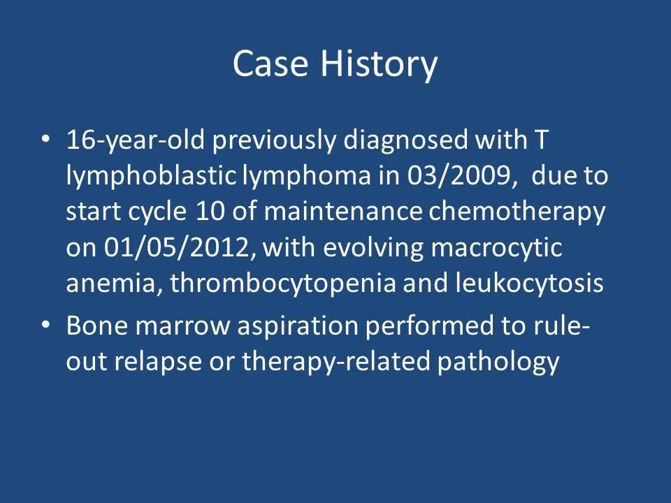 |
| Recent CBC/differential data from the months prior to bone marrow examination | 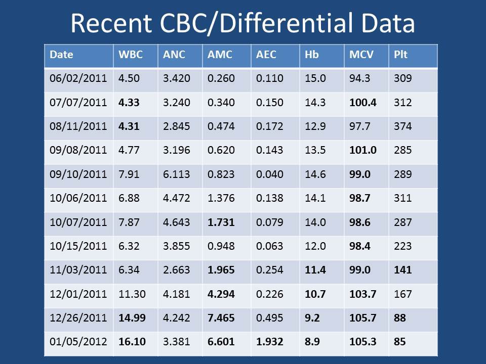 |
| 2012 peripheral blood smear showing shift to immaturity, monocytosis and eosinophilia | 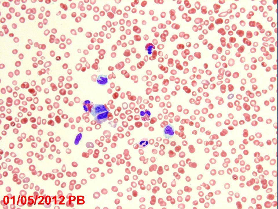 |
| 2012 bone marrow aspirate with mildly increased blasts but markedly increased eosinophils | 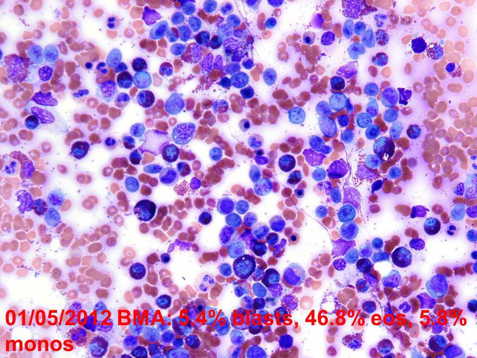 |
| 2012 flow cytometry of bone marrow aspirate with large population of eosinophils but no evidence of previously diagnosed T lymphoblastic lymphoma | 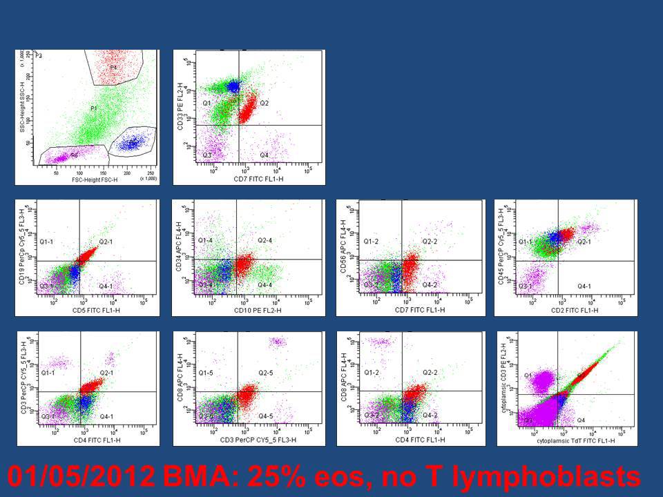 |
| 2012 peripheral blood smear (one week later) with similar findings | 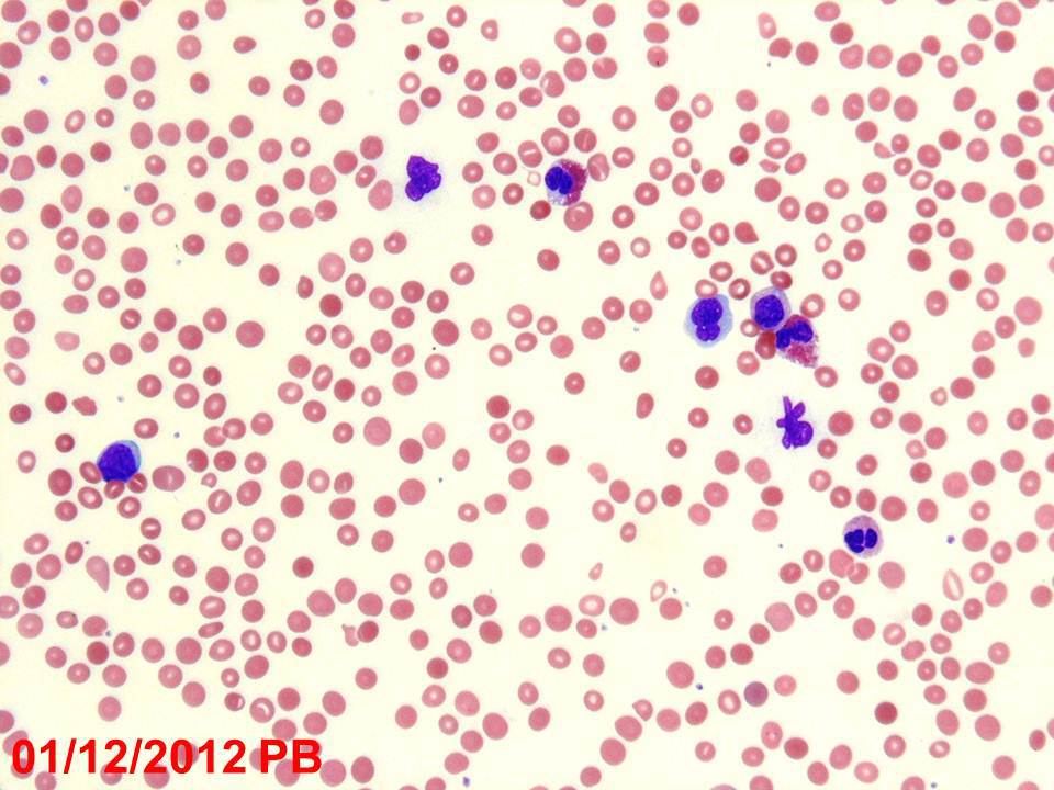 |
| 2012 bone marrow aspirate (one week later) with fewer eosinophils but a greater number of blasts | 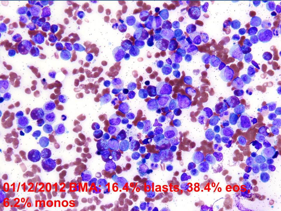 |
| 2012 bone marrow trephine biopsy (one week later) with 70% cellularity, myeloid hyperplasia, shift to immaturity and eosinophilia | 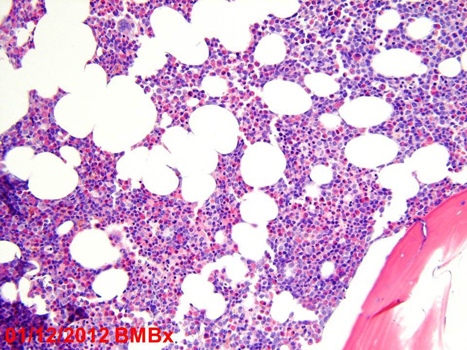 |
| Additional clinical information from initial presentation with T lymphoblastic lymphoma in 2009 | 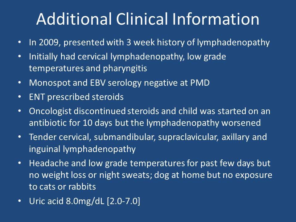 |
| 2009 touch imprint of lymph node with blasts and an eosinophil |  |
| 2009 lymph node H&E with inset showing greater histologic detail |  |
| 2009 lymph node flow cytometry results |  |
| 2009 peripheral blood smear remarkable only for eosinophilia | 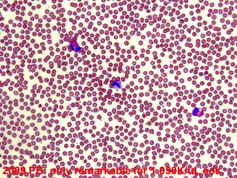 |
| 2009 staging bone marrow with 16% blasts (mostly hematogones) and 8% eosinophils |  |
| 2009 bone marrow flow cytometry with about 15% hematogones but only 3% T lymphoblasts |  |
| Karyogram showing t(7;8)(q22;p11.2), present in the lymph node and marrow in 2009, and in the marrow in 2012 | 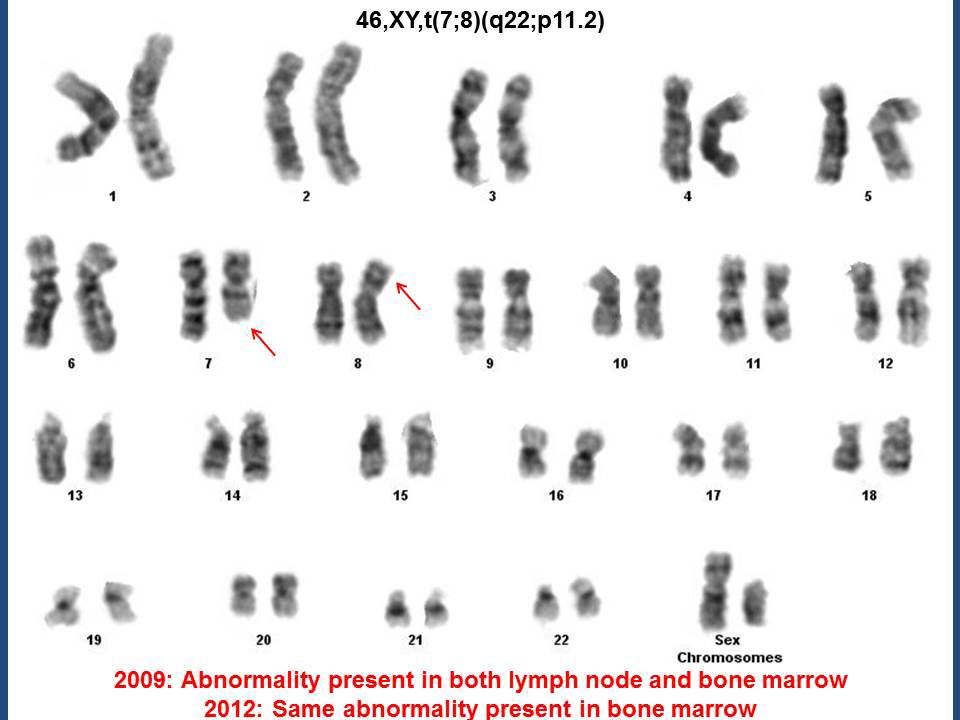 |
| FISH confirming FGFR1 rearrangement |  |