Institution: University of Michigan
Additional authors:Juan Gomez-Gelvez, M.D.
Session: Therapy-related myeloid neoplasms
HISTORY
27-year-old woman treated for an undifferentiated sarcoma (likely Ewing's) involving the soft tissue of her arm. She had the lesion excised and received 12 rounds of chemotherapy incuding vincristine and adriamycin. Eight months after treatment, unusual cells were noted in the peripheral blood (5%) with the differential diagnosis of ciculating Ewing's cells versus blasts secondary to colony-stimulating factor therapy versus monocytes. Alpha napthyl butyrate esterase on a cytospin preparation confirmed that the cells were monocytes. The cells disappeared in subsequent blood smears and the patient was observed. One month later, she developed gingival bleeding and easy bruising. Review of the peripheral blood smear revealed leukocytosis and thrombocytopenia with numerous blasts and promonocytes consistent with therapy-related acute myeloid leukemia. The sarcoma slides were re-examined and found to be CD43 negative (to rule out myeloid sarcoma).
DETAILS
Bone marrow unilateral core biopsy (formalin-fixed): Approximately 90% cellular with a predominance of blasts. Background hematopoiesis is markedly decreased. Rare megakaryocytes are present with predominantly normal morphology.
Bone marrow aspirate smear: Numerous monoblasts are present (81%). Erythroid precursors are reduced with mild dyserythropoiesis in the form of nuclear contour irregularity. Occasional dysplastic maturing granulocytes with hypolobated nuclei and abnormally condensed chromatin are seen. Blasts show evidence of erythrophagocytosis. Occasional hypolobated megakaryocytes are present.Peripheral blood smear: Leukocytosis and thrombocytopenia with circulating blasts and promonocytes.IMMUNOHISTOCHEMISTRY AND FLOW CYTOMETRY
Immunohistochemistry/flow cytometric findings: Flow cytometry demonstrated a population of cells with moderate side scatter and moderate CD45 expression. The cells expressed CD4 (dim), CD11b (moderate, subset), CD11c (dim-moderate), CD13 (dim), CD14 (moderate), CD33 (moderate), CD36 (dim-moderate), CD38 (dim-moderate), CD56 (dim, subset), and CD64 (moderate).
CYTOGENETIC FINDINGS
46,XX,t(8;16)(p11.2;p13.3)[20]
MOLECULAR FINDINGS
Positive for FLT3 codon D835 mutation.
No evidence of CEBPA mutation, IDH1/IDH2 mutations, KIT mutation, or NPM1 mutation.INTERESTING FEATURES
The patient has t(8;16), which is a rare form of therapy-related AML. She had a short latency from her therapy to diagnosis (9 months). She had unusual cells that were difficult to characterize morphologically in her blood prior to diagnosis, which raised the differential diagnosis of circulating Ewing's sarcoma cells versus atypical/immature monocytes. The fact that she had a Ewing's sarcoma is also interesting since myeloid sarcoma can express FLI-1 and CD99 and this can be a diagnostic pitfall.
PROPOSED DIAGNOSIS
Therapy-related acute myeloid leukemia.
CONSENSUS DIAGNOSIS
Therapy-related myeloid neoplasm, acute myeloid leukemia with t(8;16)(p11.2;p13.3)
| Hemophagocytosis in aspirate smear |  |
| Hemophagocytosis in aspirate smear |  |
| Dysplastic granulocytes and blasts in aspirate smear |  |
| Peripheral blood at the time of diagnosis | 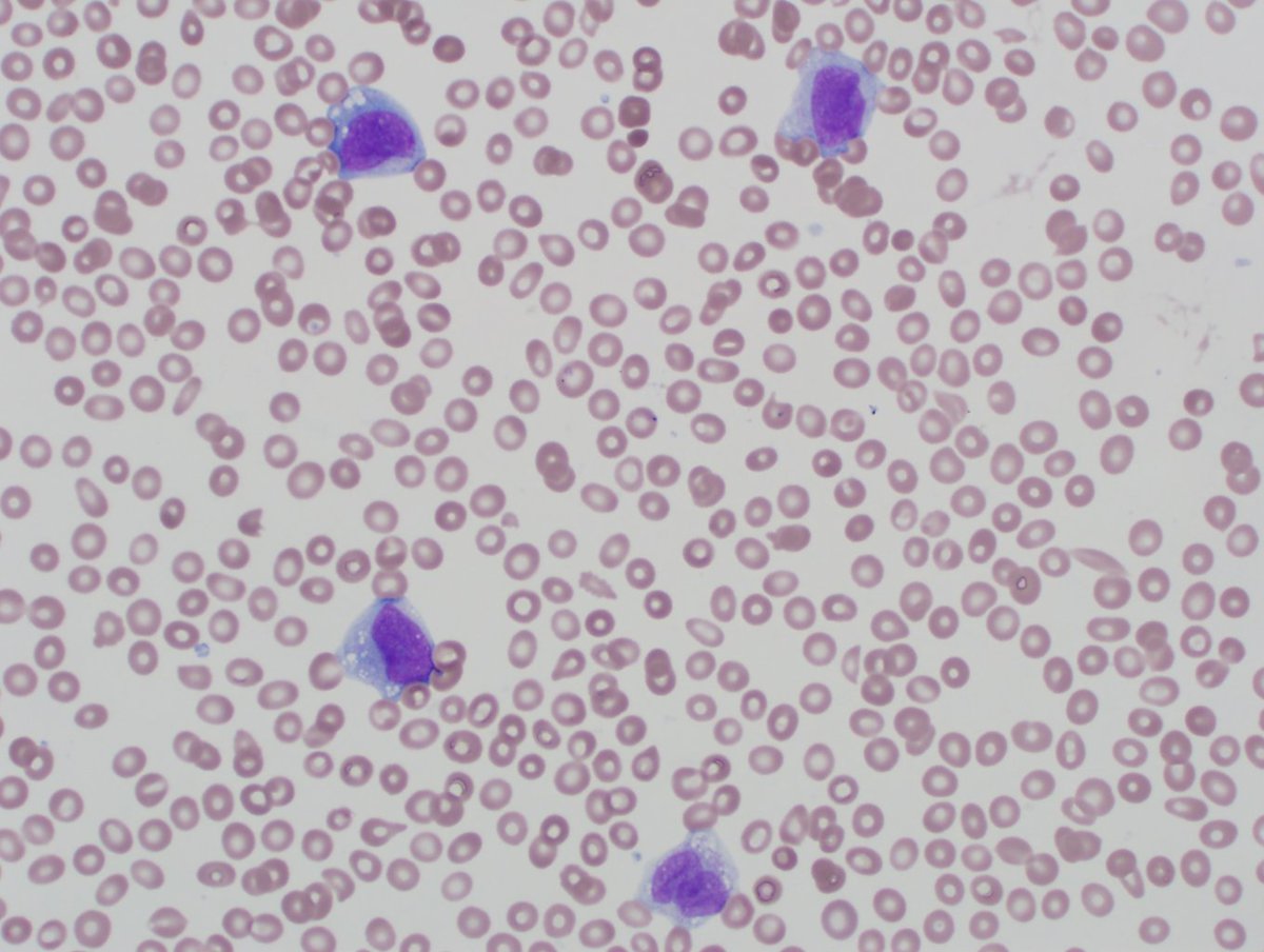 |
| Soft tissue mass from left arm diagnosed as undifferentiated sarcoma |  |
| High power sarcoma |  |
| Atypical cells in blood one month prior to diagnosis |  |