Institution: Department of Hematopathology, Moffitt Cancer Center
Additional authors: Xiaohui Zhang, Lynn Moscinski, Jeffrey Lancet and Ling Zhang
Session: B Lymphoblastic Leukemia/Lymphoma
HISTORY
64-year-old Hispanic male presented with weight loss of 25 pounds and moderate fatigue. The initial laboratory investigation noted that his white blood count was 51.3 x 10^9 /L, with platelets of 442 x 10^9 /L.. Miscroscopic examination of his peripheral blood smear showed a microcytic and normochromic anemia, left-shifted neutrophilic maturation with 2% circulating blasts, and moderate mature monocytosis. Platelets are normal in number and morphology. Imaging study exhibited mild splenomegaly. A diagnosis of B-lymphoblastic leukemia was rendered after a careful examination of bone marrow aspirate/biopsy in conjunction with the following ancillary studies.The patient was received hyper-CVAD treatment, followed by G-CSF. Two month after the initial diagnosis, his peripheral blood still showed moderate normocytic and normochromic anemia (RBC 3.48 mil/ul, Hgb 10 g/dl) with moderate anisopoikilocytosis including 1+ polychromasia and 1+ poikilocytosis. leukocytosis with left-shifted granulocytic maturation. Monocytes are increased in number, accounting for 11% of differential. However, no circulating lymphoblasts were noted. Splenomegaly was persistent. A subsequent bone marrow biopsy and additional studies were conducted to illustrate his disease process. At three month after the initial diagnosis, the patient finished last cycle of hyper-CVAD, his peripheral blood still showed moderate normocytic, normochromic anemia (RBC 3.42mil/ul, Hgb 9.7g/dl) with mild anisopoikilocytosis including 1+ polychromasia and 1+ poikilocytosis. Tear drops cells are rarely seen. Leukocytes are markedly decreased in number with occasional circulating blasts (less than 1%). We received a bone marrow biopsy for further evaluation.
DETAILS
Biopsy site: Posterior Iliac crest, bone marrow aspirates and biopsies Fixation: 10% neutroal buffered formalin The initial bone marrow aspirate smear showed 48% of lymphoblasts. The bone marrow core biopsy was 100% cellularity, the marrow was extensively infiltrated by sheets of lymphoblasts, nearly 50% of marrow space. The background of trilineage hematopoiesis was focally noted. Atypical megakaryocytic clustering was present. Reticulin stain showed moderate reticulin fibrosis (2+). Iron stain indicatedabsence of stainable iron. In contrast to his initial diagnosis, the bone marrow biopsy, performed at two month after initial diagnosis and hyper-CVAD, G-CSF treatment, showed hyperplastic marrow (85% cellularity) with overtly atypical megakaryocytic hyperplasia including hyperchromasia, disjointed nuclei and syncytial clustering, left shifted myelopoiesis and excess blasts (4%). Diffuse, moderate reticulin fibrosis was evident (3+ reticulin fibrosis). No overt lymphoblasts identified. At three month after the initial diagnosis (The patient finished last cycle of hyper-CVAD), his Bone marrow still showed hyperplastic marrow (90% cellularity). Megakaryocytes are atypical in morphology and form syncythial clustering. The number of megakaryocytes is obviously increased. Myeloid precursors are identified interstitially with slight increase in blasts (4.5%). There is erythroid hyperplasia. Reticulin stain is mild, 1+ and focally up to 2+.
IMMUNOHISTOCHEMISTRY AND FLOW CYTOMETRY
The first bone marrow biopsy showed increased lymphoblasts (approximately 50% of marrow cellularity), which were highlighted by PAX-5 and TdT immunohistochemical stains.
The corresponding flow cytometry showed approximately 30% B-lymphoblasts with expression of CD19, CD10, CD22, CD20, and TdT.
Two month after the initial diagnosis, Immunohistochemical stain revealed an increase in CD34 and CD117 positive cells (5-15%, focally formatting loose clusters up to 20%). There were rare CD19 positive B-cells and approximately 2-3% TdT positive precursors.
At three month after the initial diagnosis (The patient finished last cycle of hyper-CVAD), immunohistochemical stain revealed the CD34 and CD117 positive immature precursors are estimated 5% and 3-10%, respectively. Scattered CD10+/PAX-5+/partial CD20+ immature B-cells. MPO and Spectrin highlight both myeloid and erythroid precursors with erythroid predominance.
CYTOGENETIC FINDINGS
The cytogenetics by conventional karyotyping reported normal male karyotype, 46, XY[20] in all bone marrow biopsies.
After review of the second bone marrow biopsy (two month after initial diagnosis) a SNP array study was required for further evaluation, which showed an extended contiguous region of allele homozygosity (53 Mb) observed at the terminus of 1p which is consistent with acquired segmental uniparental disomy (aUPD) 1p with a dosage equivalence of 100% of the sample.
MOLECULAR FINDINGS
PCR studies performed on the first bone marrow specimen were positive for clonal IgH and IgK gene rearrangement. After review of the second bone marrow biopsy (two months after initial diagnosis) additional molecular study was required for further evaluation. At three month after the initial diagnosis, we received a molecular study report that showed positive for MPL W515S homozygous mutation by PCR method.
INTERESTING FEATURES
Here, we report a rare concurrence of B-acute lymphoblastic lymphoma with prefibrotic phase of primary myelofibrosis in patient with 1p uniparental disomy (UPD1p) as well as MPL (W515S) gene mutation. Acquired uniparental disomy is also called copy-neutral LOH, which is one mechanism for obtaining loss of heterozygosity (LOH) that can affect multiple chromosomes noted to be remarkably frequent in myeloid disorder patients. aUPD 1p is not uncommon in MDS, CMML or AML. In aUPD 1p, the mechanism has been linked to gene mutations in MPL or NRAS, within the homozygotic region. In our case, the aUPD 1p is indeed associated with MPL W515S mutation, which helped us make a final diagnosis. aUPD is rarely been studied in acute lymphoblastic leukemia. It is uncertain if aUPD 1p also plays a critical role in initiation and development of B-lymphoblastic leukemia in this case.
References:
1. O'Keefe C, McDevitt MA,Maciejewski JP. Copy neutral loss of heterozygosity: a novel chromosomal lesion in myeloid malignancies. Blood. 2010; 115:2731-2739.
2. Szpurka H, Gondek LP, Mohan SR, Hsi ED, Theil KS,Maciejewski JP. UPD1p indicates the presence of MPL W515L mutation in RARS-T, a mechanism analogous to UPD9p and JAK2 V617F mutation. Leukemia. 2009; 23:610-614.
PROPOSED DIAGNOSIS
Concurrent B-lymphoblastic leukemia and myeloproliferative neoplasm, favor prefibrotic phase primary myelofibrosis
CONSENSUS DIAGNOSIS
Concurrent B-acute lymphoblastic leukemia and myeloproliferative neoplasm, not otherwise specified
| Title |  |
| History |  |
| Initial BM Smear |  |
| Initial BM Biopsy HE-1 |  |
| Initial BM Biopsy HE-2 |  |
| Initial BM Biopsy Reticulin |  |
| Initial BM Biopsy Iron |  |
| Initial BM Biopsy PAX-5 | 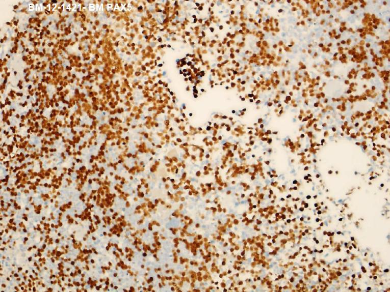 |
| Initial BM Biopsy TdT |  |
| First Diagnosis |  |
| Second BM biopsy HE |  |
| Second BM Biopsy Reticulin | 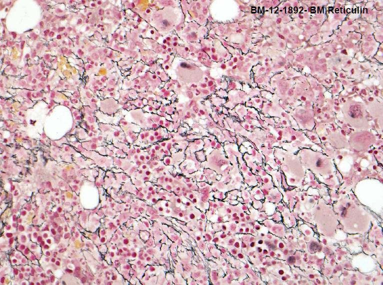 |
| Second BM Biopsy Iron | 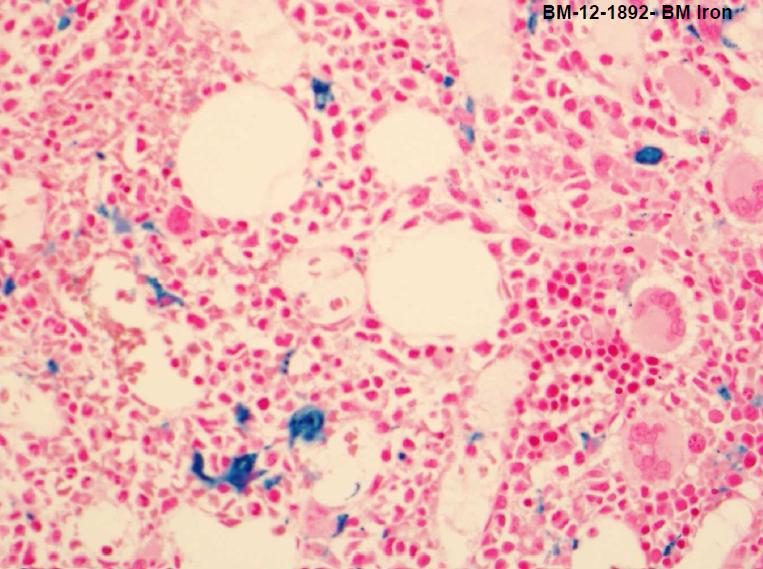 |
| Third BM biopsy HE |  |
| Third BM Biopsy Reticulin |  |
| Third BM Biopsy Iron | 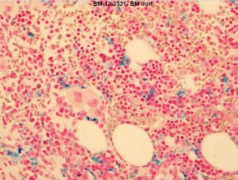 |
| Third BM Biopsy Spectrin | 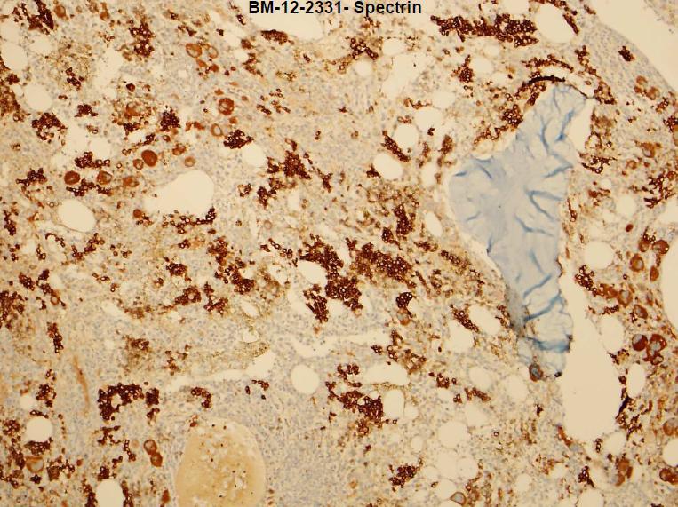 |
| Third BM Biopsy PAX-5 | 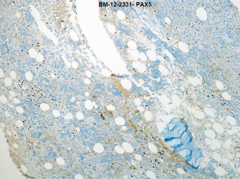 |
| Third BM Biopsy CD34 |  |
| SNP Microarray Result | 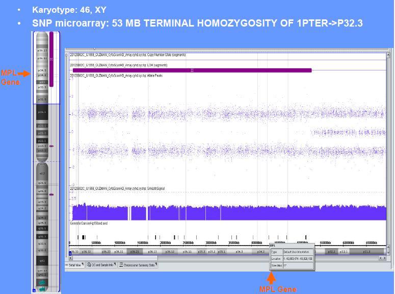 |
| Molecular study result | 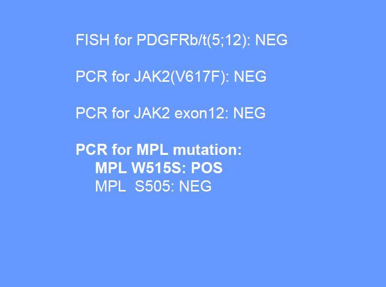 |
| Second Diagnosis |  |