Institution: The Children's Hospital of Philadelphia/University of Pennsylvania
Additional authors:Peter Papenhausen, PhD, Gerald Wertheim, MD, PhD
Session: B Lymphoblastic Leukemia/Lymphoma
HISTORY
This patient is a 10-year-old, previously healthy, boy who presented to his physician complaining of fatigue and bone pain in September 2012. A CBC was performed and he was found to have a WBC of 17.5/ uL (60% blasts) with anemia and thrombocytopenia. A bone marrow aspiration was performed. Combined morphologic, flow cytometric, cytogenetic, and single nucleotide polymorphism (SNP) array studies (see details below) demonstrated B lymphoblastic leukemia (B-ALL). Peripheral blood flow cytometry demonstrated 24.5% blasts at day 8 of therapy, while a day 29 bone marrow study was negative. The patient was referred to our tertiary care children’s hospital to discuss management options. Given his very-high risk genetic features, the recommendation was for him to proceed with allogeneic stem cell transplant immediately upon completion of consolidation therapy.
DETAILS
Bone marrow aspiration only was performed. Aspirate smears show hypercellular marrow with greater than 90% blasts with L1 morphology. A minimal amount of background hematopoiesis was present and was morphologically unremarkable.
IMMUNOHISTOCHEMISTRY AND FLOW CYTOMETRY
Flow cytometry performed on the bone marrow aspirate demonstrated a population of blasts with low side scatter and dim CD45 (97% of total events) with the following immunophenotype: Positive: CD19, CD10, CD22, CD34, CD38, HLA-DR, TdT, CD52, CD58, CD123. Negative: CD2, CD3, CD4, CD5, CD7, CD8, CD11c, CD13, CD15, CD20, CD23, CD25, CD35, CD41, CD61, CD64, CD79b, CD235a, sIgM, cIgM, MPO, kappa, lambda.
CYTOGENETIC FINDINGS
Conventional metaphase analysis revealed the following karyotype: 52,XY,+X,+Y,+14,+14,+21,+21[cp20]. There are two clues from this karyotype that this may not represent favorable-prognosis hyperdiploid B-ALL. Usually there is a gain of an X in the typical hyperdiploid, but not a Y. In addition, the standard hyperdiploid clone usually has tetrasomy 21, but no other tetrasomic chromosomes. These findings suggest evolution from near-haploid, although no such metaphases were found. FISH studies demonstrated 95% of nuclei with 4 copies of the RUNX1(AML1) gene locus on chromosome 21q22. No ETV6(TEL)/RUNX1, MLL, BCR/ABL, or TCF3 gene rearrangements were detected. No deletion of p16 was seen. Centromeric probes for chromosomes 4, 10, and 17 revealed 2 copies of each in 100% of cells. Again, no evidence of single copy signals consistent with near-haploid was detected.
MOLECULAR FINDINGS
Whole genome single nucleotide polymorphism (SNP) microarray analysis demonstrated an abnormal clone with either 26 or 52 chromosomes since all array analyses “normalize” the DNA present to a normal diploid level. The allele dosage patterns are consistent with an original clone that had 26 chromosomes including two copies of chromosomes 14 and 21 and a single copy of all other chromosomes, including the X and Y. The normal presence of heterozygosity in chromosomes 14 and 21 with 4 copy number dosage (chromosome 14 demonstrated as an example), and the loss of heterozygosity in the remaining chromosomes along with a 2 copy number dosage (chromosome 2 demonstrated as an example) are conclusive evidence of near-haploid origin. Karyotype, FISH, or flow analyses are needed to differentiate the present ploidy level.
INTERESTING FEATURES
This is an interesting case of childhood B-ALL in which application of both conventional cytogenetic and SNP array data were integral for correct sub-classification of the disease and designation of the patient to the appropriate prognostic group. On initial review, the karyotype appears hyperdiploid with 52 chromosomes and suggests that the patient would have a favorable prognosis. However, tetrasomy 14 and 21, and doubling of the Y sex chromosome are clues that suggest that the karyotype might actually represent a near-haploid clone that has doubled the chromosome number, a form of clonal evolution in neoplasia.
Severe hypodiploidy, and especially near-haploidy, confer an extremely poor prognosis and high risk of relapse in B-ALL (1, 2). Thus, the distinction between a hyperdiploid clone and a near-haploid clone with evolution is critical both for the prediction of prognosis, and more importantly for the guidance of therapy in these children.
Current recommendations are that children with near-haploid B-ALL proceed directly to allogeneic stem cell transplant at the first complete remission.
Although hypodiploidy (<46 chromosomes) can be seen in 5-8% of B-ALL, cases that fall into the near-haploid group (24-29 chromosomes) are reportedly rare, accounting for < 1% of B –ALL (2). However, this frequency has likely been underestimated due to the propensity of these clones to undergo clonal evolution by chromosomal duplication resulting in an apparently hyperdiploid karyotype (2). This genetic phenomenon highlights an important limitation of karyotyping and FISH in cases where the original clone is no longer present, as is often the case in near-haploidy and severe hypodiploidy. The use of SNP array in this case was essential to confirm the suspicion of a doubled near-haploid clone, as cells with the original 26 chromosome complement were not seen by other methods. SNP array analysis also has a resolution ~500x that of G-banding with equivalent low mosaicism sensitivity as the limited target FISH.
Of note, the retention of chromosomes in hypodiploid neoplasia is distinctly non-random. In a recent case series of near-haploid B-ALL, chromosome 21 was retained in 8/8 cases, followed by chromosome 14 in 6/8 and X/Y in 5/8 (3).
Thus, our case highlights a very typical example of the uncommon near-haploid B-ALL, made especially diagnostically challenging by the lack of the original hypodiploid clone.
Due to its rarity, relatively little is known about the specific genetic features that confer a predilection for relapse in this subgroup of B-ALL. Recent gene sequencing studies reveal that many patients with near-haploid B-ALL also have mutations in genes resulting in Ras pathway activation (1). In addition, these studies have demonstrated that near-haploid cases show mutations in a different subset of genes than those seen in other low-hypodiploid (33-39 chromosomes) cases (1). These genetic studies clearly demonstrate the potential for identification of targeted therapies, and further subclassification of this poorly understood subtype of B ALL.
In summary, this case represents a typical example of the genetic features seen in near-haploid B-ALL. It also represents a potential pitfall in properly identifying these cases, as some will present as a doubled, near-haploid clone that could be misclassified as hyperdiploid based on karyotype and FISH. In these cases, SNP array can provide critical information to aid in this distinction.
PROPOSED DIAGNOSIS
B lymphoblastic leukemia with a doubled, near-haploid karyotype
CONSENSUS DIAGNOSIS
B-acute lymphoblastic leukemia with a doubled, near-haploid karyotype
| Bone marrow aspirate showing >90% lymphoblasts. | 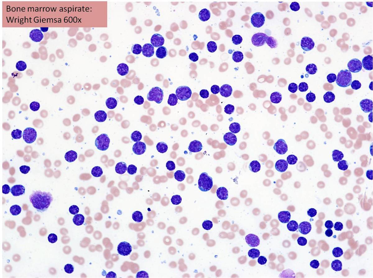 |
| Bone marrow aspirate showing >90% lymphoblasts. | 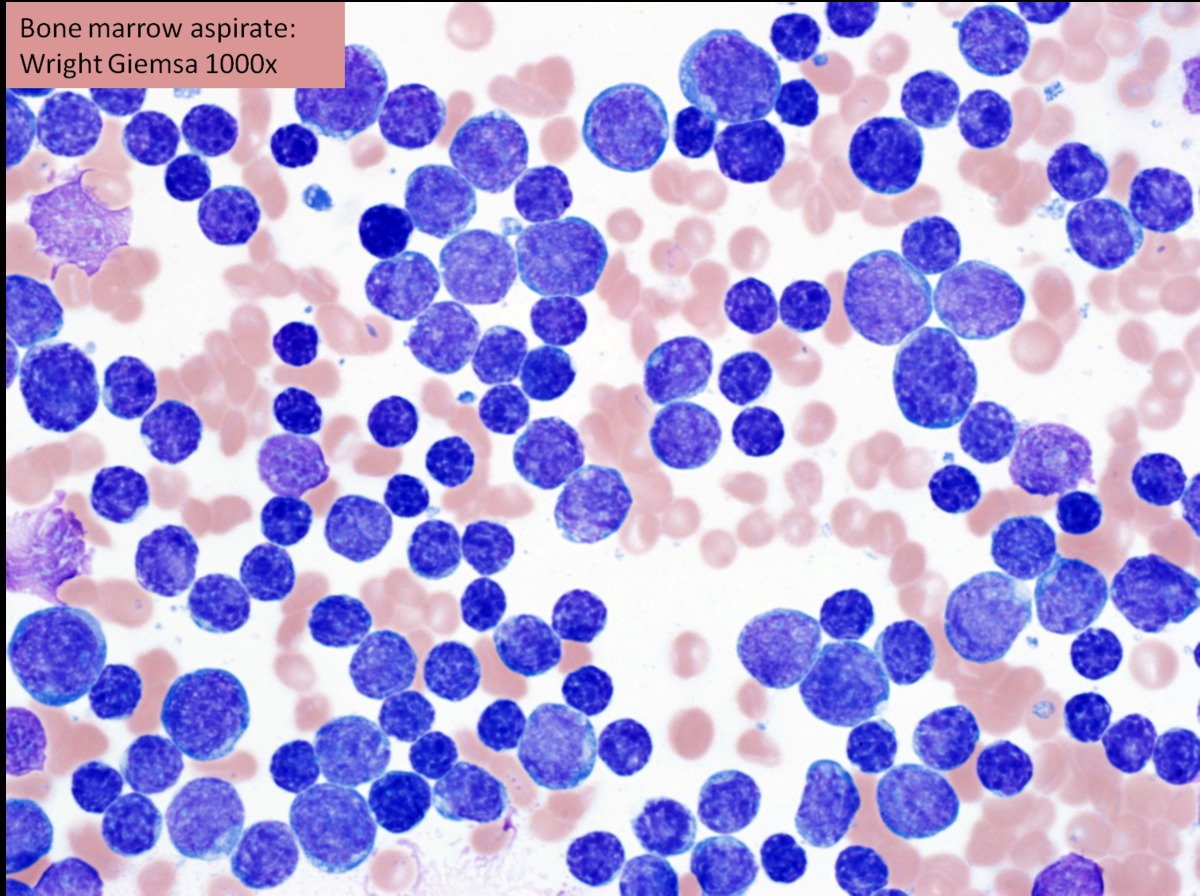 |
| Karyotype of the doubled, near haploid clone. |  |
| FISH for ETV6/RUNX1 | 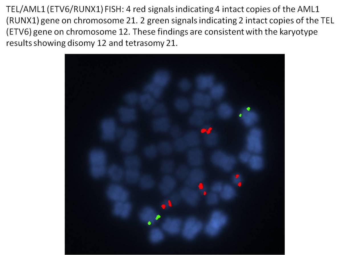 |
| SNP array of chromosome 14 | 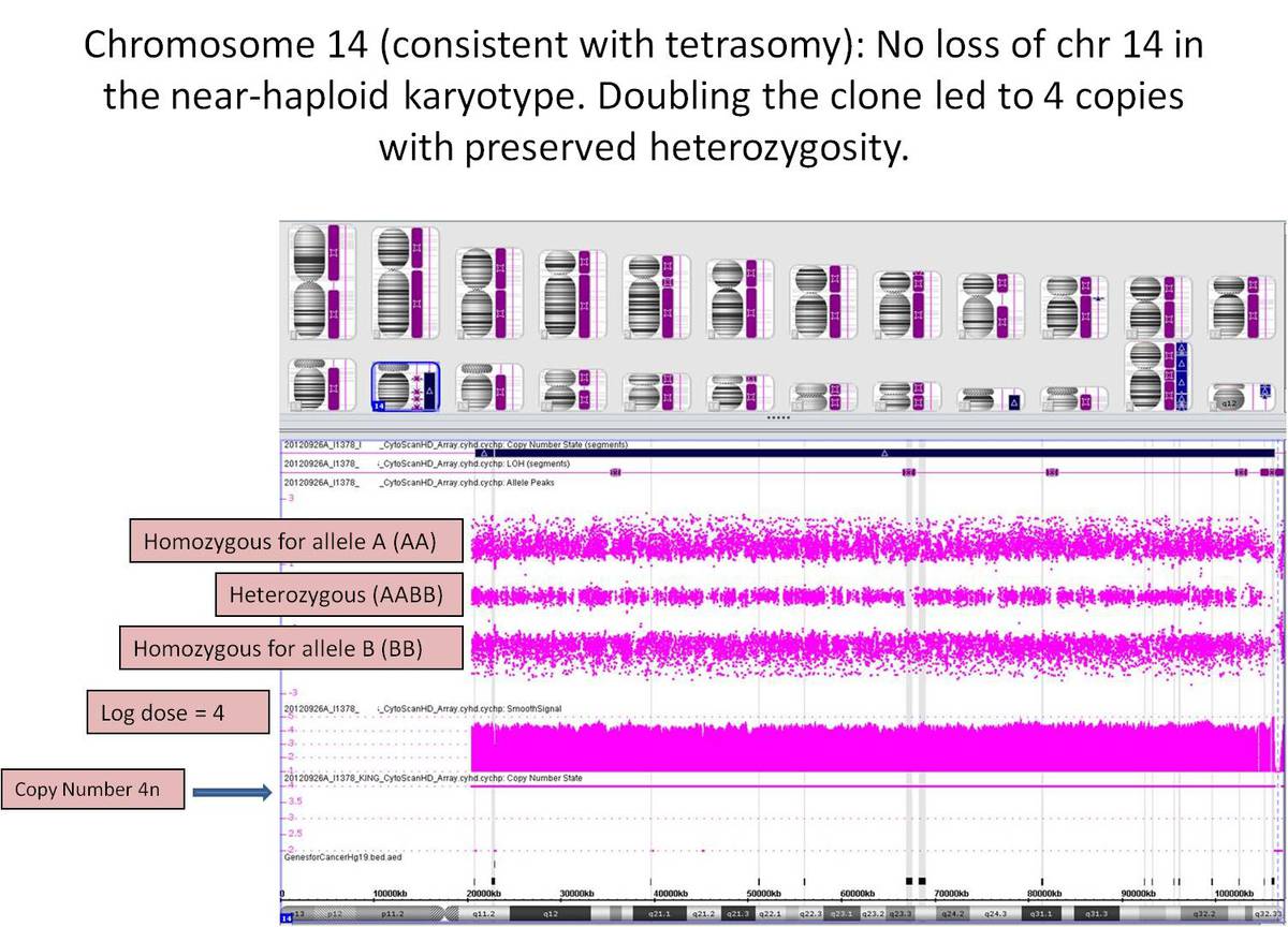 |
| SNP array of chromosome 2 | 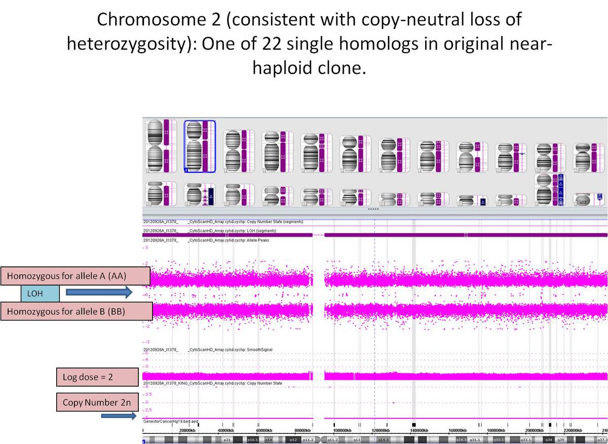 |