Institution: Orlando Health
Additional authors:John Maksem MD, Raymond B. Franklin MD PhD, Lizardo Cerezo MD, Mei Liang MD PhD
Session: AML with recurrent genetic abnormalities Part I
HISTORY
A five year old white male presented with a three day history of right knee swelling and difficulty walking that was first noted on a family road trip from New Mexico to Florida. The patient had no previous medical history with the exception of attention deficit hyperactivity disorder, which is treated with amphetamines (Adderall XR). There was no known family history of childhood cancers or leukemia. On initial presentation, his white blood cell count (WBC) was elevated to 16,600/μL with leukocytosis and 50% blasts on peripheral smear. He was also noted to have a normocytic, normochromic anemia, hemoglobin of 10 g/dL, and a platelet count of 104,000/μL. Erythrocyte sedimentation rate (ESR) was elevated to 34 mm/hr, and lactate dehydrogenase (LDH) was elevated to 282 IU/L.
DETAILS
The peripheral smear demonstrated acute leukemia with 50% blasts. The blast morphology was characterized by a high nuclear-to-cytoplasmic ratio and agranular cytoplasm. There was also normocytic anemia and thrombocytopenia. Basophilia was not identified. There was significant dysplasia of the granulocytic lineage including hypolobated and hypogranulated neutrophils. The bone marrow aspirate sample and touch imprint, stained with Wright-Giemsa, showed acute leukemia involving 40-50% of the medullary cellularity. There was decreased granulopoiesis and erythropoiesis with only rare megakaryocytes identified. There was significant dysplasia of the granulocytic lineage, as seen in the peripheral smear, and the erythropoietic lineage including nuclear blebbing of nucleated reticulocytes and nuclear-cytoplasmic dyssynchrony. Rare dysplastic megakaryocytes were also noted. The bone marrow trephine biopsy was fixed in 10% formalin and stained with H&E. The marrow was hypercellular (95% cellularity) with trilineage hypoplasia and acute leukemia involving 50% of the medullary cellularity.
IMMUNOHISTOCHEMISTRY AND FLOW CYTOMETRY
Flow cytometry identified 26% of cells as an abnormal population that was positive for CD33, CD34, CD38, CD45, CD71 (dim), CD117, HLA-DR (dim), myeloperoxidase (MPO) and terminal deoxynucleotidyl transferase (TdT); and, negative for CD3, CD5, CD10, CD11b, CD15, CD19, CD22, CD56, kappa and lambda. Immunohistochemical stains were not performed.
CYTOGENETIC FINDINGS
Cytogenetic analysis revealed an abnormal clone characterized by a 6;9 translocation in all cells analyzed. This translocation results in the fusion of two genes, CAN (NUP214) and DEK, which generate an altered fusion protein.
MOLECULAR FINDINGS
Fluorescent in-situ hybridization (FISH) for the most common AML related chromosome changes was normal. DNA probes specific for oncogenes or chromosome regions at 5q31, 7q31, RUNX1T1/RUNX1[t(8;21)], MLL (11q23), PML/RARA [t(15;17)] and CBFB (inv16) showed normal hybridization signals in all interphase cells examined
INTERESTING FEATURES
This is a classic case of AML with t(6;9) with trilineage dysplasia but without basophilia.
PROPOSED DIAGNOSIS
Acute myeloid leukemia with t(6;9)(p23;q34); DEK-NUP214.
CONSENSUS DIAGNOSIS
Acute myeloid leukemia with t(6;9)(p23;q34); DEK-NUP214
| Peripheral smear; original magnification 500X | 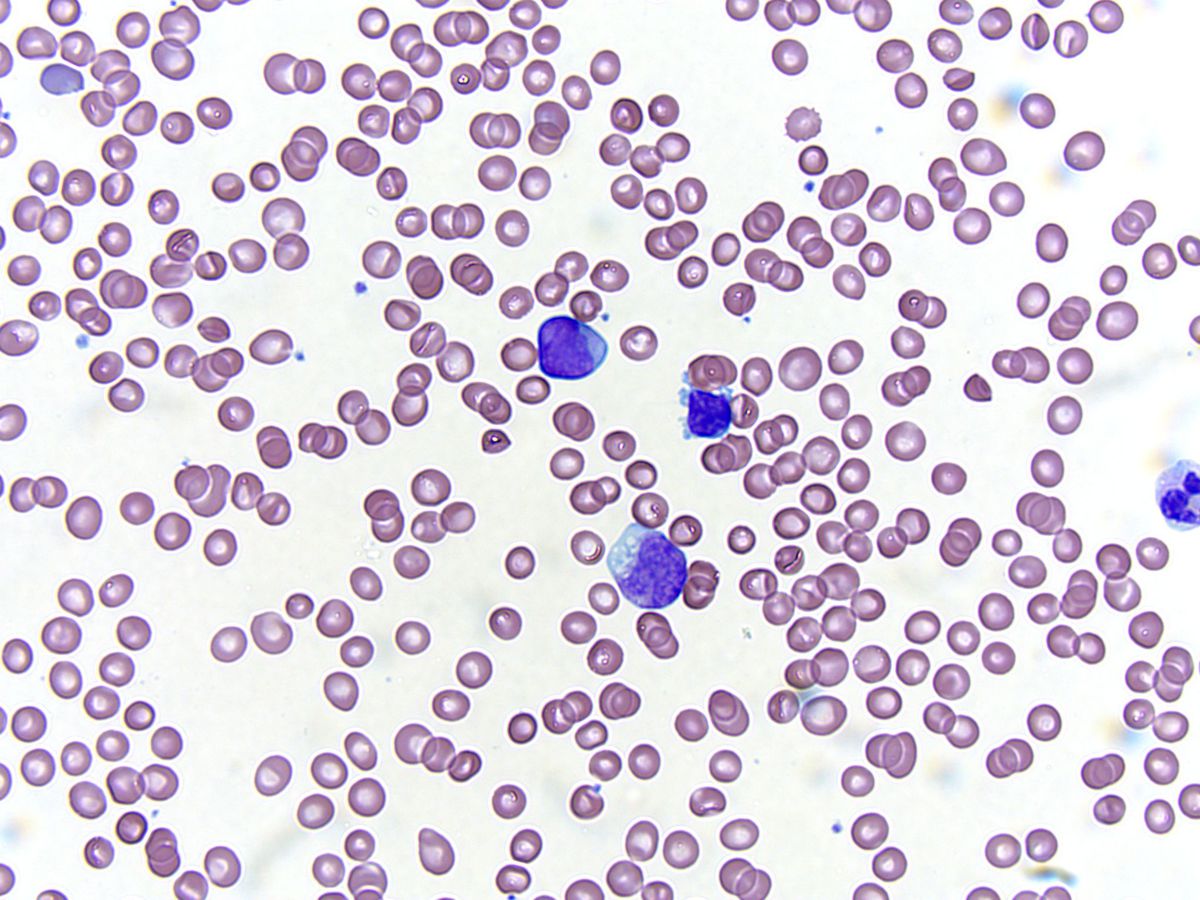 |
| Peripheral smear; original magnification 500X: Blast and dysplastic granulocytes | 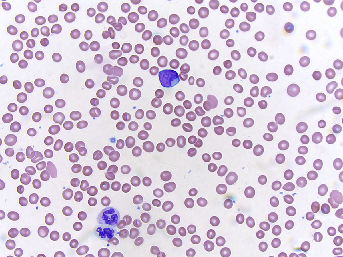 |
| Peripheral smear; original magnification 500X: Blast, monocyte and dysplastic granulocyte | 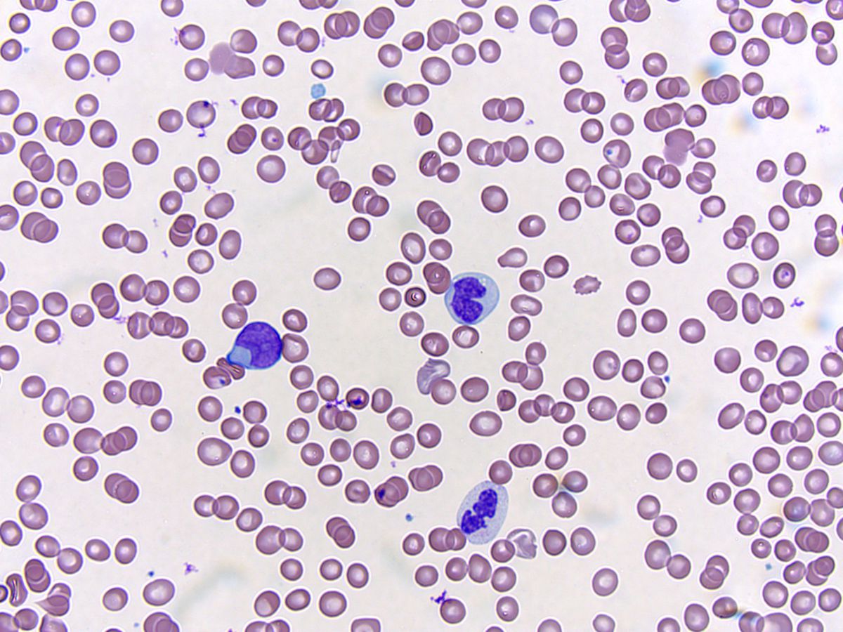 |
| Peripheral smear; original magnification 500X: Blasts and dysplastic granulocyte | 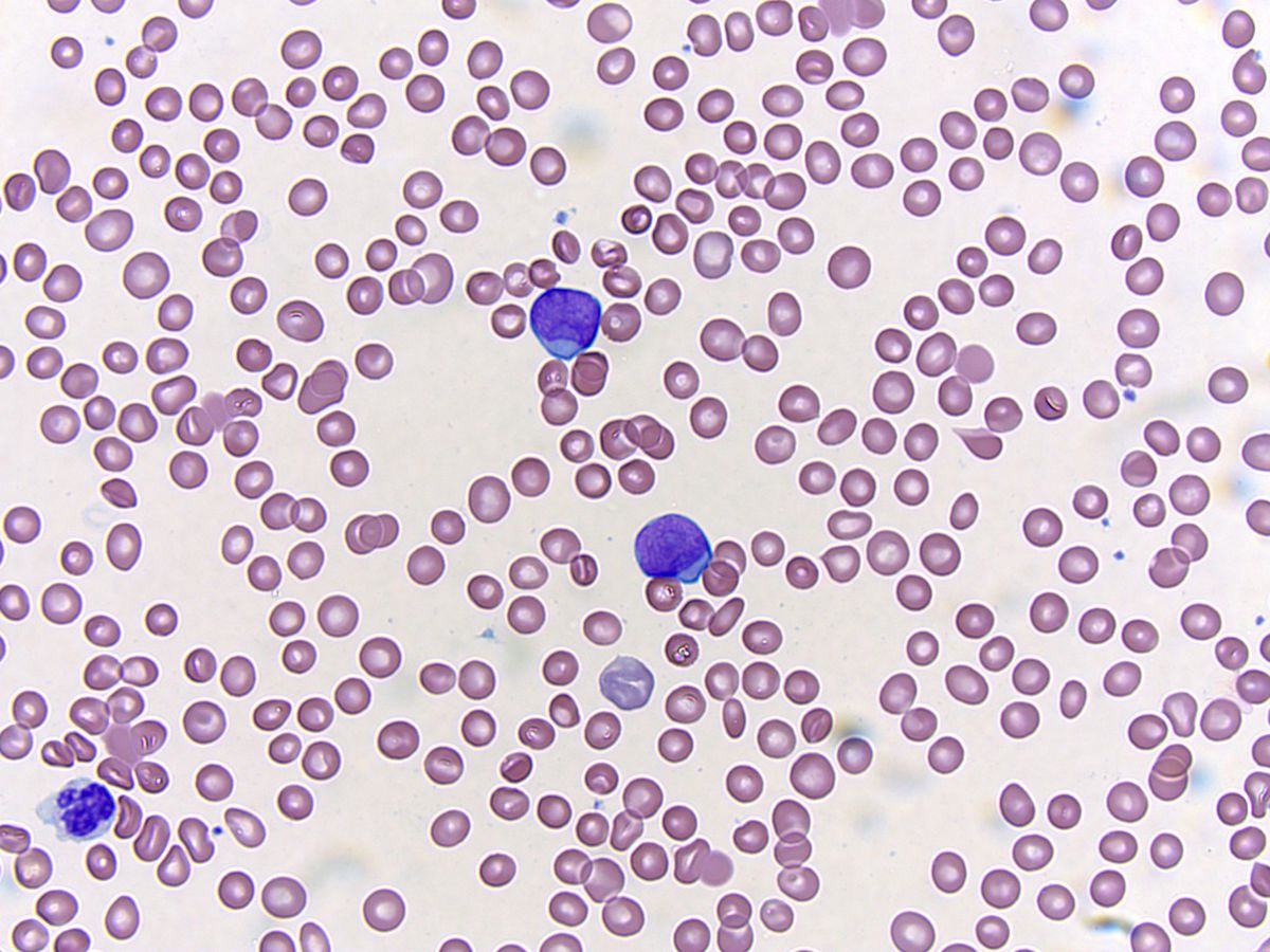 |
| Bone marrow aspirate, Wright-Giemsa; original magnification 500X: Blasts, dysgranulopoiesis, dyserythropoiesis and dysplastic megakaryocyte (center) | 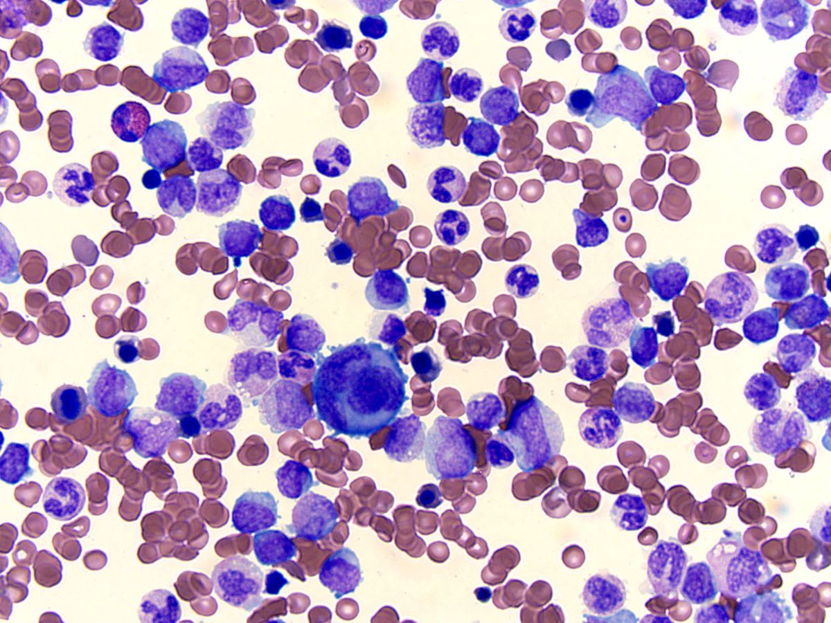 |
| Bone marrow aspirate, Wright-Giemsa; original magnification 500X: | 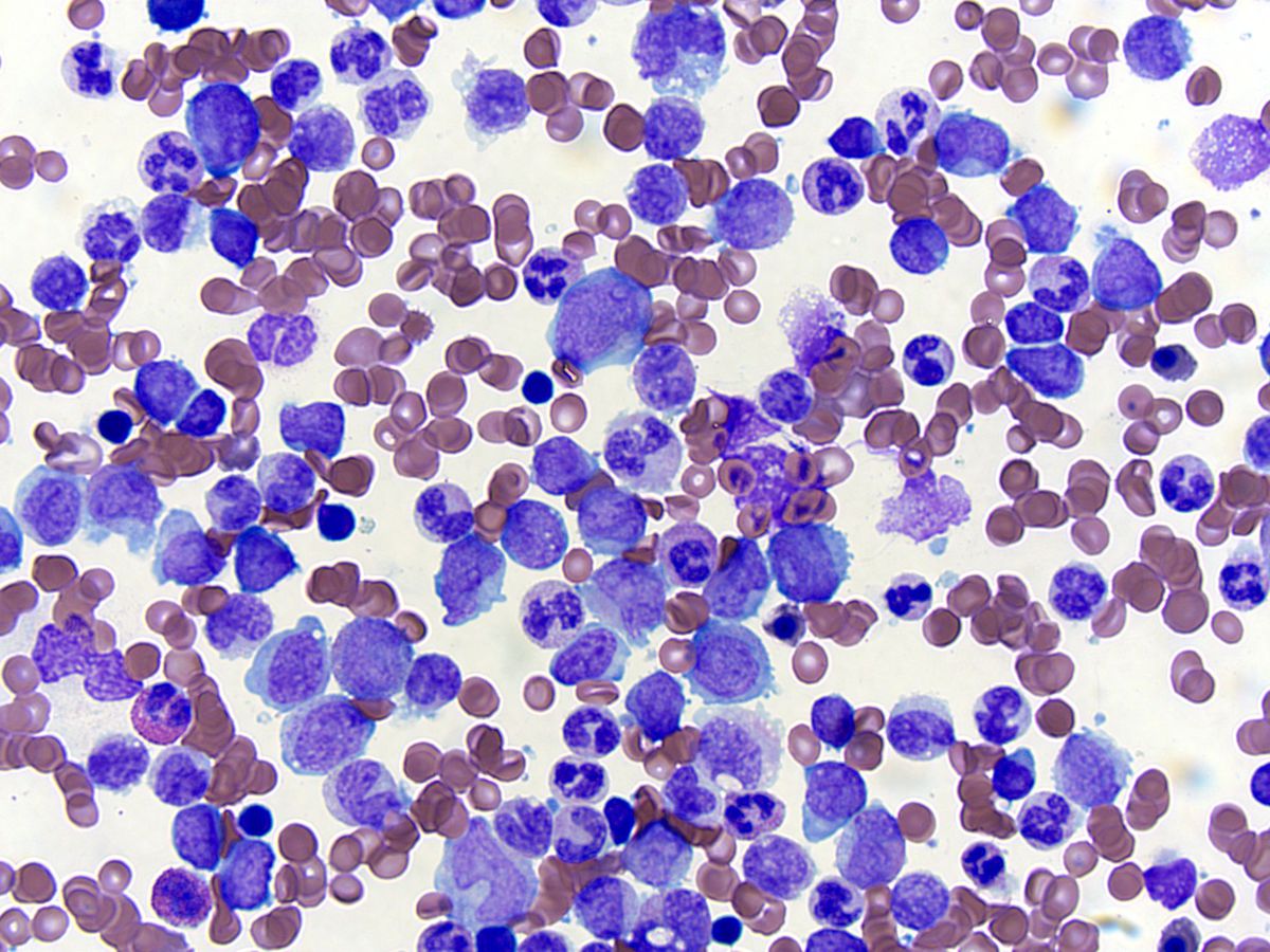 |
| Bone marrow aspirate, Wright-Giemsa; original magnification 500X: Blasts, pseudo Pelgar-Huet cell (solid arrow) and nucleated reticulocytes with irregular nuclear contours (dashed arrows) | 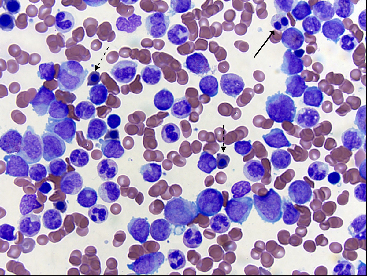 |
| Bone marrow biopsy, H&E; original magnification 200X | 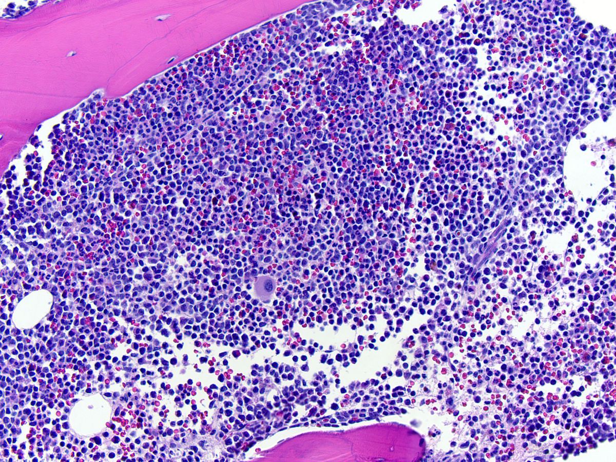 |
| Bone marrow biopsy, H&E; original magnification 500X | 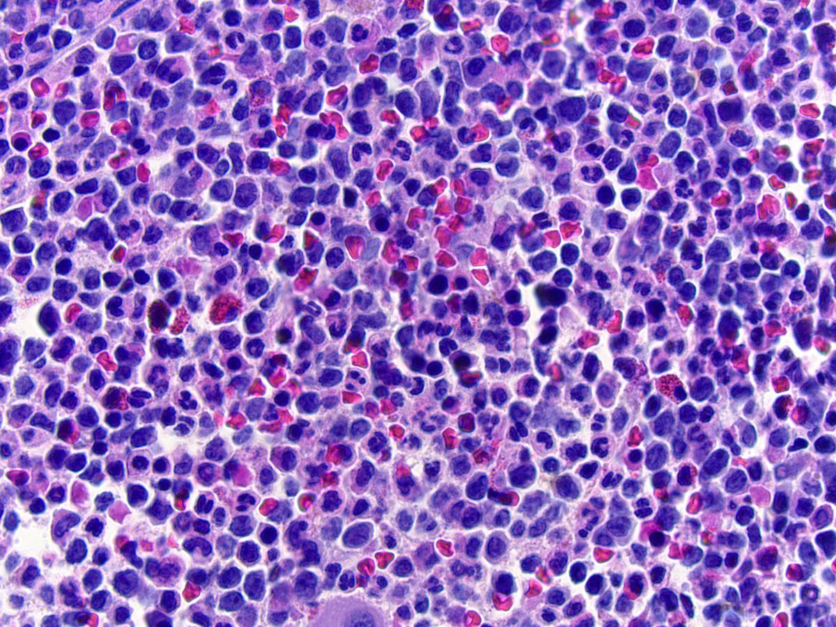 |