Institution: Vanderbilt University Medical Center
Additional authors:Ashwini Yenamandra, Ph.D., Haydar Frangoul, M.D., Mary Ann Thompson Arildsen, M.D., Ph.D.
Session: Extramedullary manifestations of myeloid neoplasms
HISTORY
15 mo old female presented with seizure and a several day history of vomiting and diarrhea without associated fever. Non contrast head CAT scan did not demonstrate evidence of acute intracranial abnormality. A lumbar puncture was performed which showed 300 cells/ul, with 98% abnormal mononuclear cells (See figure 1). Flow cytometry on the CSF sample was performed which showed increased cells in a monocyte gate (figure 2). Bone marrow biopsy the following day was negative with normal cytogenetics. CBC at the time demonstrated WBC: 12.7 K/ul with normal diff, PCV: 35%, and plts 292 K/ul .
One week later, she returned to VUMC with another seizure and repeat head CT and MRI with and without contrast demonstrated diffuse nodular leptomeningeal enhancement. Repeat CSF sample demonstrated persistent abnormal mononuclear cells and similar flow cytometric immunophenotype. An abnormal FISH study was demonstrated, (see below and figure 3), including rearrangement of the MLL locus at 11q23 in 97.5% of cells. Bone marrow performed 2 days later showed focal involvement by acute myeloid leukemia with monocytic differentiation in one of two bilateral cores (Figure 4) The patient was induced with high dose cytarabine and IT cytarabine. At Day + 21 the family noted progressive decrease in activity and acute cessation of lower extremity movement. MRI demonstrated a new large epidural lesion with compression of the conus and cauda equina extending from L1 to L5. Decompression was performed and the specimen demonstrated extramedullary AML (see figure 5 and below). At day +31 of induction chemotherapy a followup bone marrow showed extensive involvement by AML (see figure 6). The patient expired 1 week later.DETAILS
Representative sections of the formalin-fixed and decalcified biopsy of the epidural mass are submitted.
The biopsy demonstrates sheets of large pleomorphic cells with moderate eosinophilic cytoplasm, and irregular, convoluted, polylobate nuclei with dispersed chromatin and prominent red nucleoli (figure 5). There are occasional apoptotic cells and mitoses. There is no significant background hematopoiesis. There are also fragments of benign bone and cartilage. These features are consistent with involvement by this patient's leukemic blasts. Given the presence of a mass lesion, these findings are consistent with myeloid sarcoma with monocytic differentiation.IMMUNOHISTOCHEMISTRY AND FLOW CYTOMETRY
Flow cytometry performed on the initial CSF showed 96% cells with increased FSC in a CD34/SSC monocyte gate with the following immunophenotype: CD33 bright+, CD13 dim+, HLA-DR+, CD34-, CD61-, CD117-, CD64 bright+, CD14 bright+, CD15 bright+,CD11b heterogeneous+, CD16-, CD19-, CD20-, CD2-, and CD10- (figure 2).This immunophenotype was indistinguishable from normal monocytes; however, in conjunction with the abnormal blastic morphology and subsequent abnormal cytogenetics was considered to be representative of leukemic myeloid blasts.
A similar population of monocytic cells was detected in the flow cytometry of the epidural mass (though viability was reduced) and the flow cytometry of the extensively involved bone marrow biopsy.Immunohistochemistry performed on the bone marrow biopsies demonstrated that the blasts were CD68 and lysozyme positive.CYTOGENETIC FINDINGS
1). Initially FISH performed on the diagnostic CSF sample containing blasts demonstrated disruption of the MLL breakapart probe in 98.4% of cells. This sample also demonstrated 3-8 copies of the RUNX1T1 locus at 8q22 and 2-3 copies of the RUNX1 locus at 21q122 in 88% of cells, and 3-4 copies of the CBFB locus at 16q22 in 31% of cells (figure 3).
2). The bilateral bone marrow following the diagnostic CSF demonstrated focal involvement by AML on only 1 side. The non-involved side had FISH findings demonstrating an intact MLL breakapart probe and 4-5 copies of the RUNX1T1 locus in 4.5% of cells (figure 7). The involved side demonstrated the following abnormal karyotype: 50, XX, +8,+8,+8,der(10)(10pter->10q22:?:10q22->10q26::11q23-11qter), der(11)t(10;11)(q26;q13),+13,ins(19;11)(p13.1;q13q23)FISH performed on rare metaphases from this sample demonstrated the 5’MLL probe on der(19) and the 3’MLL probe on 10q, with one intact MLL probe on the normal chromosome 11 (figures 8 and 9).3). FISH performed on the epidural mass demonstrated similar metaphase results with the MLL breakapart probe. In addition, 23.5% of cells demonstrate 4 copies of MLL, 2 intact and 2 rearranged. 53% of cells demonstrate 3-5 copies of the RUNX1T1 locus. 4). The final bone marrow had an abnormal karyotype similar to the previous focally involved bone marrow, with a few cells demonstrating additional extra chromosomes.In summary, this AML has a complex karyotype with rearrangement of the MLL locus. The 5’MLL FISH probe demonstrates translocation to chromosome 19p, inserting near ELL (p13.1), a documented MLL partner. The fusion partner will be confirmed by a long range PCR strategy.MOLECULAR FINDINGS
Molecular testing for NPM1 mutations and FLT3-ITD are negative.
INTERESTING FEATURES
1) The presentation of this case is unusual. The patient presented with seizures, prompting a CSF evaluation. The morphology of the monocytic cells present in the CSF, coupled with FISH demonstrating a MLL rearrangement, was diagnostic of involvement by AML, though at the time the peripheral blood and bone marrow were negative for leukemia. A review of 1354 patients with AML treated at MD Anderson Cancer Center revealed that only 5 patients had CNS involvement at diagnosis; it is unclear whether any of these patients had CNS involvement prior to blood or bone marrow involvement (Shihadeh, F, et al, Cancer 118: 112-7. 2012).
2. Clonal evolution is evident in this case, as FISH performed on material from the non-involved side of the second bilateral bone marrow biopsy demonstrated cells with extra copies of RUNX1T1 but intact MLL loci. Leukemic cells from the CSF and the involved side of the bone marrow demonstrated a disrupted MLL locus, in addition to multiple copies of RUNX1T1.3. The MLL locus is involved in a complex rearrangement, as by FISH the 5’ MLL probe is present on der(19), most likely as an insertion at 19p13.1, and the 3’ MLL probe is on chromosome 10q. The translocation partner of 5’MLL on chromosome 19 is being identified by a long range PCR approach.PROPOSED DIAGNOSIS
Extramedullary CNS presentation of acute myeloid leukemia with complex karyotype including MLL rearrangement; negative for FLT3 and NPM1 mutations.
CONSENSUS DIAGNOSIS
Acute myeloid leukemia with complex karyotype including MLL rearrangement, with initial CNS presentation and extramedullary progression
| Figure 1. Wright stained CSF cytospin showing abnormal immature monocytic cells. | 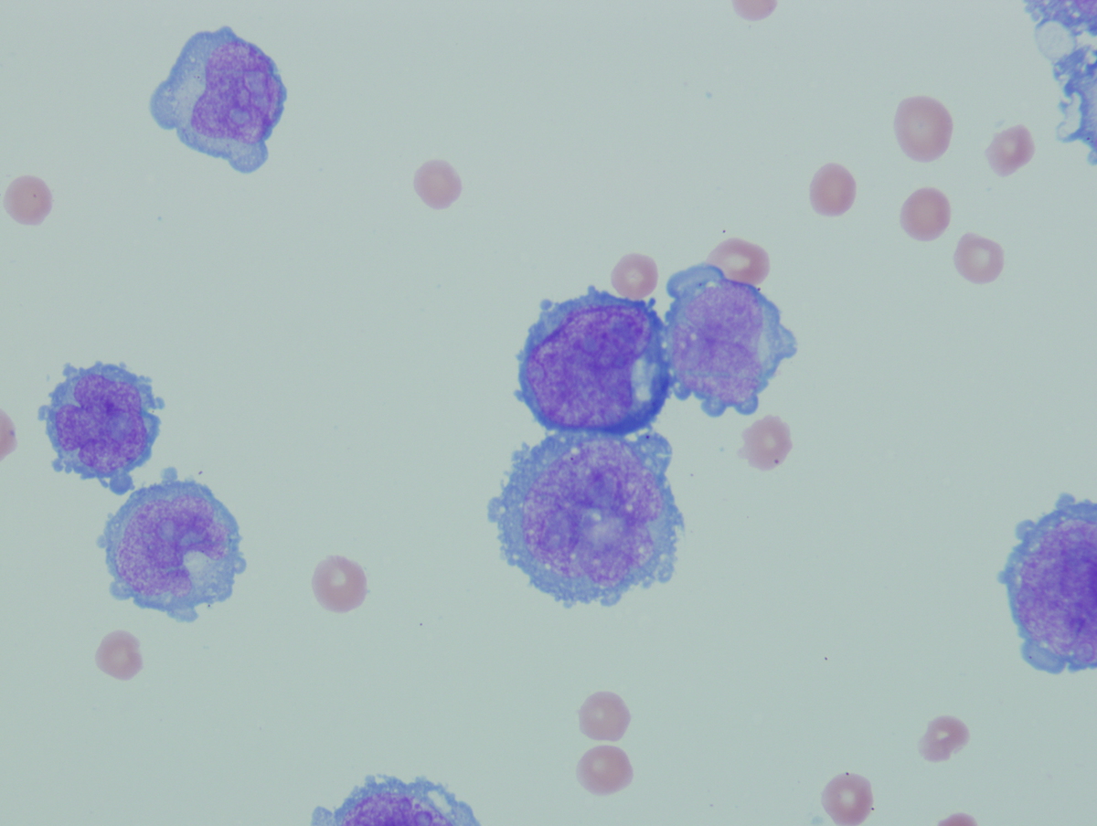 |
| Figure 2. Flow cytometry of CSF | 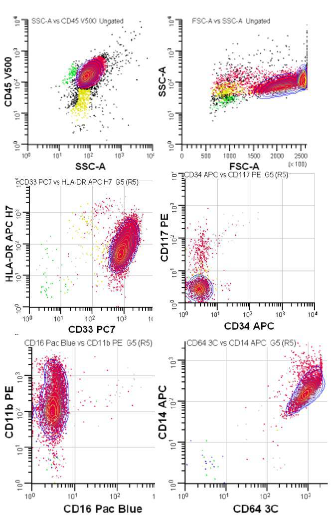 |
| Figure 3. FISH on diagnostic CSF | 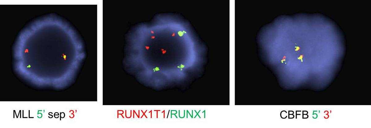 |
| Figure 4. H&E stained section of the bone marrow biopsy unilaterally focally involved by AML, 400X | 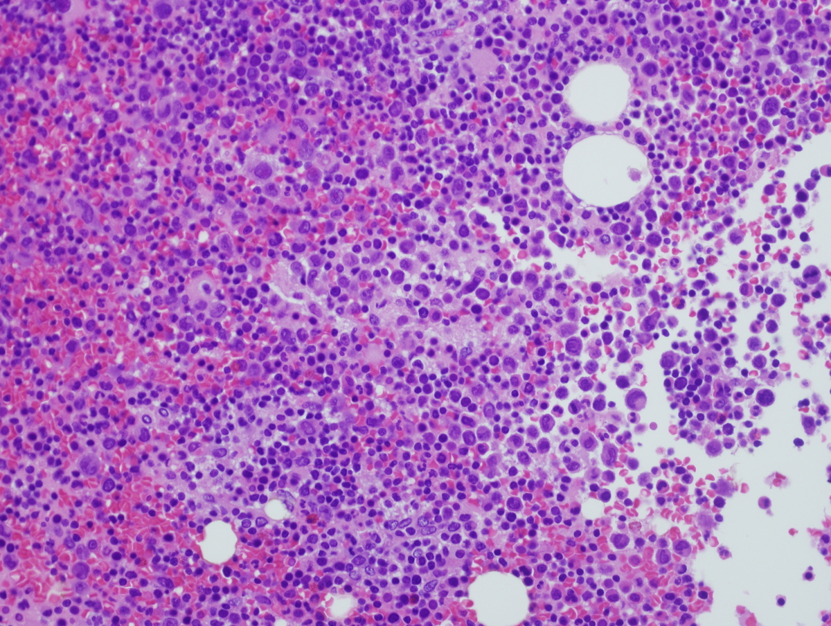 |
| Figure 5. H&E stained section of the epidural mass, 400X. | 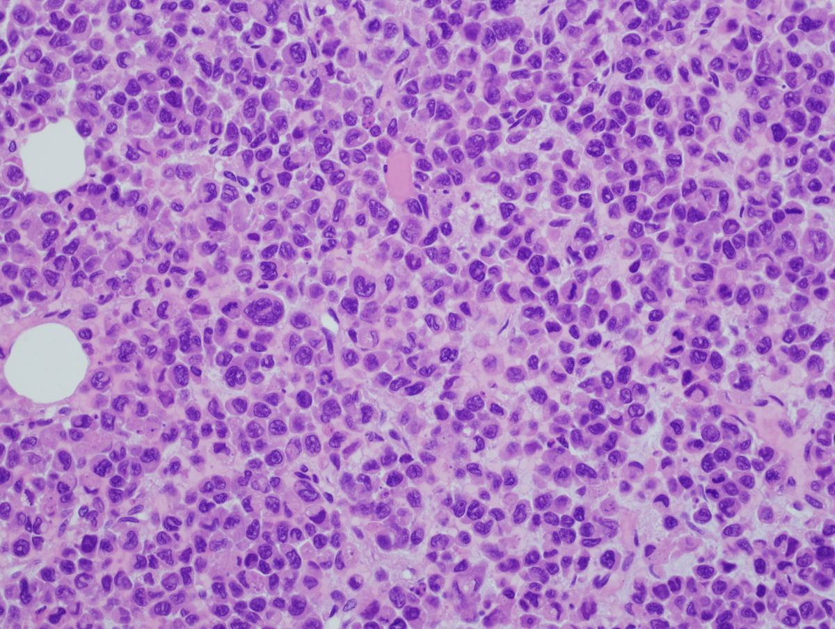 |
| Figure 6. H&E stained section of the bone marrow biopsy extensively involved by AML. | 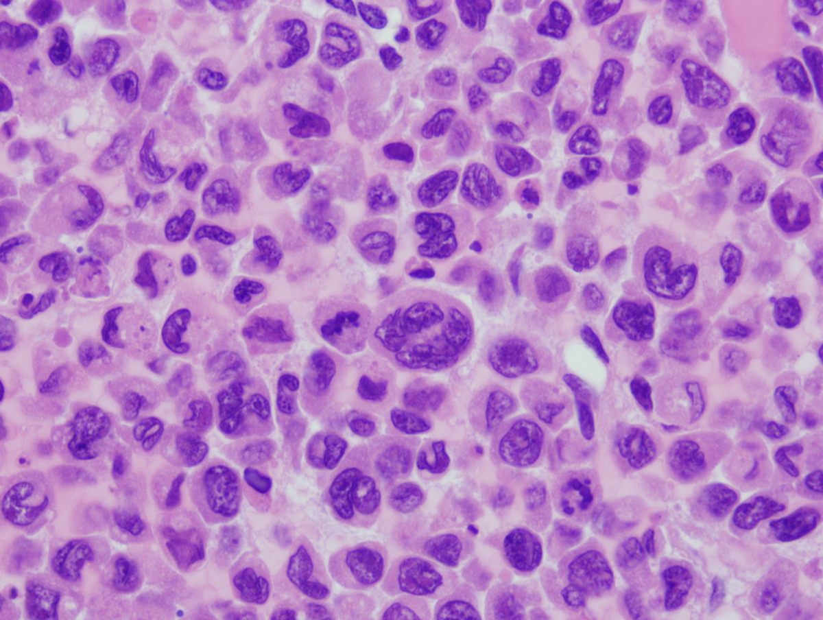 |
| Figure 7. FISH on the second bone marrow: uninvolved side. | 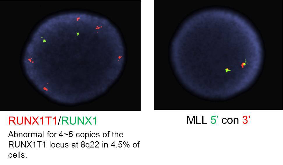 |
| Figure 8. Karyotype of second bone marrow: involved side. | 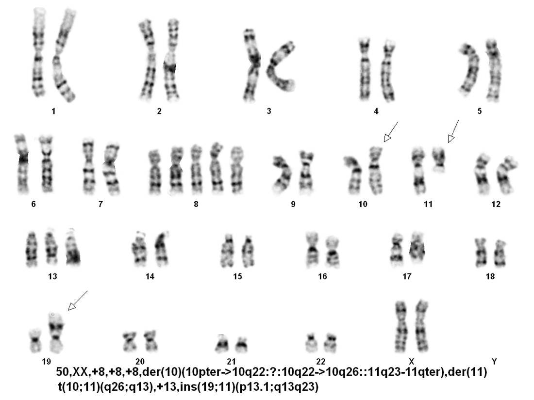 |
| Figure 9. FISH on second bone marrow: involved side. This metaphase FISH shows the complex rearrangement of MLL. | 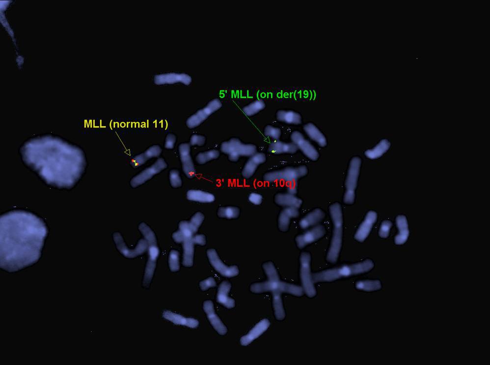 |