Institution: Department of Pathology, Indiana University, Indianapolis, IN, USA
Additional authors:Jiehao Zhou, MD, PhD
Session: T Lymphoblastic Leukemia/Lymphoma
HISTORY
15 year-old male presented with pancytopenia and peripheral blood circulating blasts in 2010. A diagnosis of acute myeloid leukemia was rendered based on bone marrow aspiration obtained on 02/27/2010. Cytogenetic study revealed t(10;16)(p15;q22) (100%), which was considered not constitutional as a normal male karyotype was obtained from the patient's peripheral blood sample. The patient received two cycles of chemotherapy per AAML0531 protocol. The chemotherapy was stopped due to the severe complications including multisystem organ failure and systemic fungal infection. However the patient was in remission with normal flow cytometric and cytogenetic studies. The patient subsequently relapsed. Bone marrow aspirate performed on 10/14/2011 showed 14% blasts with an immunophenotype of T-lymphoblast. The cytogenetic study revealed the identical translocation [t(10;16)(p15;q22)] in 35% of dividing cells analyzed. The patient underwent re-induction with hemi-Capizzi (Ara-C/PEG), which achieved remission with normal cytogenetic study. The patient was found to have peripheral blood circulating blasts while being evaluated for allogeneic stem cell transplant. The bone marrow aspirate performed on 12/30/2011 showed approximately 50% T-lymphoblasts with normal cytogenetic study.
DETAILS
Bone marrow aspiration from right posterior iliac crest was obtained on 12/30/2011. Bone marrow aspirate smears and clot sections were prepared. Portion of the marrow aspirate was sent for flow cytometric analysis, cytogenetic analysis and FISH study (AML panel). The Wright Giemsa stained bone marrow aspirate smear showed approximately 50% large blasts with high nuclear-cytoplasmic ratio, fine chromatin and prominent nucleoli.
Bone marrow diffs:
I. First bone marrow biopsy (02/27/2010):
Marrow diff: blasts 82%, lymphocytes 11%, nucleated RBCs 7%.
II. Second bone marrow biopsy (10/14/2011):
Marrow diff: blasts 15%, promyelocytes 3%, myelocytes 10%, metamyelocytes 6%, bands 13%, polys 15%, lymphocytes 16%, monocytes 1%, eosinophils 1%, nucleated RBCs 21%.
III. Third bone marrow biopsy (index case, 12/30/2011):
Marrow diff: blasts 52%, promyelocytes 1%, myelocytes 3%, metamyelocytes 5%, bands 7%, polys 11%, lymphocytes 4%, monocytes 1%, eosinophils 2%, nucleated RBCs 14%.
PB differentials:
I. First CBC (02/27/2010): WBC 39.0 k/cumm, hemoglobin 7.7 g/dL, MCV 96 fl, RDW 20.5%, platelet 22 k/cumm
diff: blasts 94%, lymphocytes 6%.
II.Second CBC (10/14/2011): WBC 9.1 k/cumm, hemoglobin 14.6 g/dL, MCV 97 fl, RDW 12.7%, platelet 217 k/cumm
diff: blasts 5%, neutrophils 52%, lymphocytes 33%, monocytes 8%, eosinophils 2%.
III.Third CBC (the “current”/index case, 12/30/2011): WBC 6.3 k/cumm, hemoglobin 11.5 g/dL, MCV 96 fl, RDW 16.3%, platelet 225 k/cumm
diff: blasts 7%, bands 1%, neutrophils 53%, lymphocytes 26%, monocytes 11%, eosinophils 2%.
IMMUNOHISTOCHEMISTRY AND FLOW CYTOMETRY
Flow cytometric analysis of the bone marrow aspirate (12/30/2011) showed a distinct blast population with expression of CD3, CD5, CD7 and CD8. The blasts were negative for CD2, myeloid, B-cell lineage markers tested. The immunohistochemical stain of the marrow clot section showed the atypical immature cells were positive for TdT.
CYTOGENETIC FINDINGS
I. First bone marrow biopsy (02/27/2010):
Karyotyping: 46, XY, t(10;16)(p15; q22)[20].
FISH: Negative for AML Panel (5p/5q EGR1, 7q ELN, RUNX1/AML1, MLL, CBFbeta, from Abbott/Vysis)
II. Second bone marrow biopsy (10/14/2011):
Karyotyping: 46, XY, t(10;16)(p15; q22)[7]/Nonclonal[1]/46,XY[12].
FISH: Negative for AML Panel
III. Third bone marrow biopsy (index case, 12/30/2011):
Karyotyping: 46, XY[20]
FISH: Negative for AML and ALL Panels (5p/5q EGR1, 7q ELN, RUNX1/AML1, MLL, CBFbeta; trisomy of chromosome 4, 10, 17, fusion BCR/ABL1, deletion of 9q34, ETV6/AML1, PML/RARA, inversion of chromosome 16, from Abbott/Vysis).
MOLECULAR FINDINGS
Not performed
INTERESTING FEATURES
1. A very rare and unusual case with different type of acute leukemia (T-ALL) developed in a teenager after the treatment of acute myeloid leukemia in a relatively short period (one year and 10 months).
2. The presence of t(10;16) in the initial myeloblasts and the absence of such translocation in the patient's most recent T-lymphoblasts suggest that most likely there were two abnormal clones, one developed AML with t(10;16), the other developed T-ALL (without this translocation) after the initial dominant clone inhibited. The most recent T-ALL could also in principle represent the relapse of acute leukemia with an immunophenotype switch or a de novo T-ALL.
3. The presence of such cases poses the question: how should we treat patients so we can eradicate (cure) the disease.
PROPOSED DIAGNOSIS
T-lymphoblastic leukemia, status post acute myeloid leukemia.
CONSENSUS DIAGNOSIS
T-acute lymphoblastic leukemia, status post acute myeloid leukemia
| Bone marrow aspirate (100X) from 12/30/2011 | 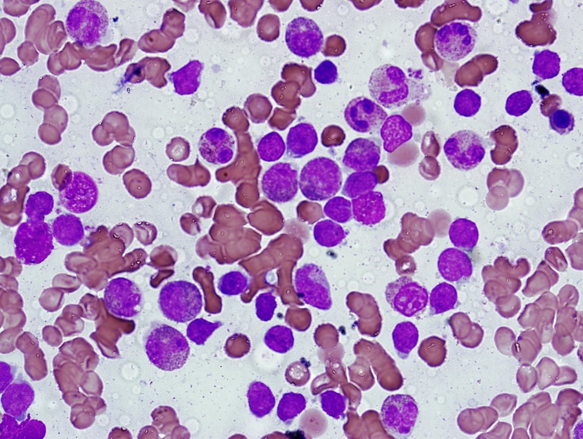 |
| Karyotyping showing t(10;16)(p15;q22) present in the initial bone marrow sample with AML, but absent in the current T-ALL. | .jpg) |
| Flow cytometry | 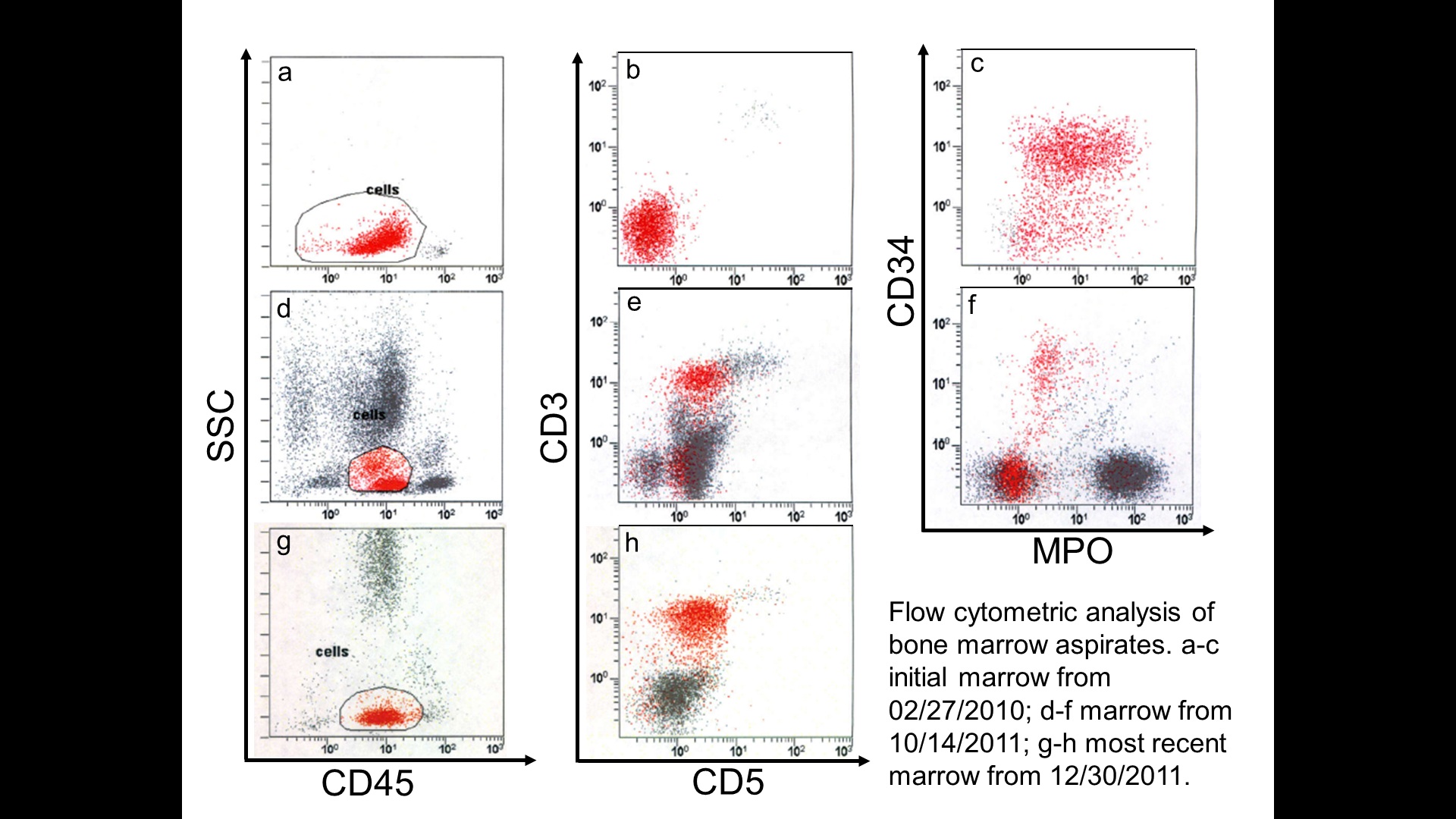 |