Institution: Penn State Hershey Medical Center
Additional authors:Michael G. Bayerl, MD, Jozef Malysz, MD
Session: Extramedullary manifestations of myeloid neoplasms
HISTORY
The patient is a 77 year old man without significant past medical history who presented to an outside hospital with a red lesion on his back of approximately 2 months duration. Biopsy of the lesion was initially interpreted as non-Hodgkin lymphoma. The patient was subsequently referred to our institution. Physical examination showed a raised red lesion with central ulceration measuring 10 x 5 cm on the back. A smaller coin-like lesion was present a few centimeters below the main lesion. His only symptoms were mild loss of appetite and an unintentional weight loss of 7 pounds. CBC showed Hgb 12.5 g/dL, platelets 35 X 109/L, and WBC 33.1 X 10⁹/L with 43% blasts, 30.8% neutrophils, 18.7% lymphocytes, 4.7% monocytes, 1.9% eosinophils and 0.9% immature granulocytes.
DETAILS
Excisional biopsy of the back lesion (FFPE) showed focally ulcerated epidermis with a dense, diffuse infiltrate extending from the base of the ulcer through the papillary dermis, deep dermis and into subcutaneous fat with no epidermotropism. The dermal component showed prominent interstitial growth pattern, but no angioinvasive or angiodestructive growth pattern. Areas of necrosis were present. The cells were intermediate to large with minimal cytoplasm, round to slightly cleaved nuclei, fine chromatin, and variably prominent nucleoli.
Peripheral blood and bone marrow were extensively involved by blasts with large, round to delicately folded nuclei, fine chromatin, one to several prominent nuclei, and moderately abundant blue-grey cytoplasm with no vacuoles, granules or Auer rods.IMMUNOHISTOCHEMISTRY AND FLOW CYTOMETRY
IHC / ISH skin:
CD45 (weak), CD43, CD4, CD5 (heterogenous), CD7, CD56 (heterogenous), TCL-1, CD123 and TdT. The following stains are negative: CD2, CD3, CD8, TIA1, ALK1, CD30, CD10, CD20, CD79a, PAX5, CD138, cyclin D1, CD34, CD117, MPO, CD15, lysozyme, pancytokeratin, CK20, AE1/AE3, EMA, and NSE. In situ hybridization for kappa, lambda and EBV (EBER) are negative.IHC marrow: Negative for CD1a.Flow cytometry peripheral blood: Blasts express CD45(dim), cCD3 (partial), CD4, CD7(dim), CD56(dim), CD71(dim), TDT (dim), TCR alpha-beta, HLA-DR, CD38 (heterogeneous), and cCD22 (equivocal). They are negative for CD34, CD2, sCD3, CD5, CD8, CD16, CD57, CD94, MPO, CD13, CD33, CD117, CD11c, CD14, CD64, CD19, CD20, cCD79a, CD10.CYTOGENETIC FINDINGS
Chromosome analysis: 46,XY,del(5)(q13q33)[2]/46-55,XY,+5,+5,del(5)(q13q33)x2,-6,+7,+8,+8,der(8)t(6;8)(p11.2;q22)x2,-15,+16,+17,+18,+21,+21[cp5]
Interphase FISH: Three copies of chromosomes 7, 8, and 16, four copies of chromosomes 5 and 21, loss of 15q22/PML and 5q-, and no MLL, CBFb or PML/RARA rearrangement.MOLECULAR FINDINGS
PCR for TCR gamma gene rearrangement showed a polyclonal pattern of T-cell receptor gamma gene amplification.
INTERESTING FEATURES
1) Though the clinical presentation, morphology and IHC of the skin lesion are diagnostic of BPDCN by current criteria, albeit with some CD5 expression, the extended immunophenotype derived by flow cytometry identified additional markers (cCD3 epsilon, sTCRab) overlapping with T lymphoblastic leukemia. This presents a rather significant diagnostic and therapeutic challenge.
2) Karyotype and FISH were highly reassuring of a myeloid neoplasm, confirming the diagnosis of BPCDN.3) More precise and widely-accepted criteria are needed to define the boundaries between the various blastic myeloperoxidase negative disorders expressing CD4 and other DC2-asscociated antigens.PROPOSED DIAGNOSIS
Blastic plasmacytoid dendritic cell neoplasm
CONSENSUS GROUP: ADDITIONAL INFORMATION/STUDIES
Additional immunohistochemical studies performed by the consensus group:
Beta F1: Mostly negative
CD3: Positive (subset)
CONSENSUS DIAGNOSIS
Blastic plasmacytoid dendritic cell neoplasm
| Skin biopsy, H&E, low power |  |
| Skin biopsy, H&E, high power |  |
| Skin biopsy, IHC | 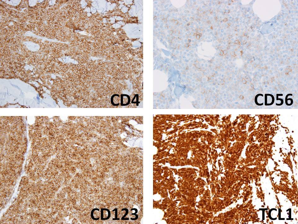 |
| Bone marrow aspirate | 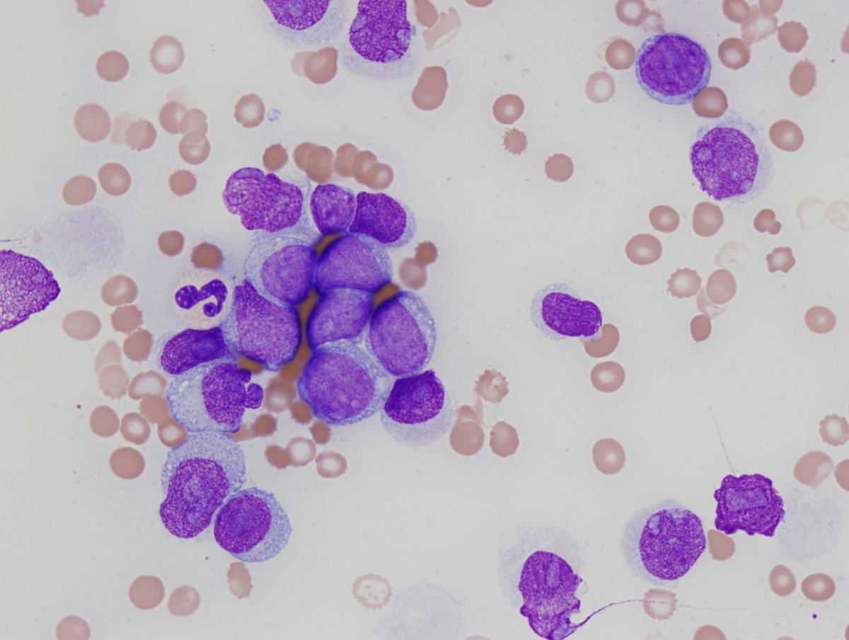 |
| Flow cytometry, peripheral blood |  |
| Flow cytometry, peripheral blood | 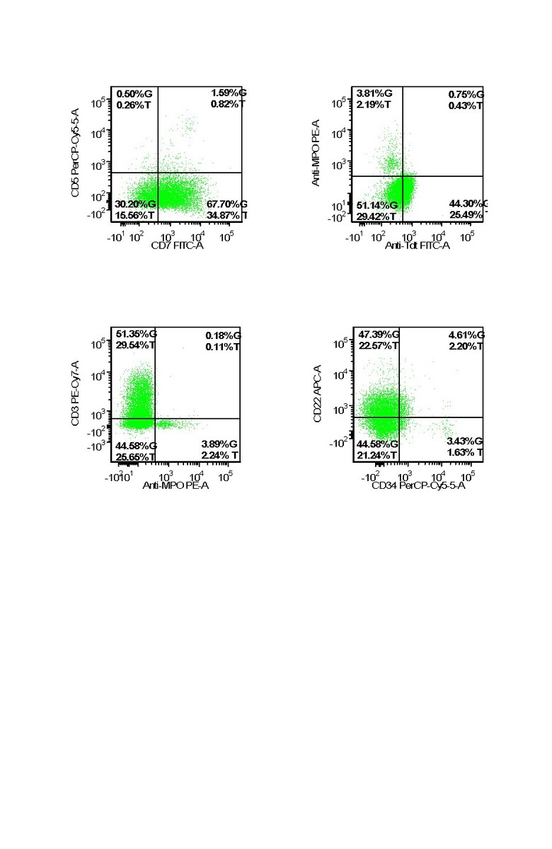 |
| Additional figure 1: Beta F1 IHC | 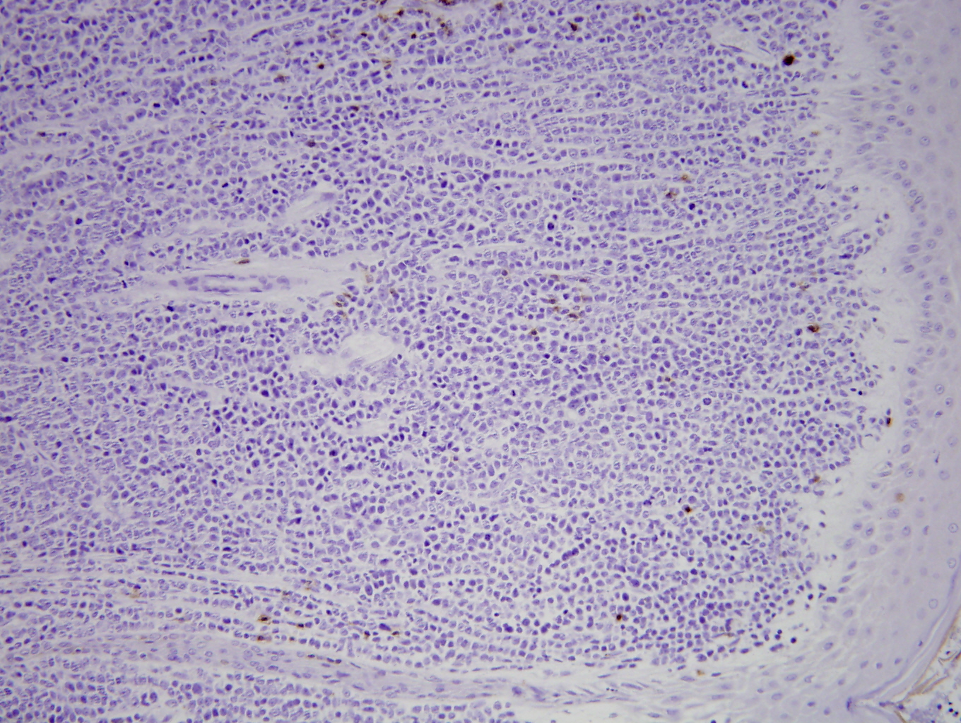 |
| Additional figure 2: CD3 IHC, 400x | 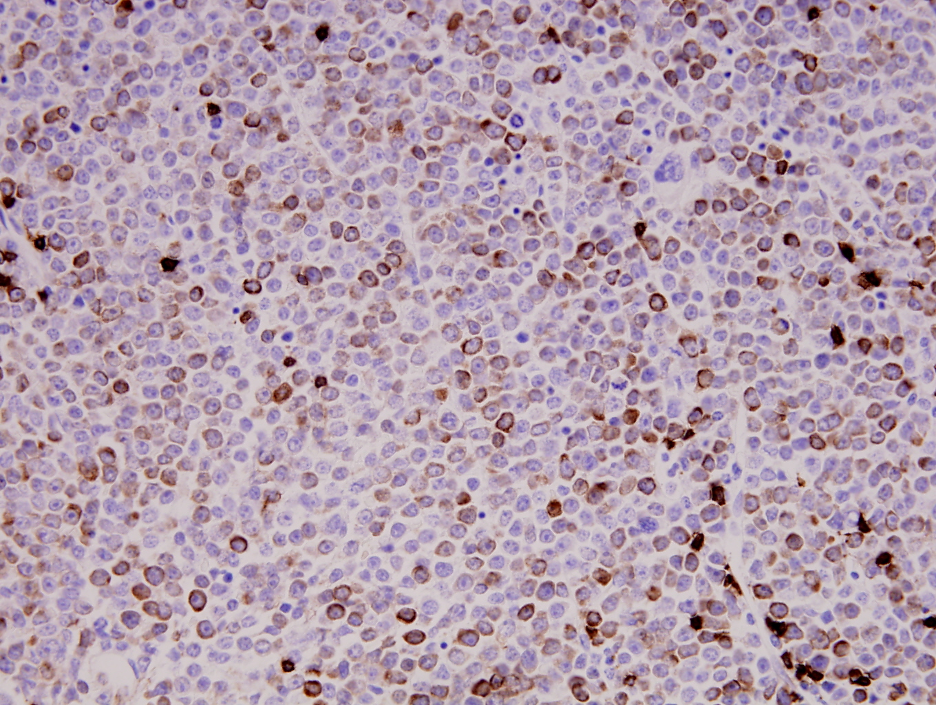 |