Institution: Penn State Hershey Medical Center
Additional authors:Robert J. Greiner, M.D., Rhett P. Ketterling, M.D.
Session: B Lymphoblastic Leukemia/Lymphoma
HISTORY
This 7 month-old, unimmunized girl was referred to Penn State Children’s Hospital for 2 weeks of tender cervical lymphadenopathy and fever following a typical viral URI. Adenopathy and fever responded temporarily to antibiotics and corticosteroids at her local hospital. On presentation here, CT scan showed bilateral, diffuse enlargement of parotid and submandibular glands, with mild, bilateral cervical lymphadenopathy and no abnormalities below the neck. Her CBC was as follows: WBC 2.39 K/dL , HGB 8.1 g/dL, HCT 24.4%, RBC 3.20 M/uL, MCV 76.3 fL, MCHC 33.2 g/dL, MCH 25.3 pg, RDW 16.1% and PLT 131 K/dL. Manual WBC differential: 34% “atypical lymphocytes,” 33% lymphocytes, 2% monocytes, 21% neutrophils, rare neutrophilic metamyelocytes and myelocytes.
DETAILS
Peripheral blood and bone marrow clot (FFPE, no decal): Tumor cells are 34% of circulating cells and 95% of marrow cells. They are mostly small to intermediate-sized with “L1” type morphology, round nuclei, homogenous chromatin, no to small nucleoli and scant blue cytoplasm; but, about one-third have larger and/or folded nuclei, more prominent nucleoli and moderate dense blue or pale blue-grey cytoplasm, occasionally with a few fine vacuoles, but no granules or Auer rods.
IMMUNOHISTOCHEMISTRY AND FLOW CYTOMETRY
Flow cytometry from blood: Tumor cells are positive for CD19, CD20, sCD22, cCD22, CD10 (dim), CD5 (dim), CD23 (dim, heterogeneous), HLA-DR and monotypic surface lambda light chains. They are negative for CD34, TdT and all myelomonocytic (MPO, CD13, CD33, CD117, CD14) and other T-cell-associated (CD2, cCD3, sCD3, CD4, CD5, CD7, CD8) antigens tested.
IHC on FFPE bone marrow: BCL2+, BCL6-CYTOGENETIC FINDINGS
Chromosome analysis: 46,XX,der(2)t(2;9)(q23;p13)t(9;11)(p22;q23)der(9)t(2;9)t(9;11),der(11)t(9;11)[4]/46,XX[16]
FISH analysis confirmed this complex translocation results in AF9/MLL fusion. Sequential FISH demonstrated AF9/MLL fusion located on the derivative chromosome 2 and the derivative chromosome 11. FISH for the following are all negative: 8q24(MYC sep), t(8;14)(MYC/IGH), t(8;22)(MYC/IGL), 14q32(IGH sep), t(1;19)(PBX1/TCF3),der(19), +4, +10, +17, 9p-(CDKN2A-),9p-x2(CDKN2A-x2), t(9;22)(BCR/ABL), t(12;21)(TEL/AML1), Amp(AML1)MOLECULAR FINDINGS
None
INTERESTING FEATURES
Mature, purely B immunophenotype without myeloid antigens is very rare in cohorts of pediatric lymphoblastic leukemia, but is associated with infants, CD10 negativity and MLL rearrangements as in this case. The morphology and immunophenotype may pose a significant differential diagnosis from other mature B-cell neoplasms, i.e. Burkitt and DLBCL, particularly when presenting in a lymphomatous clinical picture. Final diagnosis ultimately relies on cytogenetic / molecular evidence of MLL rearrangements vs. other B-cell lymphoma rearrangements, e.g. MYC.
PROPOSED DIAGNOSIS
B lymphoblastic leukemia with AF9-MLL rearrangement.
CONSENSUS DIAGNOSIS
B-acute lymphoblastic leukemia with AF9-MLL rearrangement [ t(9;11) ]
| Peripheral blood: Typical lymphoblasts (right) and a large cell with coarse chromatin and dense blue cytoplasm (left) blast vs. lymphoma cell. | 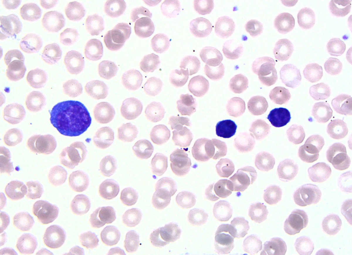 |
| Peripheral blood: Large blasts with fine chromatin, prominent nucleoli and moderate pale blue-grey cytoplasm with a few fine vacuoles. |  |
| Bone marrow: Many small to intermediate-sized blasts, a few larger blasts and occasional fine cytoplasmic vacuoles. | 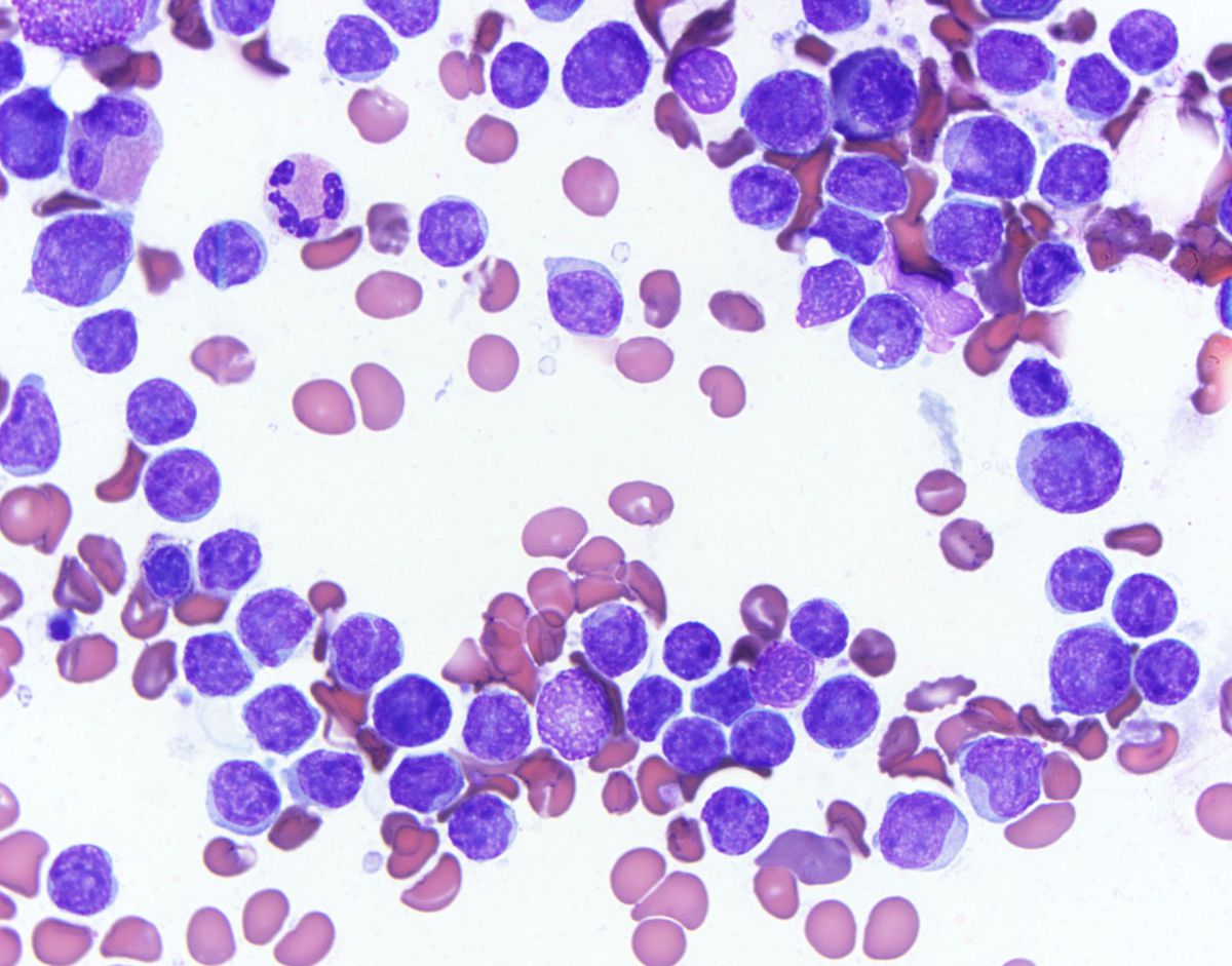 |
| Flow cytometry from blood: Mature B-cell phenotype positive for CD19, sCD22, sLambda-restricted, but only dim CD10. Notice dim CD45. | 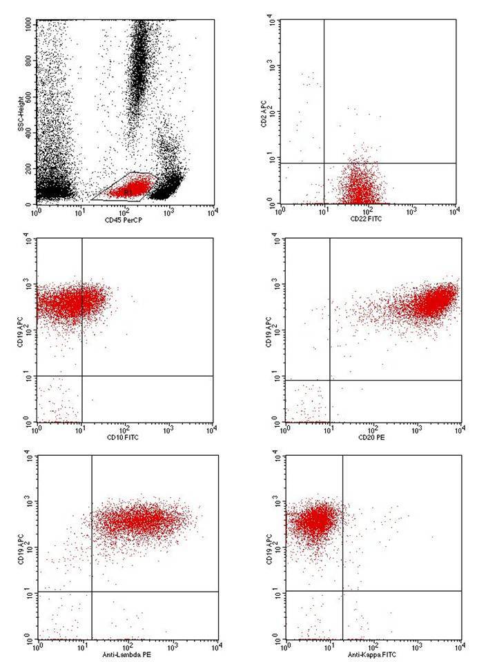 |
| Flow cytometry from blood: Tumor cells are positive for CD19 and cCD22, but negative for TdT, CD34, CD117, and cCD3. | 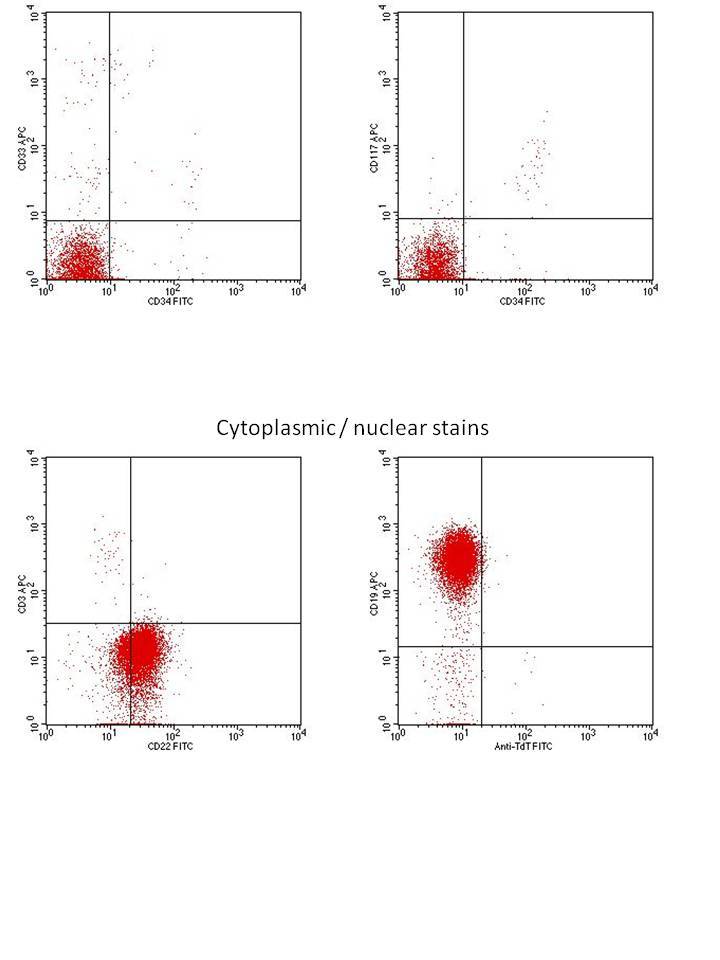 |