Institution: Hospital Universitario Marqués de Valdecilla/IFIMAV
Additional authors:Blas Meléndez Guerrero, Rocío Ramos Medina, Giovanna Roncador, Miguel A Piris
Session: Extramedullary manifestations of myeloid neoplasms
HISTORY
61 year old male with multiple cutaneous nodules in the thorax and upper extremities. The patient also had multiple lymphadenopathies in cervical, supraclavicular, axillary, inguinal regions, bone marrow and CSF involvement. After diagnosis the patient achieved CR after HYPER CVAD -MTX+ARAC DA X6 and underwent autologous stem cell transplantation. The patient is alive without evidence of disease 33 months after the initial diagnosis.
DETAILS
A skin biopsy is performed and formalin fixed paraffin embedded. HE discloses a perivascular, periadnexal and focally interstitial infiltrate of immature blastic cells. The cells have scant cytoplasm and blastic nuclei with homogeneous chromatin and inconspicuous nucleoli. Mitosis are readily seen. There is no epidermotropism nor angiotropism or necrosis.
IMMUNOHISTOCHEMISTRY AND FLOW CYTOMETRY
NEGATIVE MARKERS: CD2, CD3, CD20
POSITIVE MARKERS: CD45, CD123, TCL1, CD4, CD56, SpiB.INTERESTING FEATURES
Here we show a particular case of blastic plasmacytoid dendritic cell neoplasm with high stage at diagnosis with lymph node and bone marrow involvement in an advanced-age patient with optimal response to autologous stem cell transplantation.
We show the results with a new plasmacytoid dendritic cell associated marker (SpiB, a novel immunohistochemical marker for human blastic plasmacytoid dendritic cell neoplasms: characterization of its expression in major hematolymphoid neoplasms. Montes-Moreno S, Ramos-Medina R, Martínez-López A et al. Blood. 2013 Jan 24; 121(4):643-7) which can be used in the routine histopathological diagnosis.PROPOSED DIAGNOSIS
Blastic plasmacytoid dendritic cell neoplasm
CONSENSUS GROUP: ADDITIONAL INFORMATION/STUDIES
Additional immunohistochemical stains performed by the consensus group:
MPO: Negative
CONSENSUS DIAGNOSIS
Blastic plasmacytoid dendritic cell neoplasm
| HE (1) | 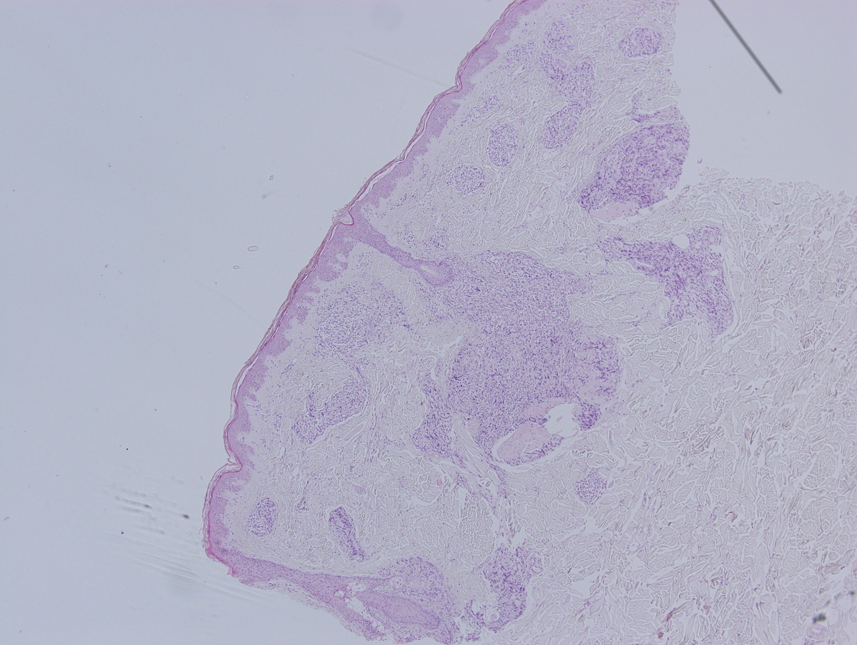 |
| HE (2) | 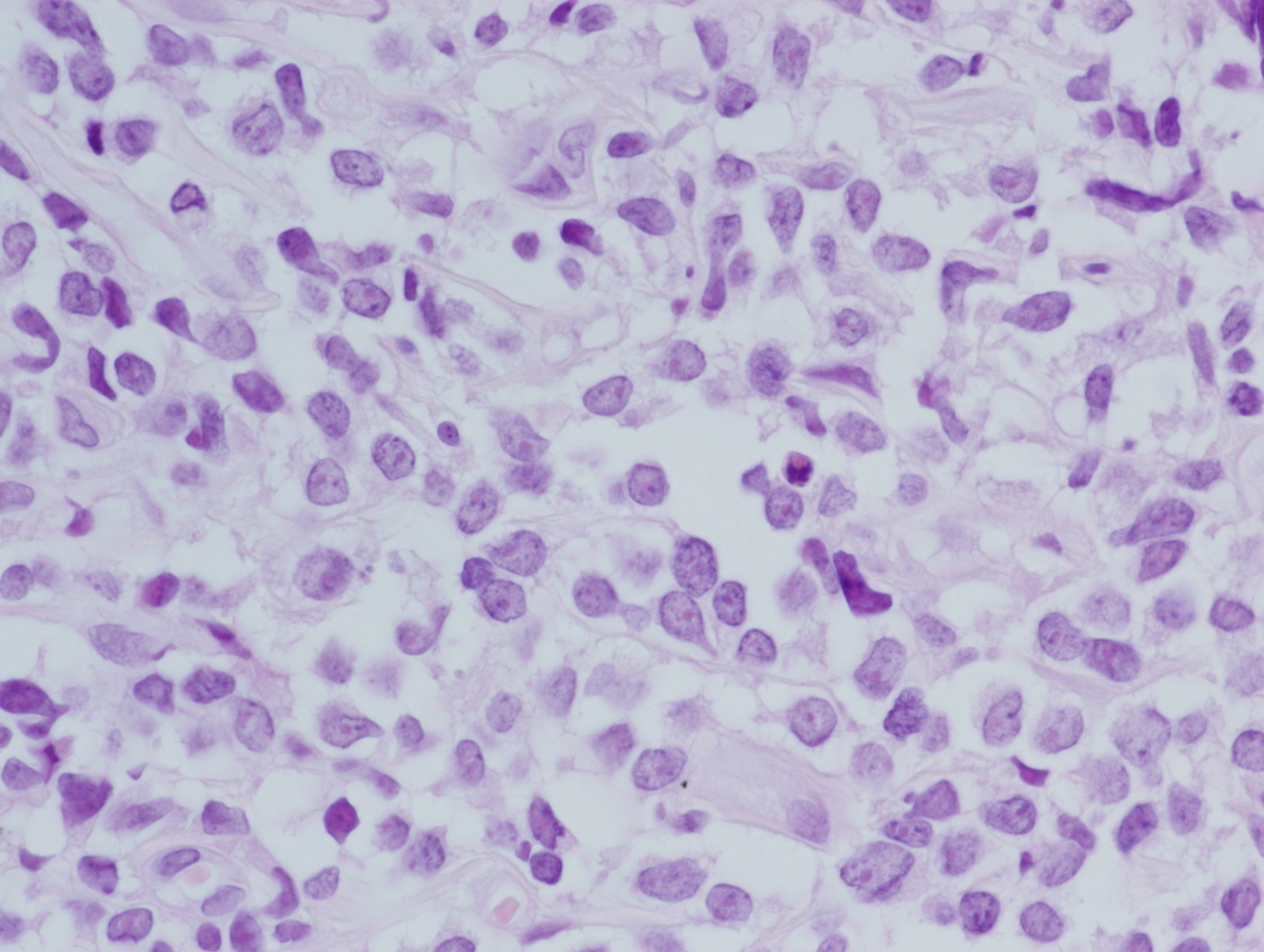 |
| HE (3) | 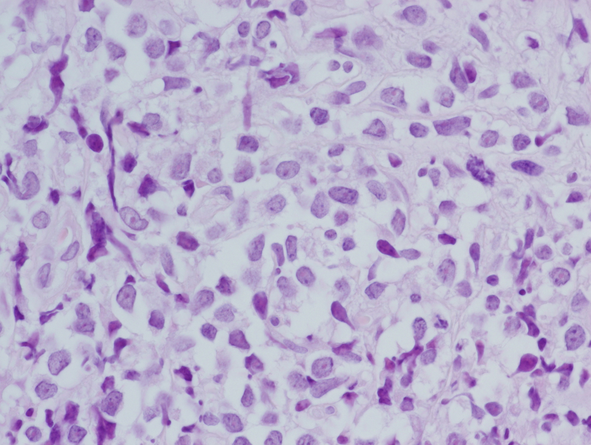 |
| CD2 | 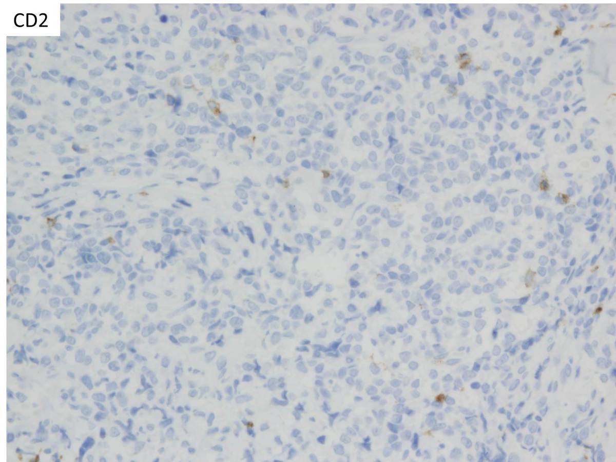 |
| CD3 | 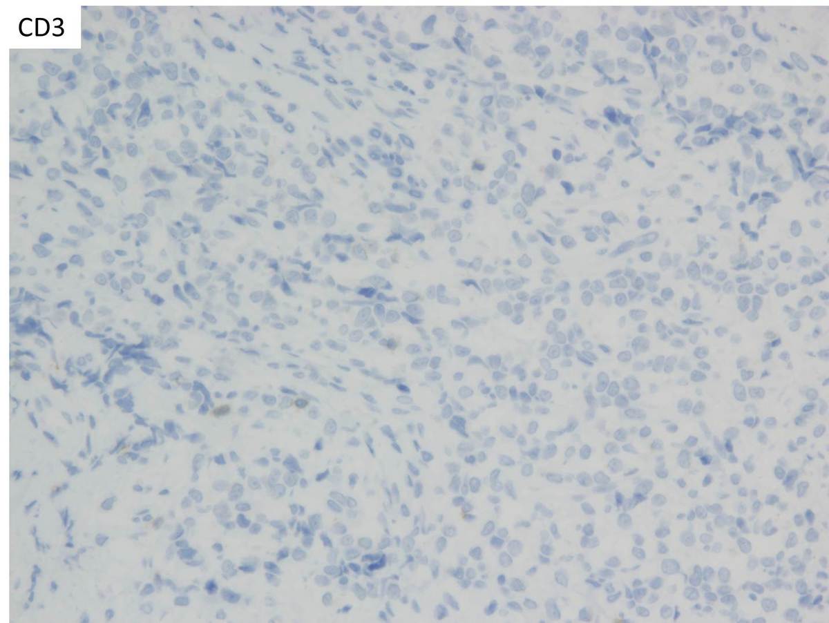 |
| CD20 | 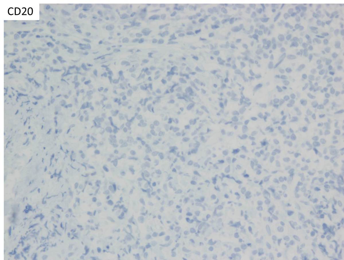 |
| CD4 | 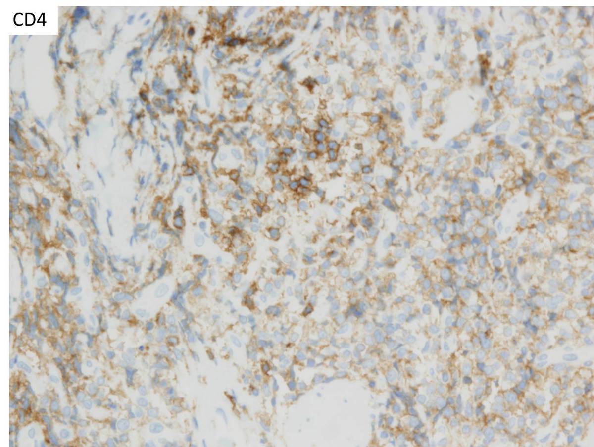 |
| CD45 | 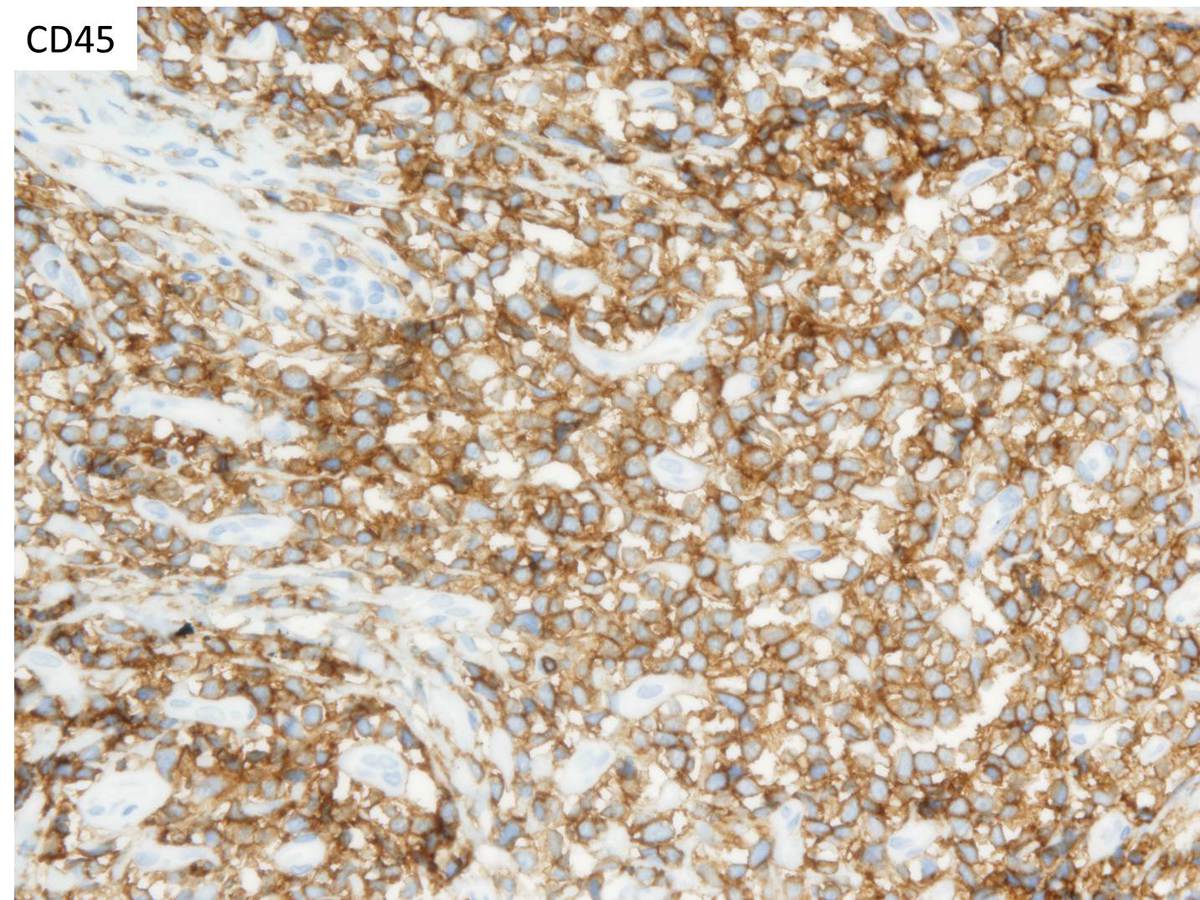 |
| CD56 | 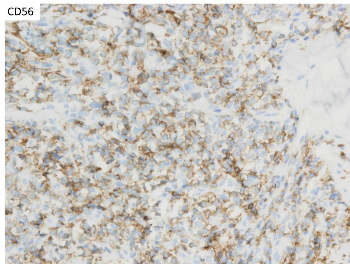 |
| CD123 | 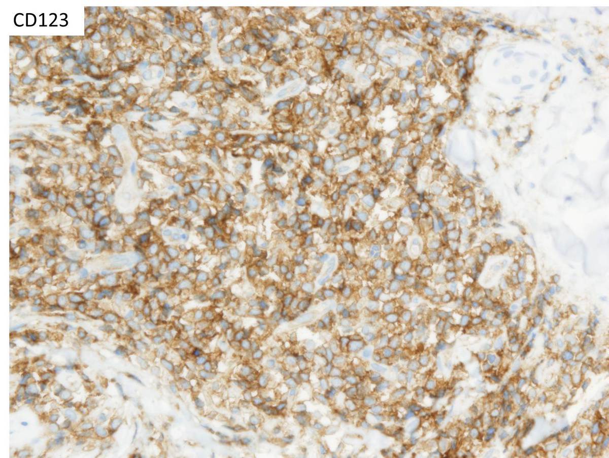 |
| TCL1 | 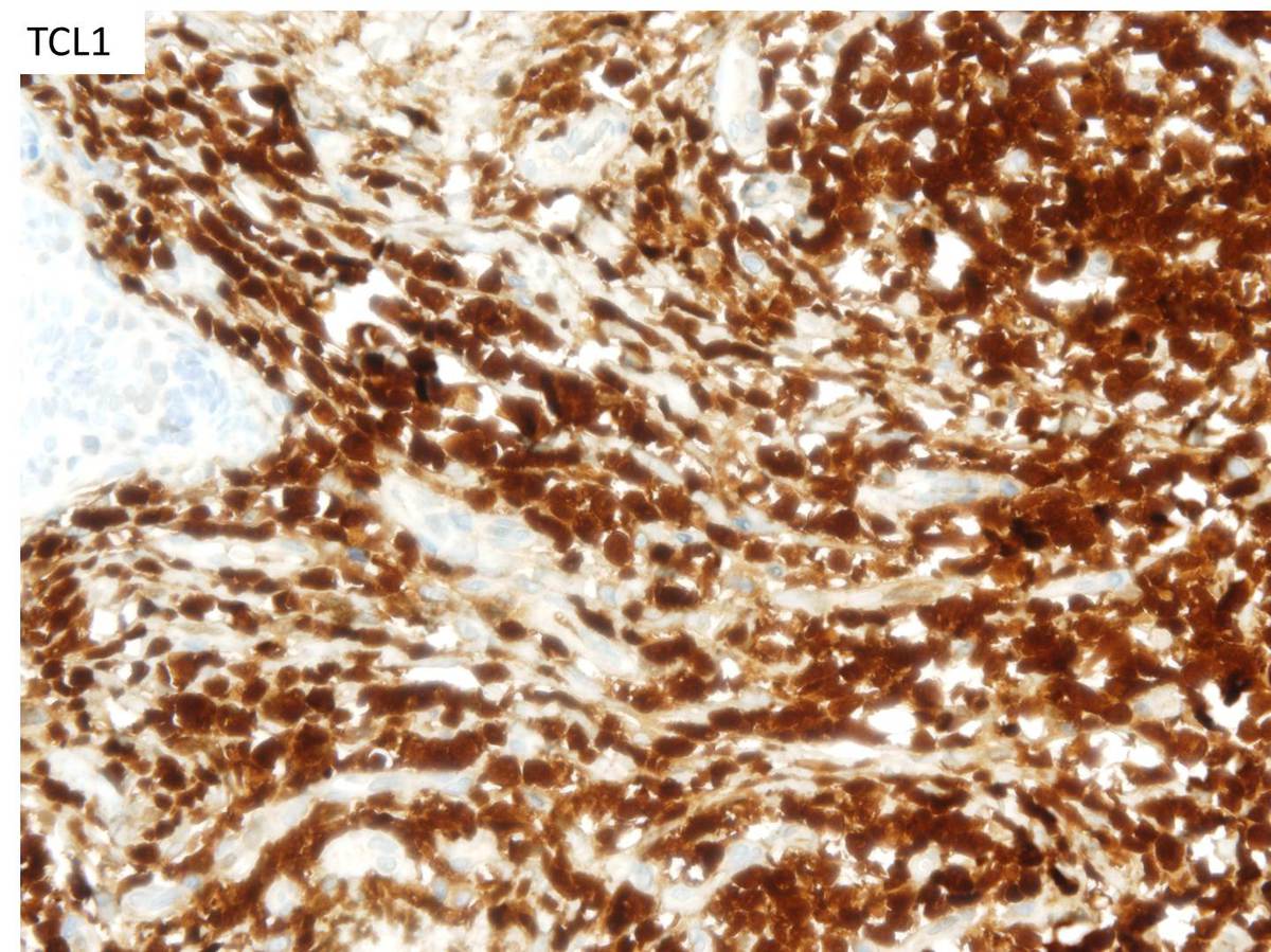 |
| SpiB | 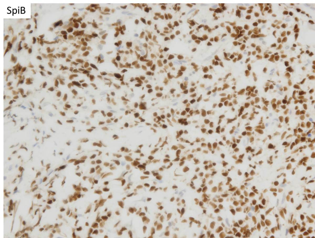 |
| Additional figure 1 | 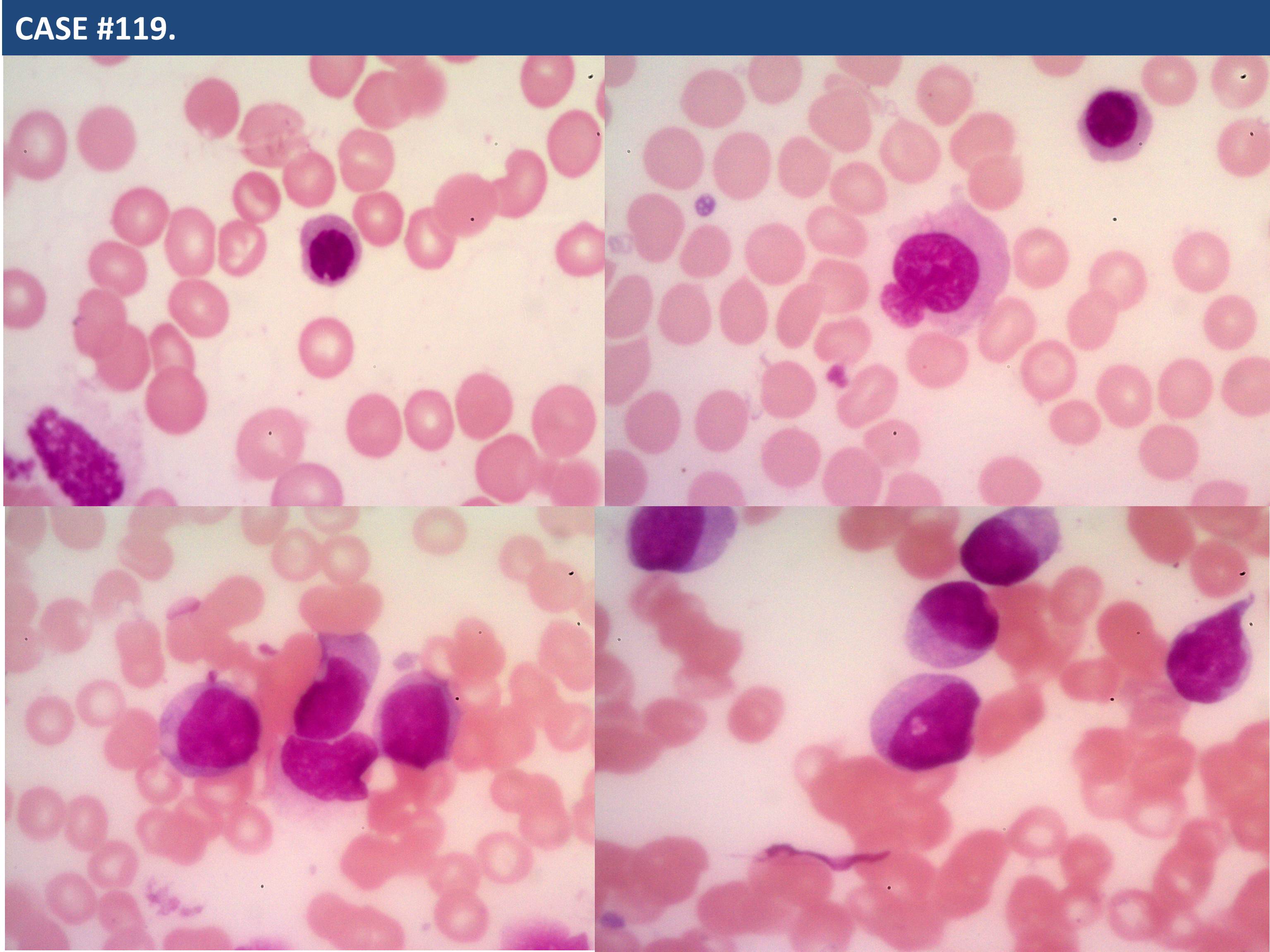 |