Institution: Cleveland Clinic
Additional authors:Megan Nakashima, Karl S. Theil
Session: Extramedullary manifestations of myeloid neoplasms
HISTORY
The patient is a 30 year-old man with chest pain. Imaging shows a 11 cm mediastinal mass. A biopsy is performed. After a diagnosis of T lymphoblastic lymphoma (T-ALL) is rendered, a bone marrow biopsy shows no evidence of malignancy. The patient receives a T-ALL targeted chemotherapy regimen (modified Larson protocol) and he is considered free of disease for 20 months.
20 months later, the patient presents with CNS involvement by a blastic process (see immunophenotype and morphology below). The bone marrow shows extensive involvement. Treatment with a myeloid regimen (7+3) results in remission. A bone marrow transplant is followed by graft-versus-host disease.A year after transplant the patient relapses again and expires during chemotherapy.DETAILS
Histologic sections of the formalin-fixed, paraffin-emdedded tissue from the mediastinal mass show diffuse sheets of small-intermediate sized cells, with irregular nuclei with fine chromatin. The cytoplasm is agranular. There are no normal structures preserved.
The bone marrow biopsies (20 and 33 months after the initial presentation) show sheets of blasts, similar to those in the mediastinal mass. The smears show small to intermediate-sized blasts, with scant, agranular cytoplasm and irregular nuclei with fine chromatin and no conspicuous nucleoli. These blasts are negative for myeloperoxidase or butyrate esterase activity.IMMUNOHISTOCHEMISTRY AND FLOW CYTOMETRY
Flow cytometry performed on cells from the mediastinal mass identified a population of abnormal cells, positive for CD7 and negative for CD3, CD4, CD5 and CD8. By immunohistochemical stains the neoplastic cells are positive for CD7, CD33, CD34 (dim), CD56. They are negative for CD1a, CD2, CD3, CD4, CD5, CD8, CD10, CD20, CD43, CD123, TCL1, myeloperoxidase, TdT, and EBER.
Flow cytometry performed on the cells from the bone marrow aspirates (recurrences) show blasts positive for CD7, CD13, CD33, CD34, CD38, CD56 and HLA-DR. These cells are negative for CD2, CD3 (surface or cytoplasmic), CD4, CD5, CD8, CD10, CD11b, CD14, CD16, CD19, CD20, CD64, CD65, CD79b, CD117, IgM (surface or cytoplasmic), myeloperoxidase, and TdT.CYTOGENETIC FINDINGS
Conventional cytogenetic analysis performed on the negative bone marrow at presentation and on the bone marrow involved by leukemia at first recurrence showed a normal male karyotype.
The test performed on the bone marrow during the second recurrence showed a complex karyotype: 45-47,XY,-Y[3],add(1)(p36.1)[11],del(3)(q12q21)[2],add(8)(p23)[15],-9[7],add(10)(p15)[14],del(11)(q2?1q2?5)[16],add(12)(p13)[4],+13[5],add(14)(q22)[4],add(16)(p13.3)[4],add(19)(q13.1)[6],+mar1[4],+1-2 mar[7][cp16]/46,XY[4].The complex karyotype at the second recurrence is probably representative for the neoplastic clone.MOLECULAR FINDINGS
Molecular analysis performed on the mediastinal mass by polymerase chain reaction, using as template DNA extracted from the fixed, paraffin-embedded tissue and primers for the T-cell receptor genes showed the presence of a population with clonally rearranged T-cell receptor genes.
Fluorescence in-situ hybridization (FISH) studies show no evidence of BCR-ABL or MLL rearrangements.INTERESTING FEATURES
This case of leukemia positive for CD7 and CD56 shows features similar to the very few other cases reported in the literature to have similar phenotype: young male predominance, presentation as an extramedullary mass, no abnormalities involving chromosome 5, and poor prognosis. In the literature, these neoplasms were diagnosed as myeloid/NK-cell acute leukemia or stem cell leukemia. Further classifying these neoplasms according to the WHO 2008 classification of hematolymphoid neoplasms is difficult, as they lack most lineage specific markers: CD3, CD10, CD11c, CD14, CD19, CD22, CD64, myeloperoxidase or lysozyme. According to the WHO 2008 classification these neoplasms could be classified as either acute myeloid leukemia with minimal differentiation (FAB M0) or acute leukemia of ambiguous lineage-acute undifferentiated leukemia. Acute myeloid/NK-cell precursor leukemia is briefly discussed in the WHO 2008 classification (page 155), but no diagnostic criteria are stated and it is recommended that the cases should be classified as acute leukemia with minimal differentiation.
PROPOSED DIAGNOSIS
Acute myeloid leukemia with minimal differentiation (M0) presenting as myeloid sarcoma.
Acute leukemia of ambigous lineage-acute undifferentiated leukemia presenting as myeloid sarcoma.Acute myeloid/NK-cell cell precursor leukemia, presenting as myeloid sarcoma.CD7+ CD56+ acute leukemia.CONSENSUS GROUP: ADDITIONAL INFORMATION/STUDIES
Additional immunohistochemical stains perfomed by the consensus group:
CD11c: subset positive, variable intensity
CD68: subset positive, paranuclear/granular
CD117: negative
CONSENSUS DIAGNOSIS
Myeloid sarcoma (minimally differentiated) involving mediastinum, relapse with only bone marrow and cerebrospinal fluid involvement
| Mediastinal Mass HE 10x |  |
| Mediastinal Mass HE 40x |  |
| Mediastinal Mass CD7 40x |  |
| Mediastinal Mass CD56 40x | 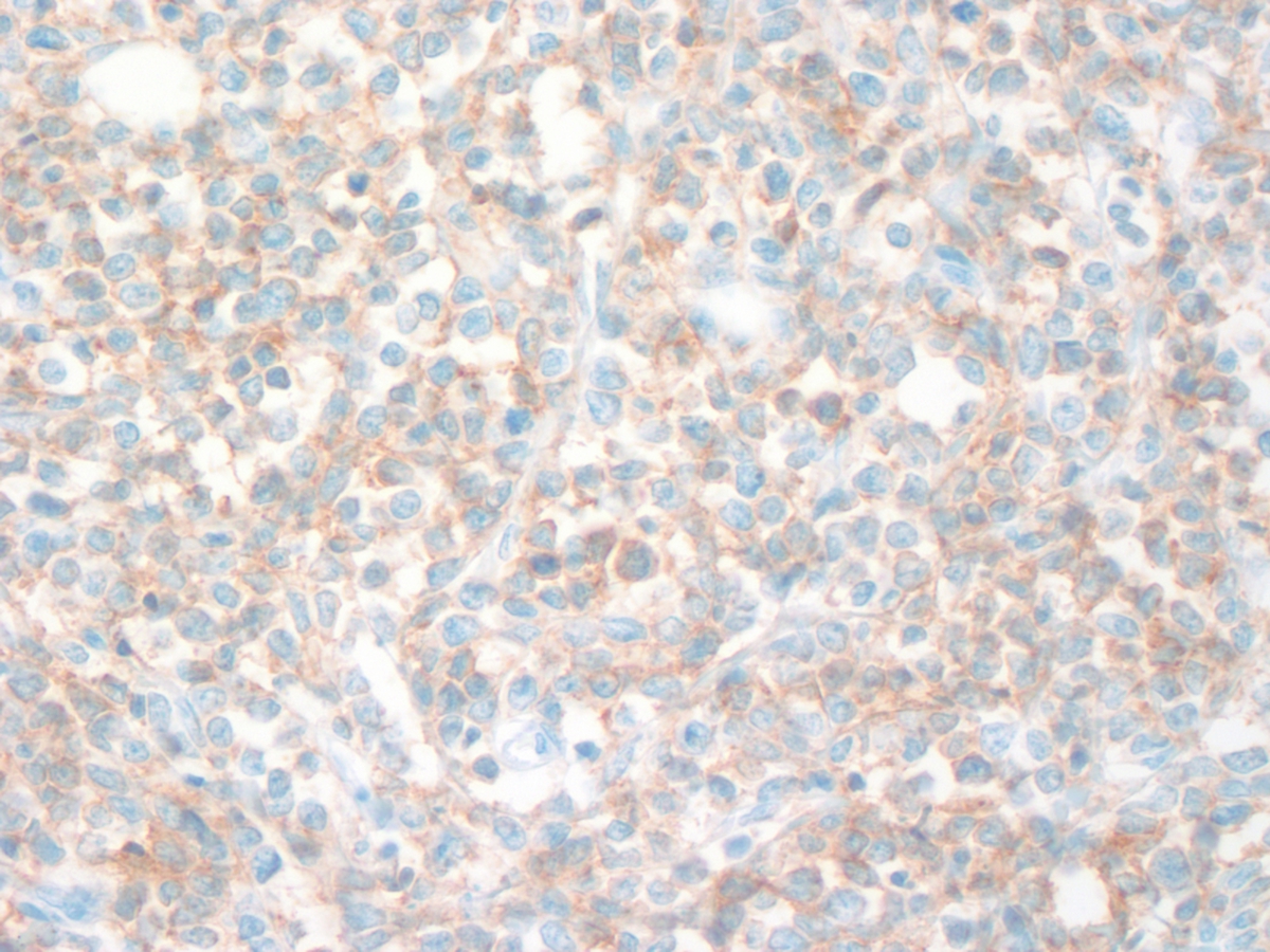 |
| Mediastinal Mass CD33 40x | 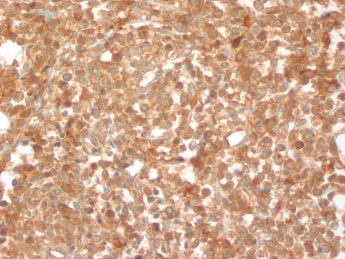 |
| Mediastinal Mass CD34 40x |  |
| Mediastinal Mass Myeloperoxidase 40x |  |
| Mediastinal Mass Ki-67 40x |  |
| Recurrence Bone Marrow Biopsy 40x |  |
| Recurrence Bone Marrow Aspirate WG 100x | 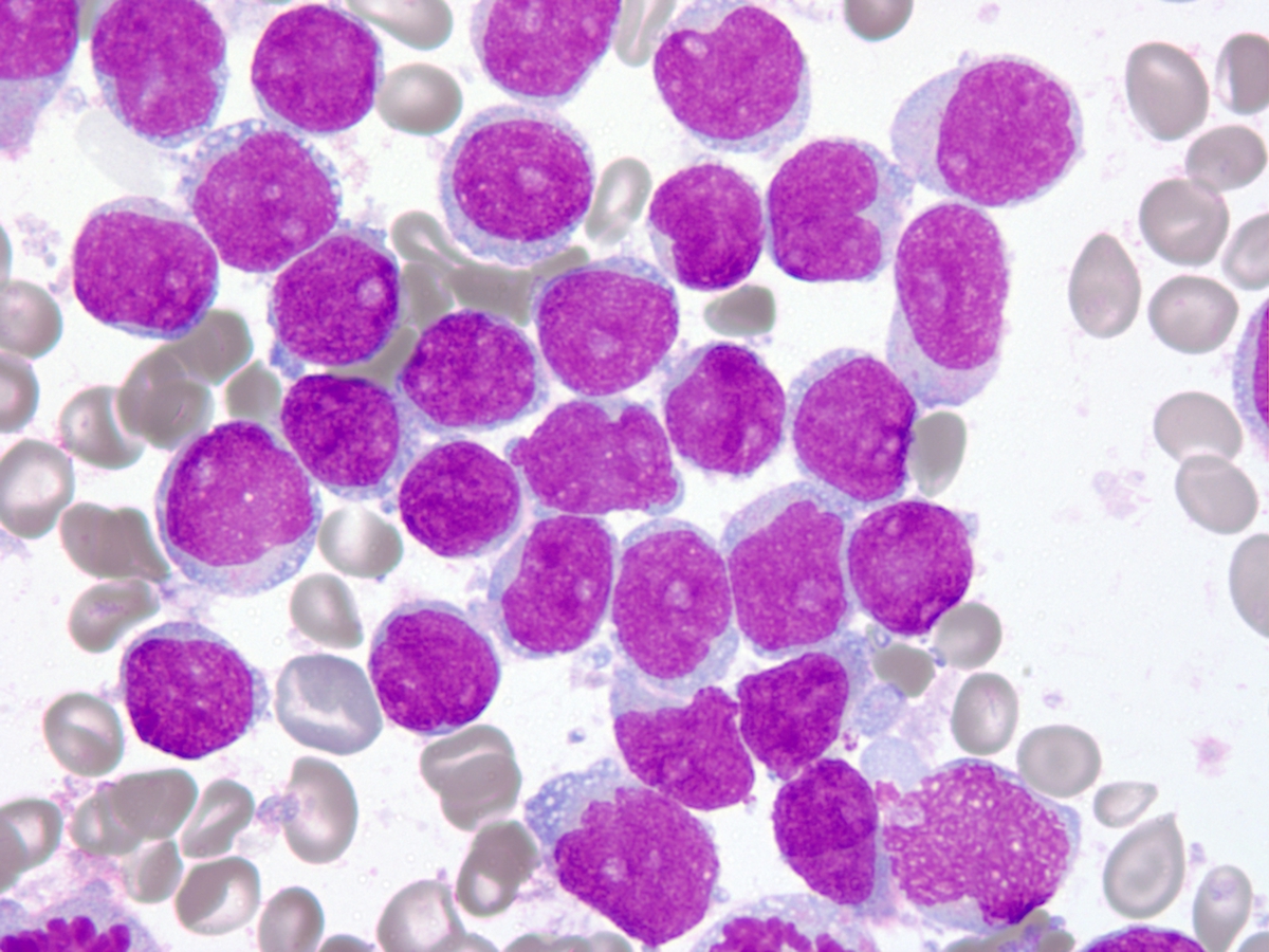 |
| Recurrence Flow Cytometry |  |
| Second Recurrence Conventional Cytogenetics | 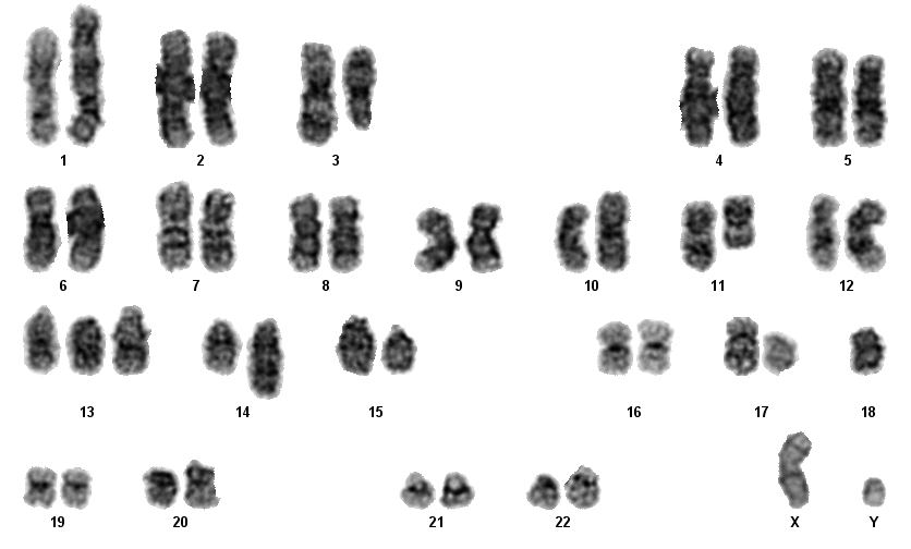 |
| Additional figure 1: CD11c IHC, 400x |  |
| Additional figure 2: CD68 IHC, 400x | 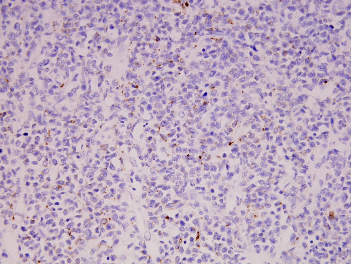 |