Institution: Stanford University Medical Center
Additional authors:Daniel Arber, MD
Session: Extramedullary manifestations of myeloid neoplasms
HISTORY
The patient is a 23 year old man with 3 weeks of left upper quadrant abdominal pain clinically suspicious for gallstone pancreatitis status-post an emergency ERCP and stent placement, although only sludge was evacuated from the common bile duct. Imaging studies demonstrated renal and pancreatic lesions with multiple enlarged retroperitoneal lymph nodes. An upper endoscopy was performed to obtain diagnostic material and to rule out IgG4 related disease. A biopsy of a gastric erosion demonstrated an extramedullary myeloid tumor with monocytic differentiation. A bone marrow biopsy was obtained 1 month later and confirmed involvement by a monocytic leukemia.
DETAILS
GASTRIC BIOPSY: The biopsy from the gastric erosion demonstrates an infiltrate of medium to large-sized cells with smooth chromatin and inconspicuous nucleoli within the mucosa. Occasional eosinophils are noted in the background. This atypical cell infiltrate is adjacent to a large reactive lymphoid aggregate. Concurrent biopsies from the stomach antrum and body demonstrated chronic active gastritis and helicobacter organisms.
BONE MARROW BIOPSY (1 month later): WBC: 15 K/uL HGB: 12.6 g/dL PLT: 119 K/uL. The peripheral blood demonstrates blasts (73%) with monocytic morphology. Occasional atypical eosinophils are noted.The aspirate smears show a monotonous population of large sized blasts (enumerated at 73%) with immature chromatin, round to irregular nuclear contours, prominent nucleoli, and abundant basophilic cytoplasm with occasional eosinophilic granules. Background trilineage hematopoiesis is reduced with no overt dysplasia. Eosinophils are increased and numerous forms with both eosinophilic and basophilic granules are appreciated. The core biopsy is effaced by blasts with irregular nuclear contours and abundant cytoplasm. Eosinophils are readily identified. Trilineage hematopoietic elements are reduced and are without significant dysplasia.IMMUNOHISTOCHEMISTRY AND FLOW CYTOMETRY
GASTRIC BIOPSY: Immunohistochemistry shows that the atypical population within the gastric mucosa is positive for CD4, CD33, CD43, CD68, CD163, and subset positive for CD34 and CD117. These cells are negative for myeloperoxidase and CD15. TdT staining is equivocal with high background, but may label a subset of the atypical cells. Notably, CD4 stains more cells than CD3 and CD5. CD8 labels small scattered T cells. CD20, CD10, CD79a, PAX5 and CD21 highlight a well-circumscribed germinal center. CD56 labels only rare small lymphocytes.
BONE MARROW: Flow cytometry performed on the bone marrow aspirate showed that blasts are positive for CD34, CD117, HLA-DR, CD38, CD13, CD33, CD15, MPO, partial CD64, partial CD4, and partial TdT. The monocyte gate shows a mixture of aberrant blasts and maturing monocytes with partial aberrant CD2 expression.CYTOGENETIC FINDINGS
46,XY,inv(16)(p13q22)[20]; nuc ish(CBFBx2)(5'CBFB sep 3'CBFBx1)[180/200]
MOLECULAR FINDINGS
KIT gene sequencing analysis demonstrated the following mutations: c.1249A>C, c.1251T>C, c.1252T>G, c.1253A>G, c.1254C>A, c.1256A>G, c.1257C>G, c.1258A>T (p.T417P, p.Y418G, p.D419G, p.R420W)
Positive for NRAS c.38G>A p.G13D and NRAS c.182A>G p.Q61R mutationsNegative for other mutations identified in the cancer somatic mutation panel: APC exon 16; BRAF exons 11, 15; CTNNB1 exon 3; IDH1 exon 4; IDH2 exon 4; DNM3TA exon 23; EGFR exons 18, 19, 20, 21; ERBB2/HER2 exon 20; KRAS exon 2; MYD88 exon 5; NOTCH1 exon 26; NRAS exon 2; PIK3CA exons 10, 21; PTEN exons 5,6,7; TP53 exons 7,8; SF3B1 exon 15Negative for NPM1, FLT3, and CEBPA mutationsINTERESTING FEATURES
1) We describe an atypical presentation of AML with inv(16) initially diagnosed in a stomach biopsy of a young man with symptoms related to constriction of the common bile duct and pancreatitis. The stomach biopsy showed an atypical large cell population positive for CD4, but negative for CD3 and CD5. These cells were further revealed to be immature monocytic cells. A subsequent bone marrow biopsy demonstrated involvement by AML with inv(16), also with NRAS and KIT mutations. Although granulocytic sarcoma is rare, it is associated with AML with inv(16) and is more likely to present with abdominal symptoms (85% present in the GI tract, Zhang et al., Leukemia Research, 2010).
2) Molecular: Additional mutations in NRAS and KIT were identified that likely contributed to leukemogenesis. Mutations in exon 8 of the KIT gene detected in AML often consist of small insertion-deletion events affecting the highly conserved amino acid position Asp419 (Gari et al., British J Haematology, 1999). In this case, a novel balanced 10-bp insertion-deletion event alters amino acid positions 417-420 with preservation of distal coding sequence. This particular insertion-deletion in exon 8 of the extracellular domain of KIT has not been previously reported in the literature. Some studies show that exon 8 mutations are associated with worse clinical outcomes in adults with AML with inv(16)/t(16;16) (Paschka et al., J Clin Oncology, 2006) while others have not shown a difference in overall survival (Boissel et al., Leukemia, 2006). NRAS mutations are frequently identified in AML with inv(16)/t(16;16) with no significant effect on prognosis (Boissel et al., Leukemia, 2006).PROPOSED DIAGNOSIS
Stomach biopsy: extramedullary myeloid tumor with monocytic differentiation.
Bone marrow biopsy: acute myeloid leukemia with inv(16)(p13.1q22); CBFB-MYH11CONSENSUS DIAGNOSIS
Acute myeloid leukemia with inv(16)(p13.1q22), CBFB-MYH11, NRAS and KIT mutations, involving bone marrow and stomach mass (myeloid sarcoma)
| gastric erosion, H&E, 4x, demonstrating atypical large cell infiltrate within gastric mucosa with an adjacent reactive lymphoid aggregate |  |
| gastric erosion, H&E, 10x, atypical large cell infiltrate within gastric mucosa | 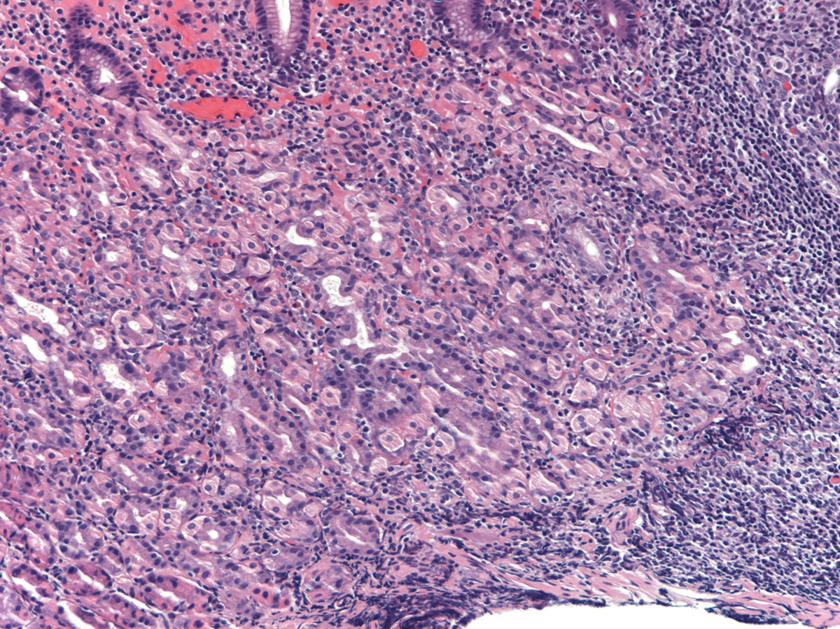 |
| gastric erosion, H&E, 40x, atypical large cell infiltrate within gastric mucosa admixed with eosinophils | 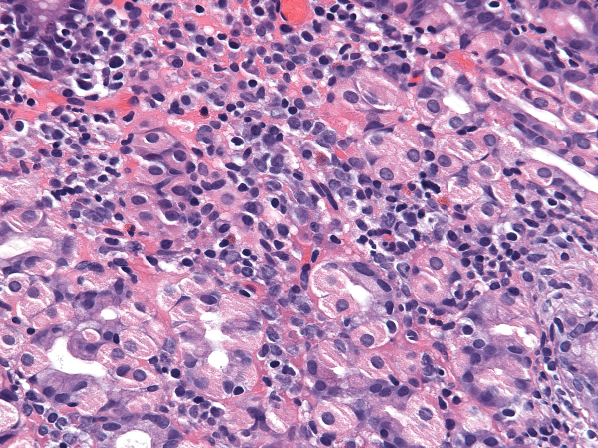 |
| gastric erosion, CD4, 40x, demonstrating positivity for CD4 on atypical cells |  |
| gastric erosion, CD33, 40x, demonstrating positivity for CD33 |  |
| gastric erosion, CD117, 40x, demonstrating clusters of CD117 cells | 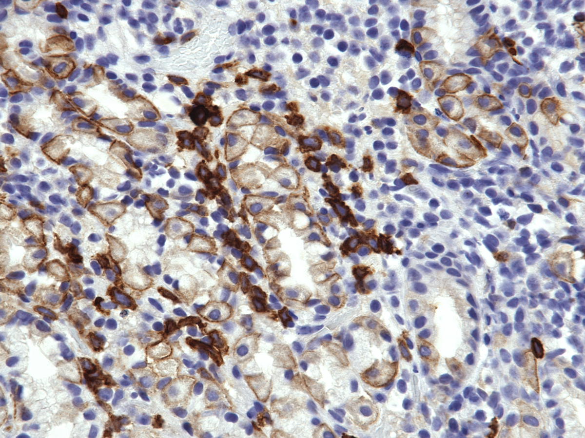 |
| gastric erosion, CD34, 40x, demonstrating clusters of CD34 positive cells | 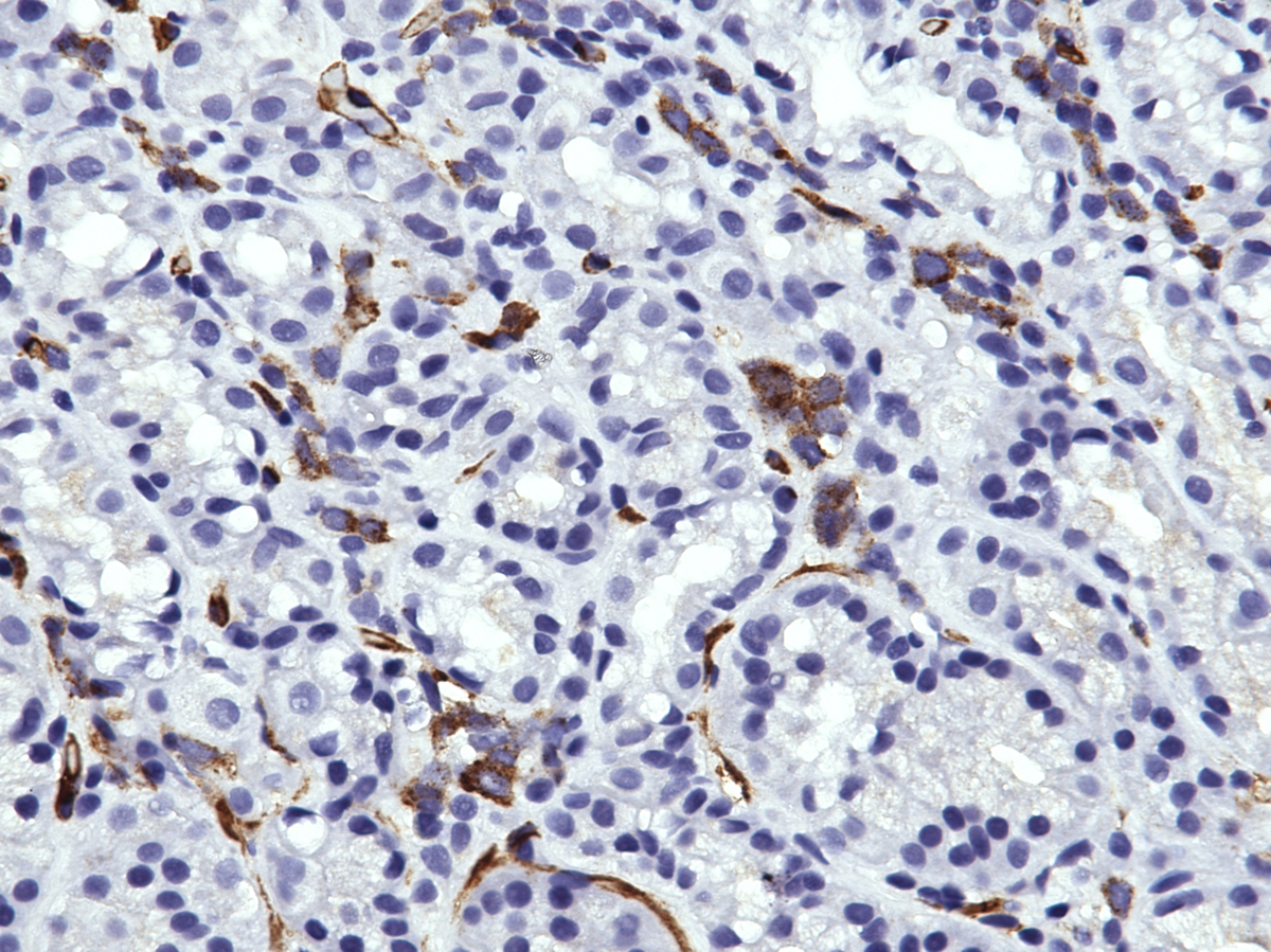 |
| peripheral blood with blasts, 60x, 1 month after gastric biopsy | 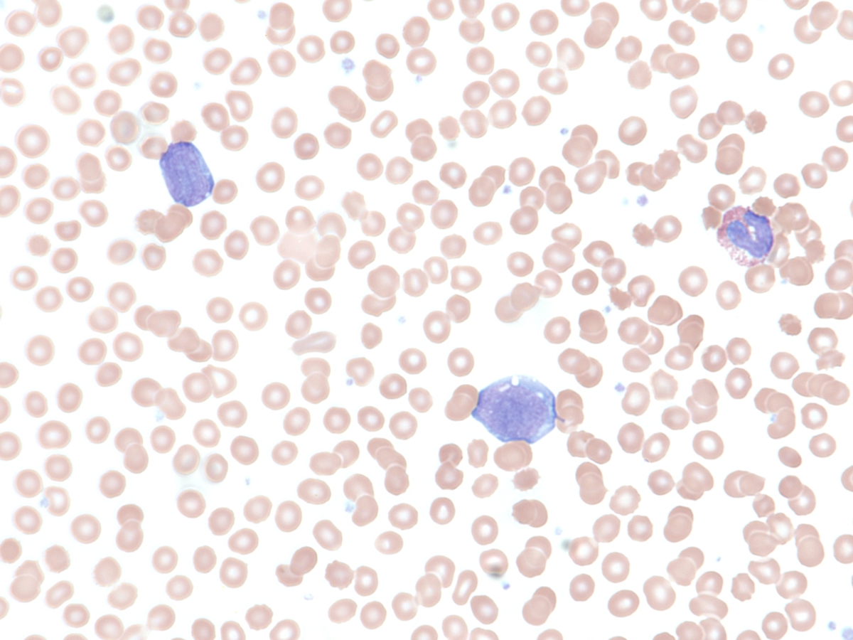 |
| bone marrow aspirate with blasts, 60x, 1 month after gastric biopsy | 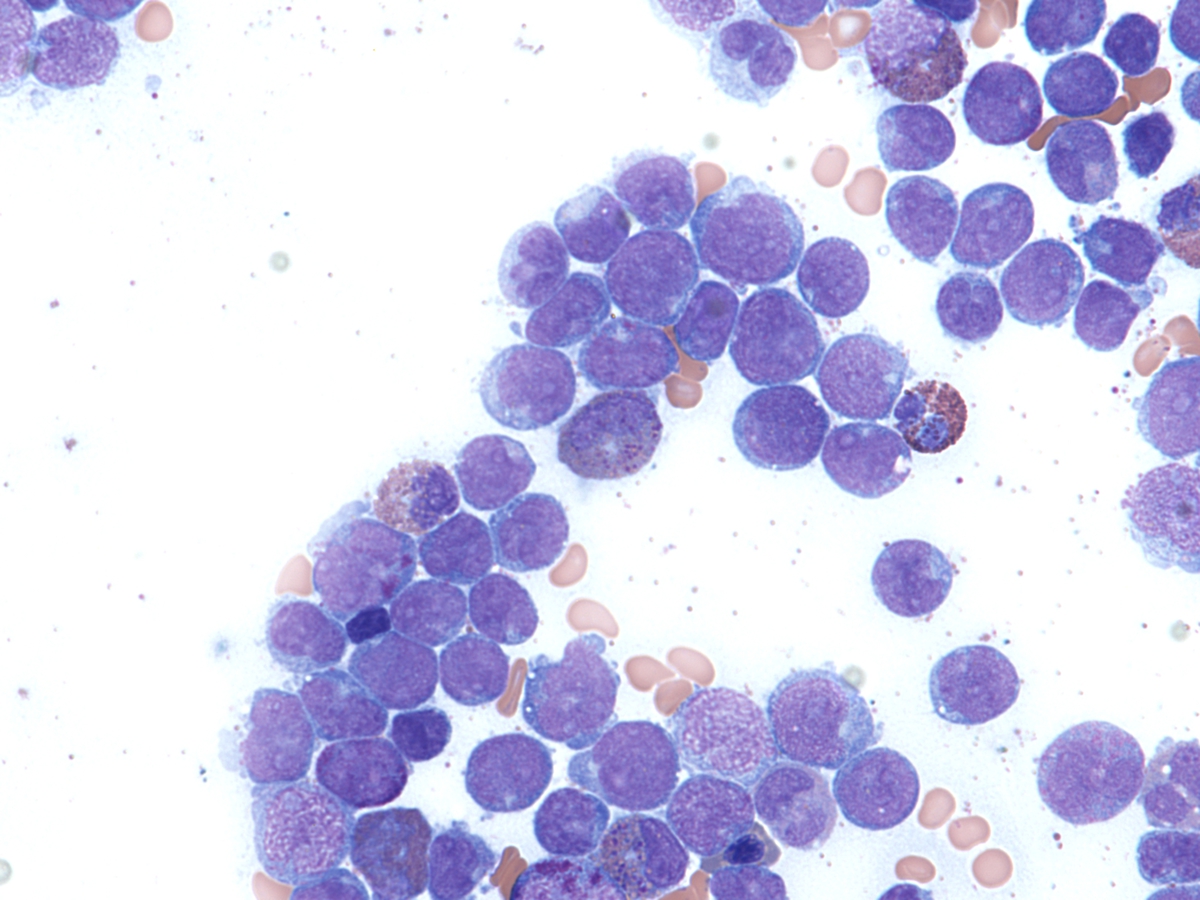 |
| core biopsy with blasts and eosinophils, 1 month after gastric biopsy | 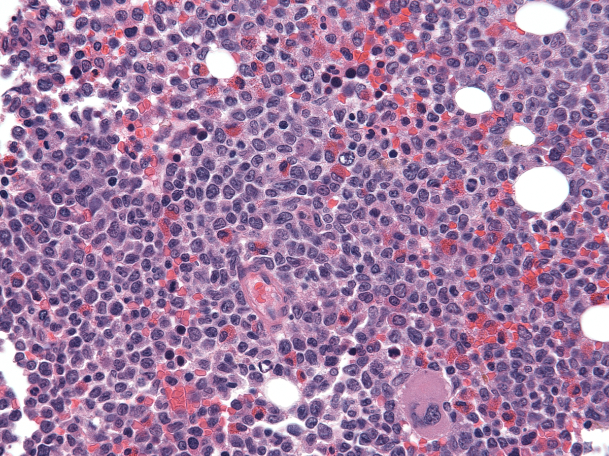 |