Institution: Western General Hospital & University of Edinburgh
Session: Extramedullary manifestations of myeloid neoplasms
HISTORY
This male patient was diagnosed with chronic myelomonocytic leukaemia in March 2011, aged 66 years. At presentation his blast cell count was 12% (CMML-2). He was initially considered for intensive chemotherapy but had no matched sibling for transplant and worsening angina. He was treated conservatively with occasional blood transfusions. Triple artery coronary artery bypass grafting was undertaken in October 2011. The patient was subsequently investigated for suspected renal colic, at which time CT scans revealed splenomegaly and generalised lymphadenopathy of moderate size. An enlarged left axillary lymph node was excised in July 2012 (submitted specimen). Treatment with Azacytidine was initiated in September 2012 and the patient has currently received three of the planned six cycles of therapy.
DETAILS
Two left axillary lymph nodes were received received fresh. These measured 28 x 20 x 10 mm and 12 x 12 x 8 mm. Samples were taken for flow cytometry and cytogenetic analysis. The remaining tissue was fixed in 10% buffered formalin then routinely processed.
Microscopic examination revealed residual reactive lymphoid follicles but much of the paracortex was replaced nodular aggregates of intermediate size cells with moderate amounts of cytoplasm, round or ovoid nuclei and inconspicuous nucleoli. Numerous apoptotic bodies were identified in association with these cells. The nodular aggregates were surrounded by a more polymorphic population of cells. This comprised many cells of large size with abundant cytoplasm and slightly cleaved or horseshoe-shaped nuclei, admixed with mononuclear cells containing eosinophilic cytoplasmic granules.IMMUNOHISTOCHEMISTRY AND FLOW CYTOMETRY
Immunohistochemistry was performed on sections cut from formalin fixed paraffin embedded tissue blocks of the lymph node biopsy. The cells forming the nodular aggregates stained positively for CD4, CD45, CD68, CD303 (BDCA2) and granzyme B. They were, at most, only weakly positive for CD33 and did not express CD3, CD7, CD20, CD34, CD56, CD61, CD117, TdT, glycophorin A and myeloperoxidase. The cells forming the more polymorphic population surrounding the nodules were positive for CD33 with a proportion reacting with antibodies to CD117, TdT and possibly CD4.
CYTOGENETIC FINDINGS
A normal male karyotype (46XY[20]) was found in both the presenting bone marrow specimen and in the lymph node biopsy.
MOLECULAR FINDINGS
Not done.
INTERESTING FEATURES
Nodular proliferations of plasmacytoid dendritic cells (PDC) are frequently seen in bone marrow biopsies of patients with CMML. This case illustrates the less common phenomenon of tumour forming plasmacytoid dendritic cell proliferations that are occasionally reported at extramedullary sites. There is some evidence that these proliferations are clonally related to the associated CMML. It has also been suggested their presence is associated with a poor outcome, but the numbers of cases studied to date precludes a definitive statement on the prognostic significance of such a finding.
PROPOSED DIAGNOSIS
Tumour forming accumulation of plasmacytoid dendritic cells in association with chronic myelomonocytic leukaemia.
CONSENSUS GROUP: ADDITIONAL INFORMATION/STUDIES
Additional immunohistochemical stains performed by the consensus group:
TCL1: Labels groups of cells located in the nodules
CONSENSUS DIAGNOSIS
Chronic myelomonocytic leukemia with nodular plasmacytoid dendritic cell proliferation in lymph node
| Fig 1: H&E x4 |  |
| Fig 2: H&E x20 PDC nodule | 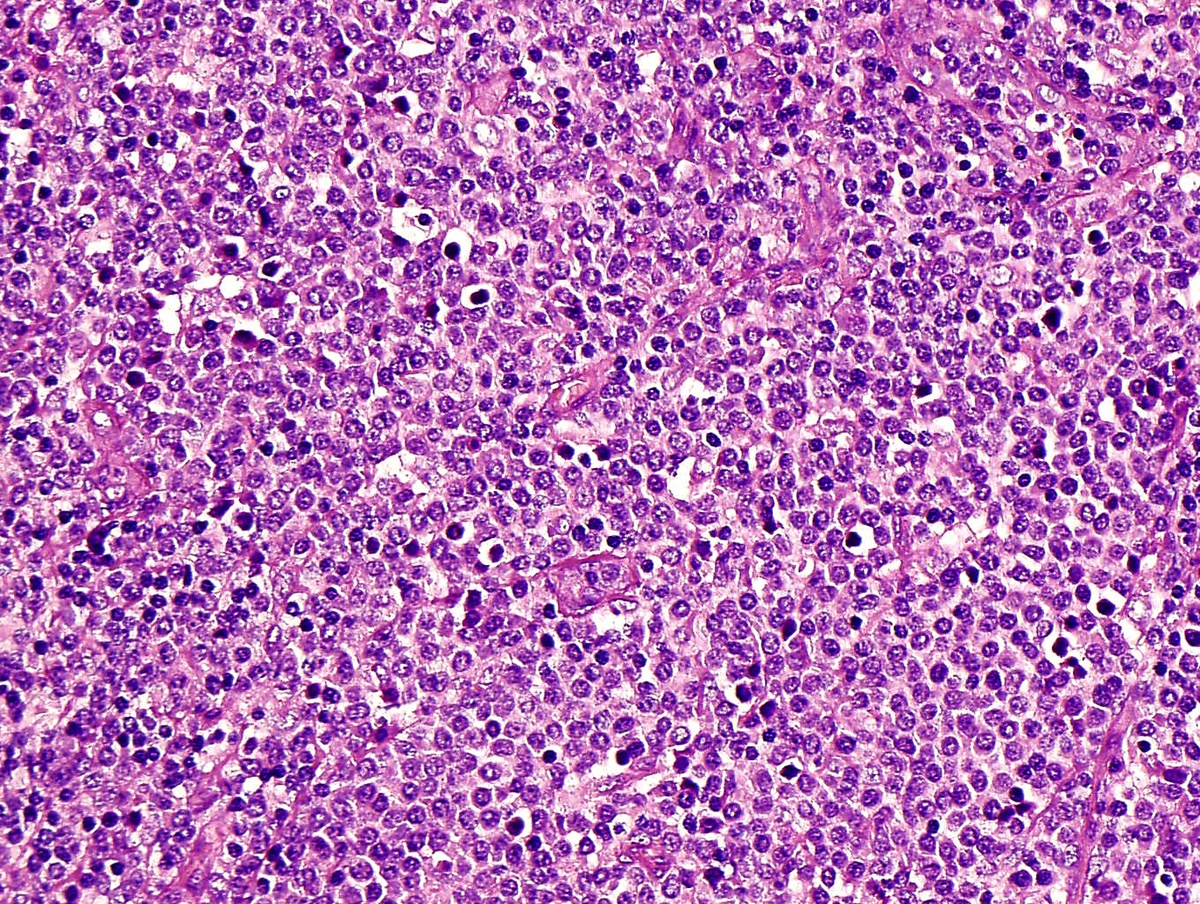 |
| Fig 3: CD4 |  |
| Fig 4: CD33 |  |
| Fig 5: CD56 | 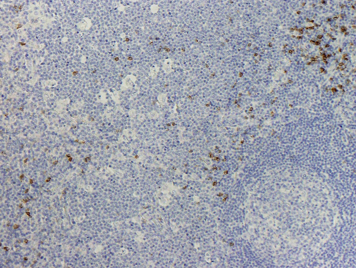 |
| Fig 6: CD303 / BDCA2 | 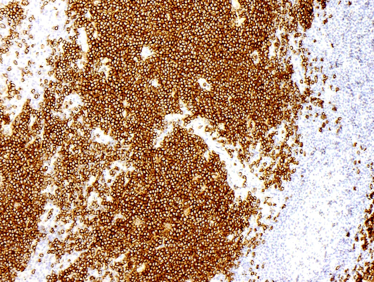 |
| Fig 7: Granzyme B | 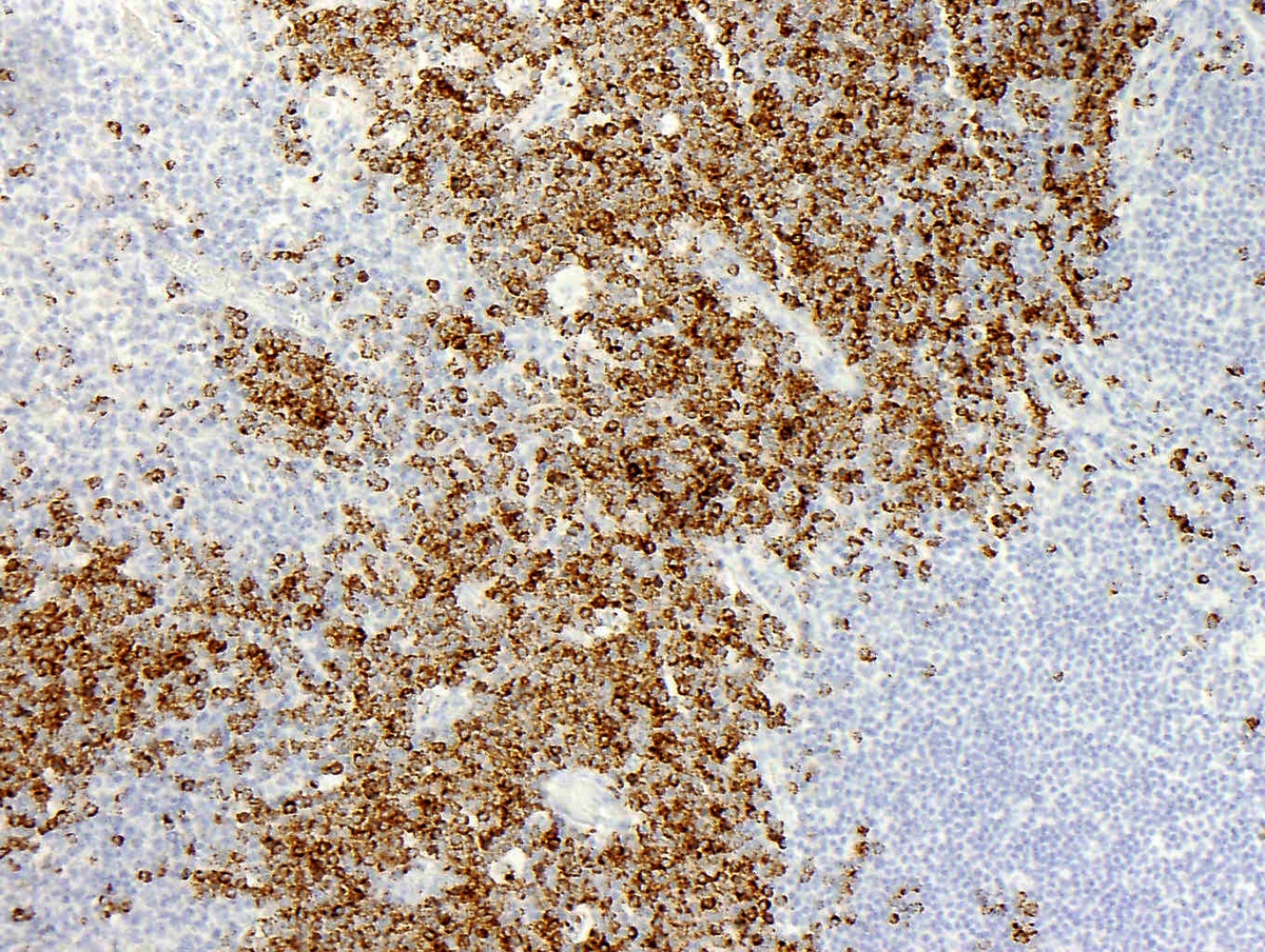 |
| Additional figure 1: TCL1 IHC, 20x | 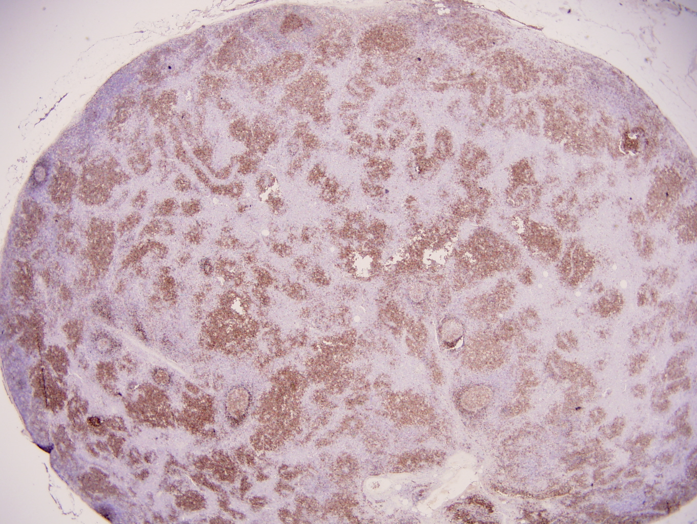 |
| Additional figure 2: TCL1 IHC, 200x |  |