Institution: Columbia University, New York, NY
Additional authors:Bachir Alobeid, Govind Bhagt, Daniela Hoehn
Session: B Lymphoblastic Leukemia/Lymphoma
HISTORY
The patient was a 17-year-old previously healthy female with a vague history of microcytic anemia. She presented with 10-day duration of abdominal pain, nausea, headache, weakness, dizziness, 2-day of fever and emesis, and sudden onset of hypotension, tachypnea and hypoxia.
CBC revealed a markedly increased WBC with hypereosinophilia, anemia and thrombocytopenia (WBC: 136, Hb: 8.8, Hct: 27.5, Platelets: 153). Differential showed 25% neutrophils and 60% of eosinophils. LDH was markedly elevated at 1135. CT scan showed thickened terminal ileum, cecum and splenomegaly. Echo showed diminished ventricular function. Peripheral blood flow cytometry was suspicious for B-cell lymphoblastic leukemia. Bone marrow biopsy/aspirate and flow cytometry were diagnostic of B-cell lymphoblastic leukemia with hypereosinophilia. Biopsy of the right ventricle of the heart showed eosinophilic myocarditis. Following the bone marrow examination, chemotherapy was initiated. The patient subsequently developed cardiac failure, renal and hepatic insufficiency, respiratory failure and retroperitoneal hemorrhage. The patient expired 2 weeks after initial presentation.DETAILS
Bone Marrow Examination
A bone marrow trephine biopsy was fixed in Bouin’s solution. The bone marrow aspirate was evaluated by morphology, cytogenetics and flow cytometry.H&E stained sections of the biopsy showed extensive infiltration by blast cells with eosinophilia. Giemsa-Wright stained aspirate smears revealed a large population of lymphoblasts (70% of all nucleated marrow elements) with numerous eosinophils.Right Ventricle of Heart ExaminationBiopsy from the right ventricle of the heart was fixed in formalin and evaluated by morphology.H&E stained slides showed myocardium with interstitial edema, prominent capillary endothelium, and scattered interstitial eosinophils.IMMUNOHISTOCHEMISTRY AND FLOW CYTOMETRY
Bone Marrow Examination
Immunohistochemical analysis of the bone marrow biopsy showed the blasts: CD79a+, CD10+, CD34+, TdT+, CD20-/+. Peripheral Blood ExaminationFlow cytometry analysis detected a very tiny population (<<0.5% of total events) of CD45dim blast cells variably expressing the following markers: CD19, CD20dim, CD10, CD34, HLA-DR, CD43, CD13dim, Cyt. CD22, Cyt. CD79a, and TdT. Bone Marrow ExaminationFlow cytometry analysis detected a population (>30% of total events) of CD45dim blast cells variably expressing the following markers: CD19, CD20 (partial and dim), CD10, CD34, HLA-DR, CD52, CD38, CD43, CD13dim, Cyt. CD22, Cyt. CD79a, and TdT. Cyt. IgM is negative.CYTOGENETIC FINDINGS
Peripheral Blood Examination:
No abnormality seen in one metaphase available for analysis .FISH analysis using 4q12 (PDGFRA) tricolor probe showed no evidence of rearrangement/deletion.Bone Marrow ExaminationG-banding karyotype analysis demonstrated the following karyotype: 47,XX,t(5;14)(q31;q32),+10,add(19)(q13.3),-20,+21[7]/46,XX[13]FISH analysis using 4q12 (PDGFRA) tricolor probe showed no evidence of rearrangement/deletion.FISH analysis using IGH break apart probe is positive for rearrangement in 46.4% cells. Consistent with t(5;14) seen by karyotype analysis. FISH analysis using CEP 4, CEP 10, and CEP 17 probe panel showed trisomy 10 in 39.4% cells.FISH analysis using MDS probe (CSF1R, D20S108, CEP 8) panel showed deletion 20q in 40.1% cells.MOLECULAR FINDINGS
Bone Marrow Examination
Clonal TCR-beta gene rearrangement, but polyclonal for IgH gene rearrangement.INTERESTING FEATURES
We believe this case is an example of B-cell lymphoblastic leukemia/lymphoma with hypereosinophilia, associated with t(5;14)(q31;q32).
PROPOSED DIAGNOSIS
B-cell lymphoblastic leukemia with t(5;14)(q31;q32) and hypereosinophilia.
CONSENSUS DIAGNOSIS
B-acute lymphoblastic leukemia with t(5;14)(q31;q32) and hypereosinophilia
| Peripheral blood smear, 60X | 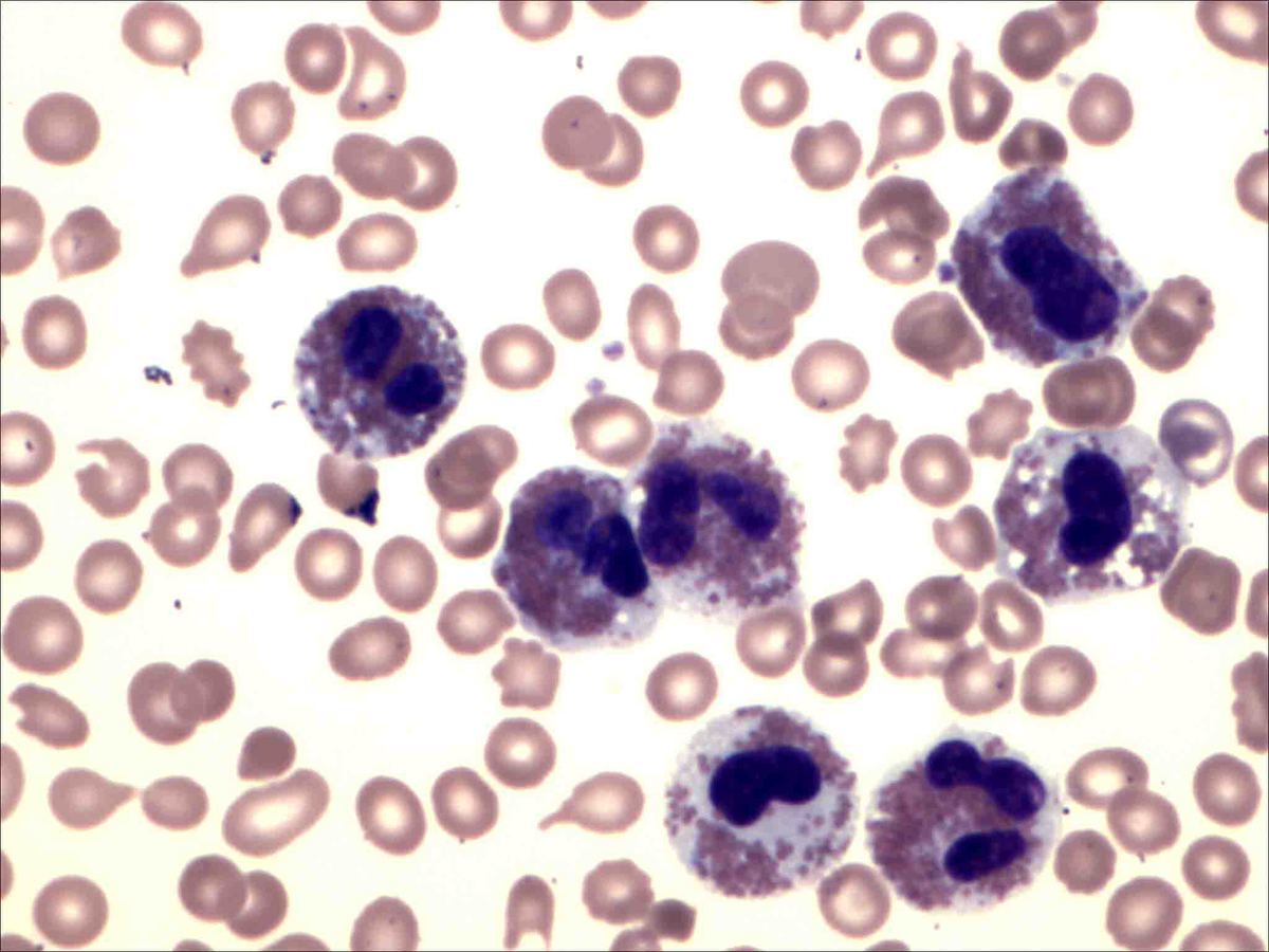 |
| Bone marrow aspirate, 60X | 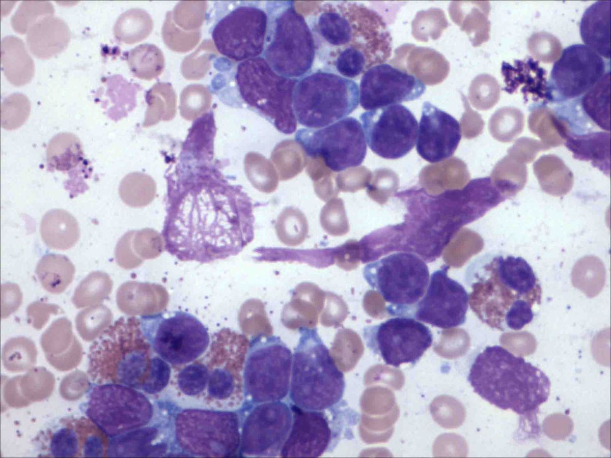 |
| Bone marrow biopsy, 20X | 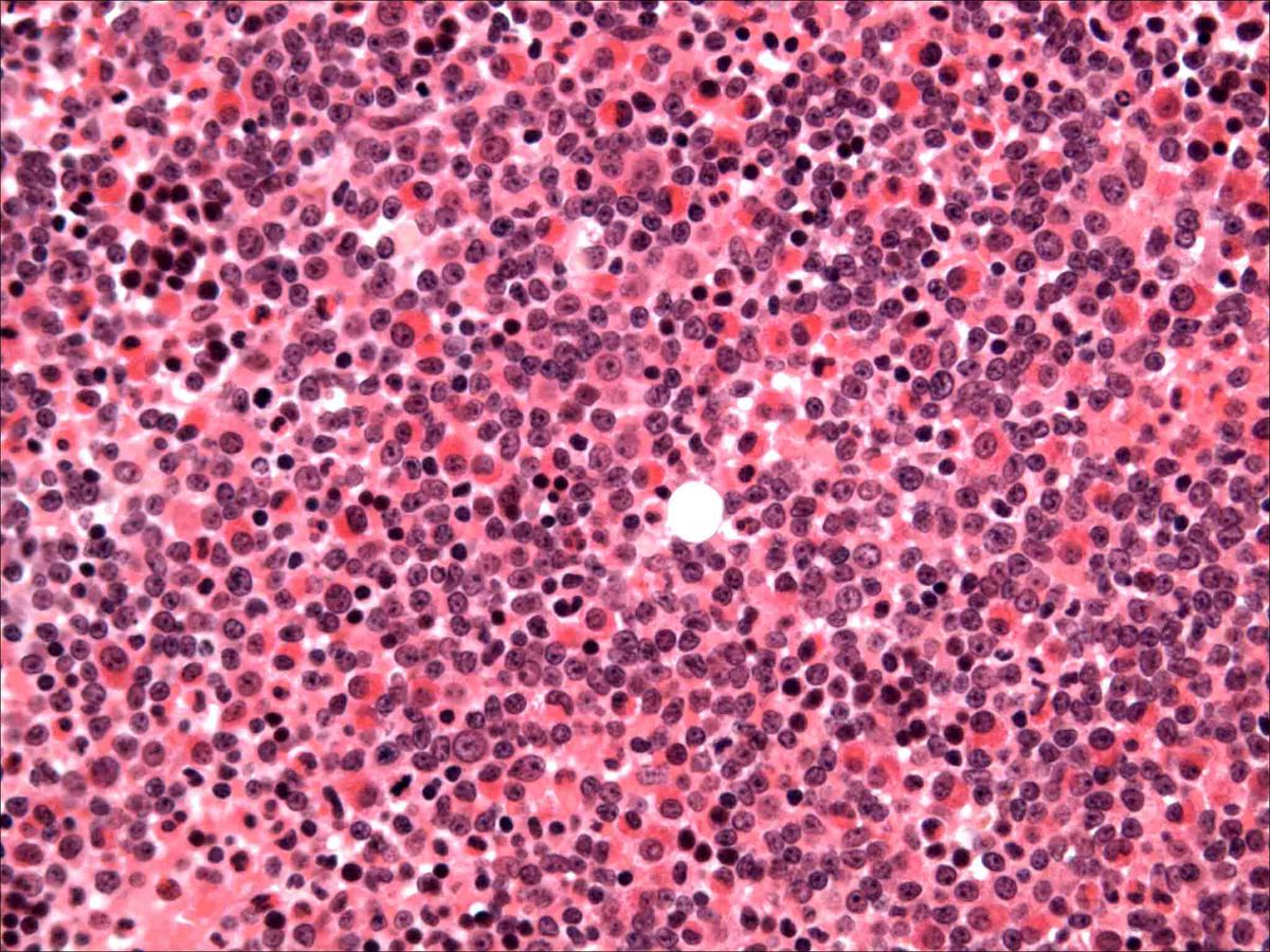 |
| Bone marrow biopsy, 60X | 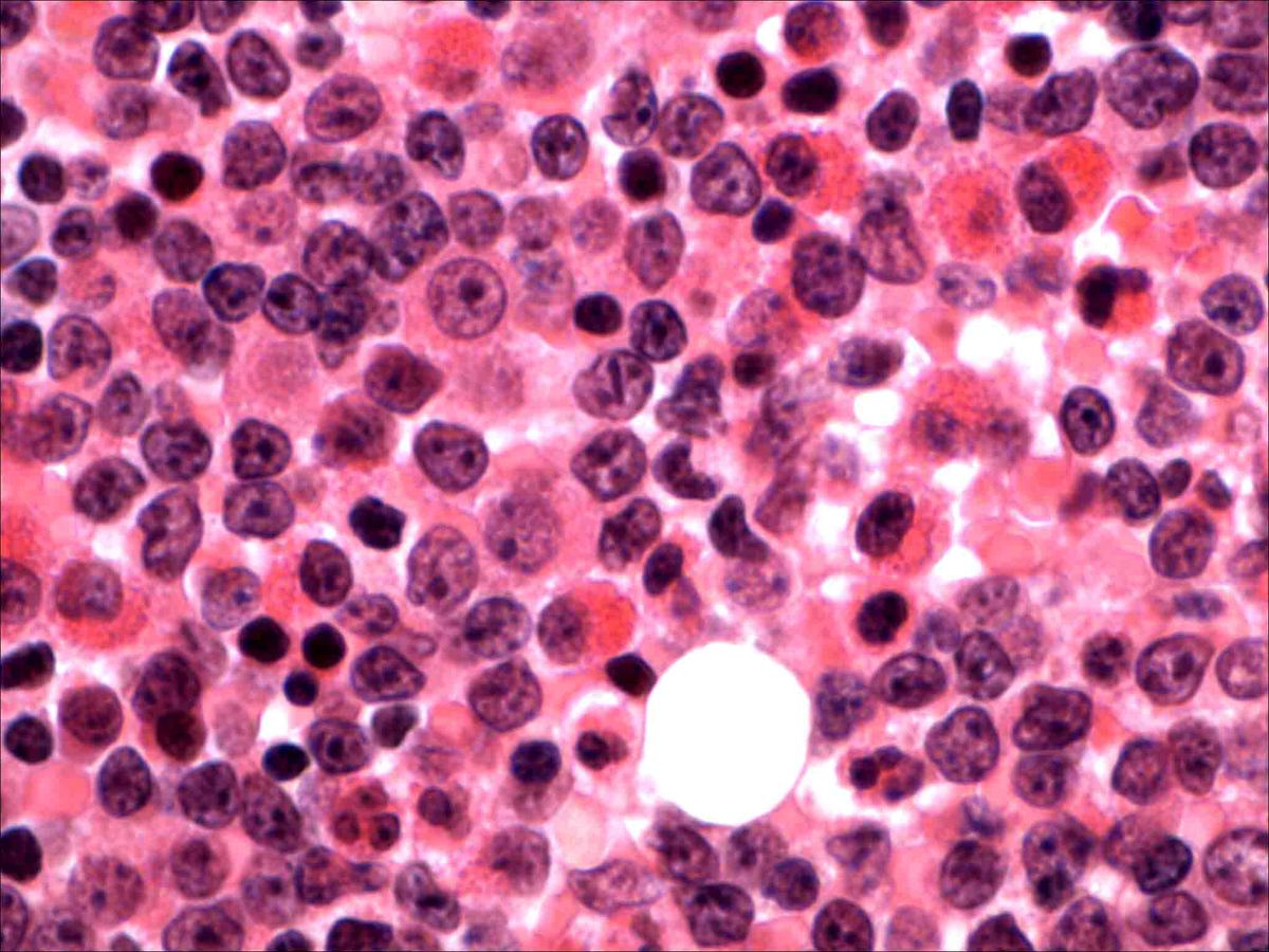 |
| CD45 vs SSC | 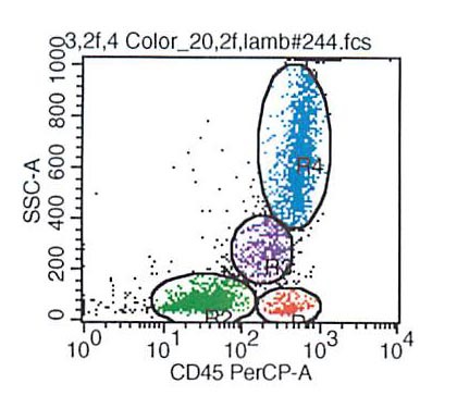 |
| CD19 VS CD3 | 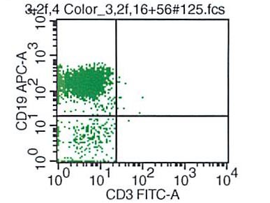 |
| CD19 vs CD20 | 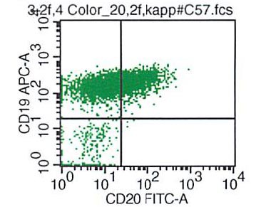 |
| CD19 vs CD10 | 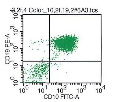 |
| CD34 vs CD10 | 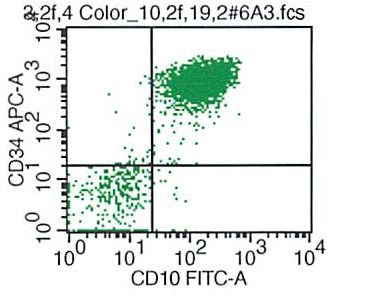 |
| CD34 vs CD19 | 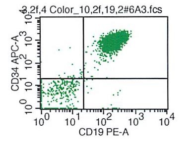 |
| CD34 vs HLA-DR | 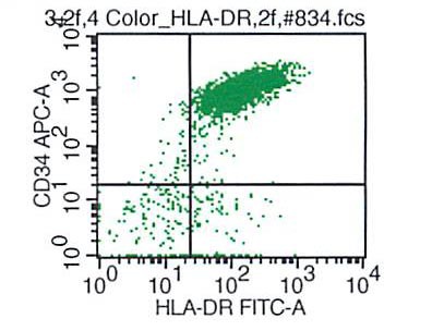 |
| CD34 vs CD13 | 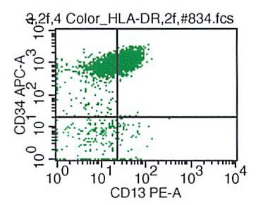 |
| TdT | 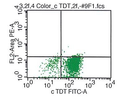 |
| CD34 vs CD20 | 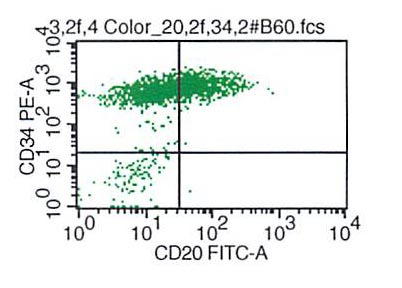 |
| CD79a | 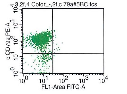 |
| CD19 vs CD43 | 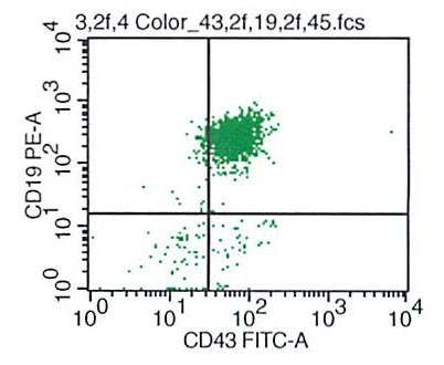 |