Institution: Mayo Clinic Arizona
Additional authors:Ryan S. Robetorye, M.D., Ph.D.
Session: Extramedullary manifestations of myeloid neoplasms
HISTORY
A 72 year old man presented with a solitary skin lesion on the forehead. There was no associated lymphadenopathy. A skin biopsy was performed elsewhere, but the slides were not available for review.
A bone marrow biopsy was performed in our institution.DETAILS
Peripheral blood smear, unilateral iliac crest bone marrow biopsy and bone marrow aspirate smears were received.
The peripheral blood smear and bone marrow aspirate smears were stained with Wright Giemsa. The bone marrow core biopsy was fixed in Acetic Zinc Formalin (AZF fixative). Core biopsy slides were stained with hematoxilin eosin.The peripheral blood smear showed mild red blood cell macrocytosis without anemia and mild neutropenia. There were no circulating blasts.The bone marrow aspirate smears were adequately cellular and showed increased numbers of mononuclear cells with high nuclear/cytoplasmic ratios, slightly irregular nuclear contours and finely dispersed chromatin, suggestive of blasts. Trilineage hematopoietic maturation was present without dysplastic features. The megakaryocytes showed unremarkable morphology.The 2.2 cm core biopsy contained hypercellular bone marrow with 70% cellularity. There were several large collections of mononuclear cells with irregular nuclei, dispersed chromatin and visible small nucleoli. The mononuclear cells comprised 40-50% of total cells. Maturing trilineage hematopoietic elements were also present.IMMUNOHISTOCHEMISTRY AND FLOW CYTOMETRY
Flow cytometric immunophenotypic analysis performed on the bone marrow aspirate revealed a cell population with features of low side scatter and dim CD45 staining (blast gate) showing the following immunophenotype: CD2, CD4, dim CD45 and CD56 positive. These cells were negative for expression of all myeloid markers, CD3, CD5, CD10, CD16, CD19, CD20 and CD34.
Immunohistochemistry with a TCL1 antibody performed on the core biopsy was positive on 40-50% of the bone marrow cells. A CD123 immunostain was attempted on the core biopsy but it was technically unsuccessful.CYTOGENETIC FINDINGS
Normal male karyotype: 46, XY [20].
AML FISH panel showed normal results with all probes tested.MOLECULAR FINDINGS
Not done.
INTERESTING FEATURES
This case shows a classic presentation of the blastic plasmacytic dendritic cell neoplasm. The patient, an elderly male, presents with a solitary skin lesion. The skin biopsy performed elsewhere was reported as blastic plasmacytic dendrititc cell neoplasm. There was no lymphadenopathy, but the bone marrow shows an extensive involvement by the neoplasm. The immunophenotype is classic for this entity. Normal cytogenetic findings may be seen in one third of cases.
PROPOSED DIAGNOSIS
Blastic plasmacytoid dendritic cell neoplasm
CONSENSUS GROUP: ADDITIONAL INFORMATION/STUDIES
Additional immunohistochemical stains performed by the consensus group:
CD123 (bone marrow): positive
CONSENSUS DIAGNOSIS
Blastic plasmacytoid dendritic cell neoplasm
| Bone marrow aspirate smear 20x |  |
| Bone marrow aspirate smear, 50x |  |
| Bone marrow aspirate smear, 100x |  |
| Core biopsy, 10x | 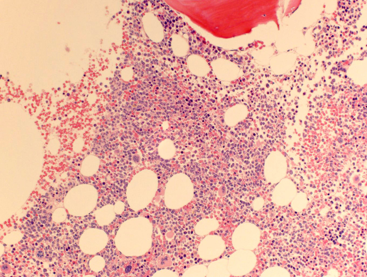 |
| Core biopsy, 100x |  |
| Core biopsy, TCL1 immunostain, 100x |  |
| Additional figure 1: 400x CD123 |  |