Institution: University of New Mexico Health Sciences
Additional authors:Frank Moore; Tracy I George
Session: AML secondary to myeloproliferative neoplasms and other types of disease progression in MPN
HISTORY
The patient is a 69-year-old male who presented with leukocytosis, thrombocytopenia, anemia, and increasing shortness of breath with a large right pleural effusion in January of 2013. His past medical history includes congestive heart failure, myocardial infarction, diabetes, renal failure and reportedly a myelodysplastic syndrome diagnosed in July of 2012. The patient has been followed by an oncologist for his myelodysplastic syndrome; however, the records and details of his treatment are not available to us. He was admitted and underwent a thoracentesis and a bone marrow aspiration and biopsy was performed. Review of a peripheral blood smear revealed leukocytosis due to an absolute monocytosis, dysplastic neutrophils, occasional blasts and rare mast cells. The bone marrow aspirate smears and trephine biopsy revealed numerous mast cells, as well as a significant increase in blasts. The patient was not considered to be a candidate for further conventional treatment due to multiple medical problems. The patient expired approximately one week following admission.
DETAILS
A complete blood count performed at admission revealed WBC count of 32X10^9/L, hemoglobin of 8.7 g/dL, hematocrit of 32%, MCV of 93fL, and a platelet count of 35 X10^9/L. Manual differential count on peripheral blood includes: 29% neutrophils, 8% lymphocytes, 53% monocytes, 4% metamyelocytes, 1% myelocytes, 2% blasts, 3% mast cells, and 2 NRBC/100 WBC.
Wright-stained bone marrow aspirate smears reveal decreased erythropoiesis, decreased neutrophilic myelopoiesis, variably increased mast cells (30%), and increased blasts (41%). The majority of the mast cells are atypical type II mast cells (promastocytes) showing indented or bilobed nuclei and well-granulated to hypogranular cytoplasm with very few spindle-shaped mast cells. Blasts contain large oval to indented nuclei with fine chromatin, frequent distinct nucleoli and moderate amounts of pale blue cytoplasm with occasional intra-cytoplasmic small azurophilic granules without Auer rods. The residual maturing neutrophilic myeloid cells include a subset of dysplastic forms. No significant dysplasia is seen in erythroid lineage. The H&E-stained clot and trephine biopsy sections show a hypercellular for age bone marrow with patchy loose aggregates of blasts and sheets and large aggregates of atypical mast cells including spindled forms particularly around trabecular bone as well as forms with oval nuclei and abundant pale to clear cytoplasm distributed interstitially. Residual trilineage hematopoiesis is present but significantly decreased. Small numbers of dysplastic megakaryocytes including small hypolobated forms and megakaryocytes with separated nuclei are seen.IMMUNOHISTOCHEMISTRY AND FLOW CYTOMETRY
Flow cytometric analysis performed on bone marrow aspirate revealed an increased population of myeloid blasts (16% of total events) expressing dim CD45, CD13, CD33, CD34, CD117, HLA-DR, myeloperoxidase, and subset CD7. No monoclonal B-cell or aberrant T-cell population was detected.
Immunohistochemical stains performed on paraffin embedded trephine biopsy sections reveal that the patchy distributed blastic appearing cells are positive for CD34, CD117, and myeloperoxidase and negative for CD19 and TdT. The sheets and large aggregates of mast cells are positive for tryptase, bright CD117, and aberrantly co-express CD25 antigen.CYTOGENETIC FINDINGS
Karyotype: 46,XY[20]. FISH analysis revealed no PML/RARA fusion.
MOLECULAR FINDINGS
A D816V mutation identified by pyrosequencing analysis of PCR amplified products of exon 17 of the KIT gene.
INTERESTING FEATURES
Systemic mastocytosis, including mast cell leukemia, may be accompanied by associated hematologic disorders. These disorders are typically myeloid in origin and include acute myeloid leukemia (AML), myelodysplastic syndrome (MDS), myelodysplastic/ myeloproliferative neoplasms (i.e. chronic myelomonocytic leukemia), and myeloproliferative neoplasms. Of AMLs seen with systemic mastocytosis, many are AML with t(8;21)(q22;q22) which previous studies have shown to be clonally related. This case represents a rare occurrence of an aleukemic mast cell leukemia with a concurrent cytogenetically normal acute myeloid leukemia. The prior history would suggest the AML is an AML with myelodysplasia-related changes, although prior material was not available for review. The mast cells in this case are promastocytes (atypical type II mast cells), which have been reported to be more commonly seen in mast cell leukemia, associated with a poor prognosis and short survival.
PROPOSED DIAGNOSIS
Mast cell leukemia (aleukemic variant) associated with cytogenetically normal acute myeloid leukemia.
CONSENSUS DIAGNOSIS
Mast cell leukemia (aleukemic variant) associated with acute myeloid leukemia with myelodysplasia-related changes (by history and morphology)
| Peripheral blood smear demonstrating increased monocytes and a circulating mast cell |  |
| Wright-stained bone marrow aspirate smear showing increased mast cells and blasts |  |
| H&E-stained bone marrow trephine biopsy showing increased interstitial blasts and peri-trabecular aggregate of spindled mast cells | 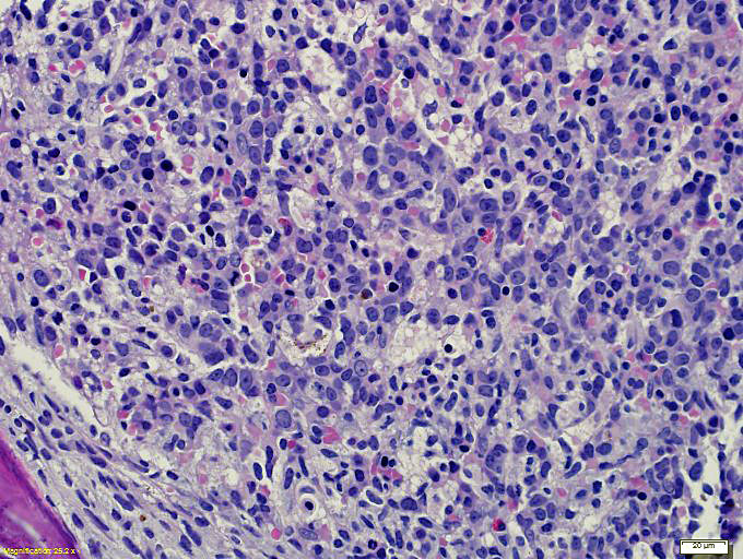 |
| H&E-stained marrow trephine biopsy showing sheets of atypical mast cells with abundant clear cytoplasm |  |
| Immunohistochemical stain highlighting increased CD34+ blasts adjacent to a large aggregate of mast cells | 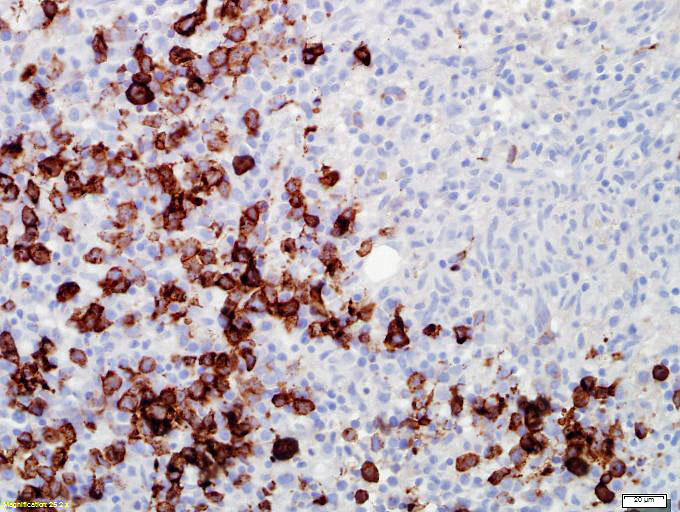 |
| Immunohistochemical stain for CD117 highlighting mast cells and blasts |  |
| Immunohistochemical stain for tryptase highlights large numbers of mast cells |  |
| Immunohistochemical stain for CD25 demonstrates aberrant co-expression of this antigen by mast cells | 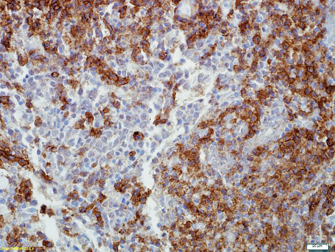 |
| Molecular analysis of exon 17 of KIT gene by pyrosequencing demonstrating a GAC>GTC mutation resulting in D816V |  |