Institution: University of Maryland Medical Center
Additional authors:Nahid Nanaji MD, Qing Chen MD/PhD
Session: Erythroleukemia and megakaryoblastic AML and mimics
HISTORY
The patient was a 73 year old woman, who was initially diagnosed with IgG kappa plasma cell myeloma. She underwent autologous stem cell transplant approximately seven months after the initial diagnosis. She was received thalidomide briefly, which was discontinued and replaced with lenalidomide and dexamethasone. This regimen was discontinued after several months due to renal failure, and reinstituted at a lower dose. She also transiently received bortezomid, which was discontinued due to peripheral neuropathy. Eventually she joined a clinical trial for the agent Carfilzomib. Approximately 55 months after initial diagnosis, she was noted to be developing worsening anemia (hemoglobin 7.4 g/L) and thrombocytopenia (53,000 x 10^9/L). In addition, a very high level of LDH was noted (3844 u/L). A bone marrow biopsy was performed. Ultimately, a diagnosis of therapy related myeloid neoplasm, in the form of pure erythroid leukemia, was made. A course of decitabine was initiated, but after thirty-three days the patient had profound pancytopenia and a bone marrow biopsy revealed hypercellular marrow with cellularity consisting almost exclusively of leukemic erythroid blasts. The patient elected to receive palliative and supportive treatment.
DETAILS
Bone marrow aspirate - Wright Giemsa stain Bone marrow core biopsy, iliac crest - formalin fixation, brief decalcification, hematoxylin and eosin stain. Gross pathology - Unremarkable appearing bone marrow aspirate and core biopsy Microscopic pathology - 55 months prior to the case submitted here the patient had a bone marrow biopsy which was positive for plasma cell myeloma, plasma cells mainly mature in morphology, with otherwise unremarkable trilineage hematopoiesis. A bone marrow core biopsy showed moderately hypercellular marrow for age, with multiple intrasinusoidal clusters of large atypical cells with round nuclei, open to slightly condensed chromatin, prominent nucleoli, and relatively scant basophilic cytoplasm, representing approximately 30% of marrow cellularity. In addition to morphologic overlap with plasmablasts, the intrasinusoidal location raised concerns of metastatic non-hematopoietic neoplasm. Granulocytic maturation and megakaryocytes were present but normal erythroid maturation was markedly decreased. The malignant cells were strongly positive for CD71 and focally positive for glycophorin A. Based on these results, the patient was given a trial of decitibine. After 33 days of decitibine, a marrow aspirate and biopsy showed a markedly hypercellular marrow for age (>90%). Cellularity was composed almost entirely of erythroid blasts.
IMMUNOHISTOCHEMISTRY AND FLOW CYTOMETRY
Flow cytometry identified atypical cells with very dim CD45 expression and high side scatter. These cells were positive for CD71 (bright), CD235a (subset), and HLA-DR, and showed subset expression of CD33, but were negative for other standard lymphoid and myeloid markers. The patient's plasma cell myeloma population, at presentation to UMMC, was composed of mature CD38 positive plasma cells. In the case submitted, The malignant cells were strongly positive for CD71 and focally positive for glycophorin A. They were weakly positive for CD45 and strongly positive for CD43. CD138, kappa, and lambda were negative. Markers for major non-hematopoietic neoplasms, a possibility raised by the clustered, intrasinusoidal distribution, were negative.
CYTOGENETIC FINDINGS
Cytogenetic analysis in the submitted case revealed the following karyotype, in 20/20 metaphases examined: 57,X –X, +1, +2, +6, +8, +10, +13, +15, +15, +21, +21, +2mar
MOLECULAR FINDINGS
PCR for clonal IgH rearrangement was negative. (PCR on prior marrow specimen containing plasma cell myeloma was performed and showed a strong clonal band.)
INTERESTING FEATURES
Pure erythroid leukemia (PEL) is a potential morphologic mimic of plasma cell myeloma with plasmablast morphology. It is relatively rare for therapy related myeloid neoplasm to occur in the form of PEL. However, several cases of PEL in chemotherapy treated plasma cell myeloma have been described. In addition to the morphologic overlap with plasma cell myeloma, in this case, a highly unusual clustered sinusoidal distribution of the malignant erythroblasts added to diagnostic challenge. The karyotype was also unusually complex.
PROPOSED DIAGNOSIS
Therapy related myeloid neoplasm (in the form of pure erythroid leukemia), in a patient treated for plasma cell myeloma.
CONSENSUS GROUP: ADDITIONAL INFORMATION/STUDIES
PAS stain showed globule-like granules in the erthroblasts. The patient received supportive care only.
CONSENSUS DIAGNOSIS
Therapy-related myeloid neoplasm, consistent with acute erythroid leukemia (pure erythroid leukemia)
| Eryrthroblasts in bone marrow aspirate (1000X) | 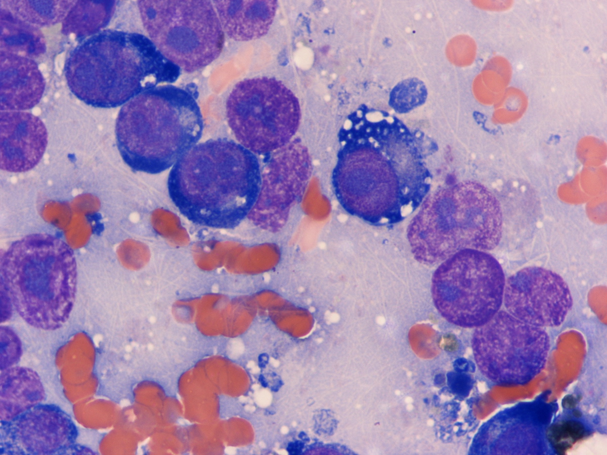 |
| Malignant erythroblasts showing intrasinusoidal distribution (400X) | 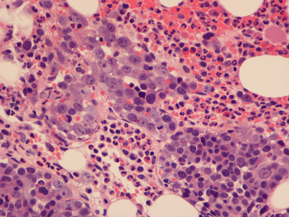 |
| CD71 stain highlights malignant erythroblasts | 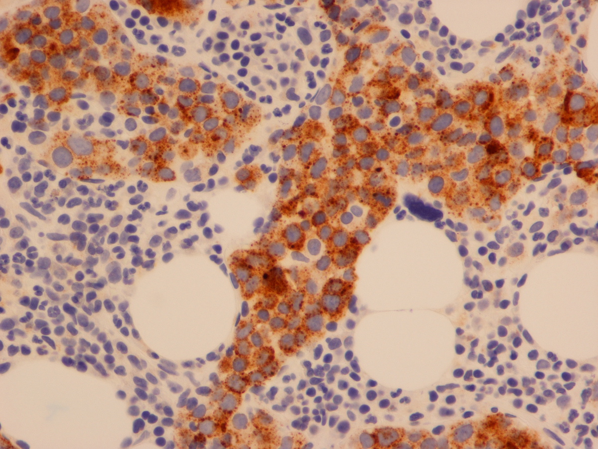 |
| Marrow replaced by malignant erythroblasts (after 33 day trial of decitabine) | 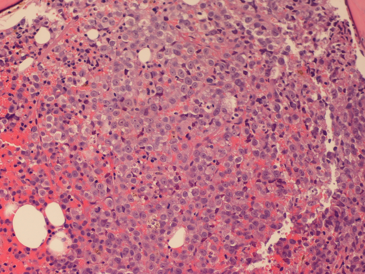 |
| Additional figure 1: PAS |  |
| Additional figure 2: Smear | 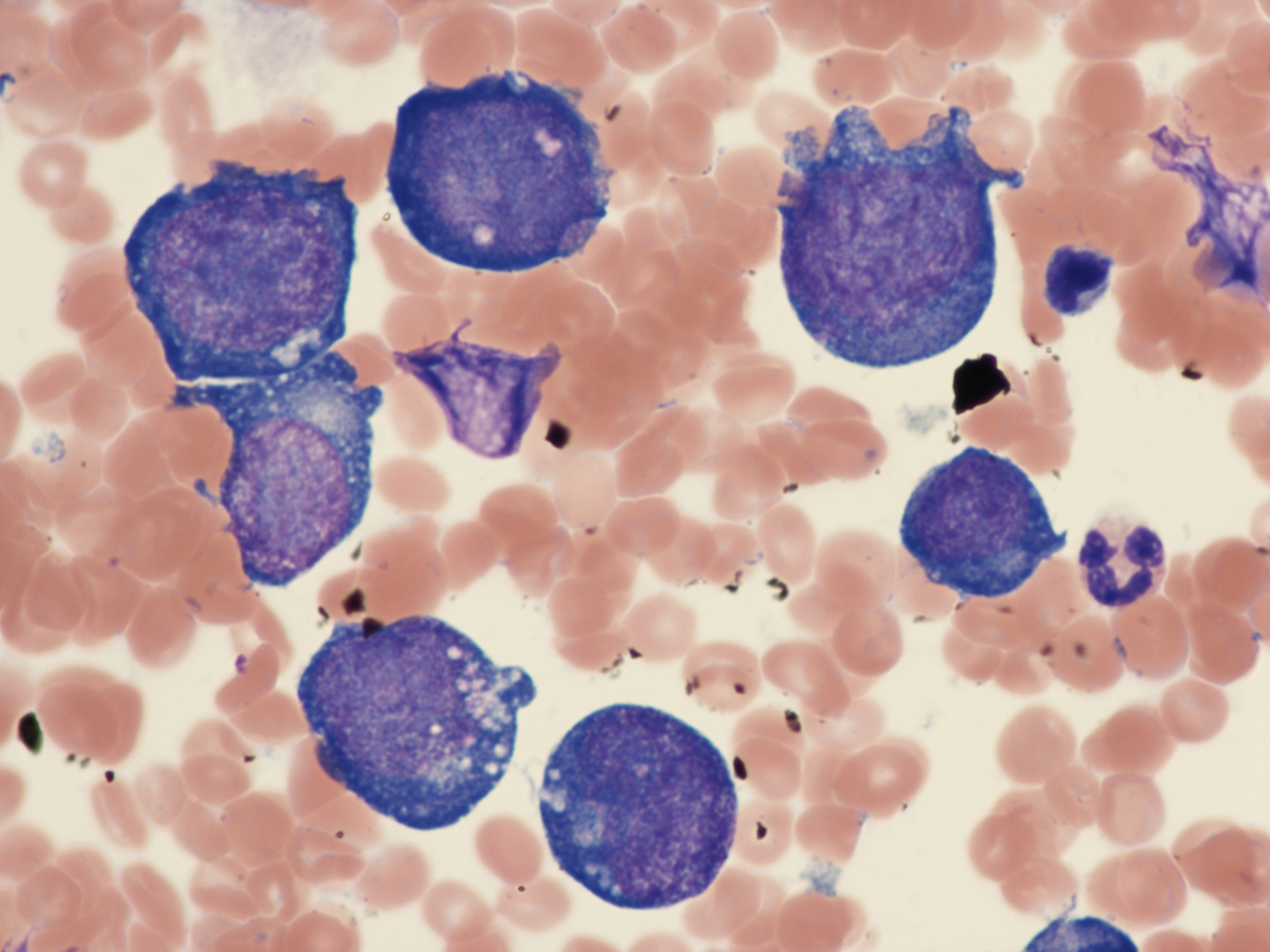 |