Institution: Vanderbilt University Medical Center
Additional authors:Jennifer Giltnane, Juliann Chmielecki, Katherine Hutchinson, Rebecca S. Dean, Ashwini Yenamandra, David Head, Madan Jagasia, Utpal Dave, William Pao
Session: Myeloid and lymphoid neoplasms with eosinophilia and abnormalities of PDGFRA, PDGFRB, or FGFR1
HISTORY
A 38 year old man initially presented with right inguinal lymphadenopathy. His CBC at diagnosis showed a white count of 10.44 K/uL, a hematocrit of 37.2%, and platelet count of 180 K/uL, with mild eosinophilia (2300 cells/uL). A complete set of CT scans revealed diffuse lymphadenopathy, including significantly enlarged retroperitoneal lymph nodes at 2.7 cm, enlarged pelvic lymph nodes and a right extremity iliac chain conglomerate of 5 cm and a right inguinal lymph node of 5 cm. In addition, the spleen measured 14.4 x 8 cm. Several of the lymph nodes were metabolically active by positron emission tomography.
The patient underwent excisional biopsy of the right inguinal lymph node (LN1) and staging bone marrow biopsy (BM1) at an outside institution. Subsequently, the patient received chemotherapy with methotrexate, cytarabine, cyclophosphamide, vincristine, adriamycin, and dexamethasone. However, two months after completion of the chemotherapy, the patient returned with recurrent right inguinal lymphadenopathy, leukocytosis and thrombocytopenia, more than one month after+ the last dose of filgrastim. The patient’s counts at that time were: WBC 64.5 K/uL, hematocrit 30.1%, platelets 39 K/uL with 31% eosinophils. A bone marrow biopsy (BM2) was performed at that time as was a second lymph node biopsy (LN1). The lymph node biopsy demonstrated recurrence of the patient’s original diagnosis.
The patient underwent re-induction chemotherapy with vincristine, prednisone, peg-asparaginase, and daunorubicin, with intrathecal methotrexate, which was followed by additional treatment for residual lymphadenopathy (etoposide, cytoxan, methotrexate, and high dose cytarabine). A follow-up bone marrow biopsy (BM3) was then performed.
The patient subsequently was treated with imatinib for 7 days with excellent response to therapy. A matched related allogeneic peripheral blood stem cell transplant was subsequently performed.
DETAILS
Excisional biopsy of the right inguinal lymph node (LN1, formalin) demonstrated partial involvement by sheets of monotonous small to intermediate sized lymphoid cells with slightly irregular nuclei, moderately condensed chromatin, occasional small nucleoli, and scant cytoplasm. Scattered tingible body macrophages were identified within the sheets of neoplastic cells. Immunophenotypically, the neoplastic cells were diagnostic of a precursor T lymphoblastic lymphoma which expressed CD2, cytoplasmic CD3, CD4, CD38 (bright), CD43, and TdT. The neoplastic cells were negative for CD10 and CD34. The karyotype of the lymph node tissue demonstrated a normal male karyotype: 46,XY[20]. The initial staging bone marrow evaluation (BM1) was not available for review: by report, the karyotype of the marrow was significant for t(5;6) in 10 of 20 metaphases, despite the absence of morphologic or immunophenotypic involvement by the patient’s known T lymphoblastic lymphoma.
A bone marrow biopsy (BM2) performed two months after completion of chemotherapy and reviewed at Vanderbilt University Medical Center (BM #2) demonstrated a markedly hypercellular marrow (95% cellularity) with left-shifted myeloid hyperplasia and eosinophilia. Concern was raised for a possible FGFR or PDGFR gene rearrangement, although there was no evidence of lymphomatous involvement.
After additional re-induction therapy and additional chemotherapy, a follow-up bone marrow biopsy performed at Vanderbilt University Medical Center (fixed in B-plus, BM3) was performed which again demonstrated a hypercellular marrow (95% cellularity) with myeloid hyperplasia and eosinophilia, without involvement by lymphoma. This marrow was diagnosed as a myeloproliferative neoplasm with eosinophilia, associated with a rearrangement of PDGFRB.
IMMUNOHISTOCHEMISTRY AND FLOW CYTOMETRY
Flow cytometry on BM3 demonstrates 1% of total cells are CD3+ T lymphocytes that are negative for CD1a and CD61. CD45 dim, CD33+, and CD19- myeloblasts make up 1.2% of total cells. This sample is negative for blasts with the patient's previous phenotype (CD3+/CD1a+).
CYTOGENETIC FINDINGS
Karyotype analysis of the BM3 revealed t(5;6)(q33-34;q23) in 20 of 20 cells with additional loss of the Y chromosome in 3 metaphases. Confirmation of the involvement of the PDGFRB locus on chromosome 5 in the rearrangement was obtained by FISH which demonstrated a split red signal (CSFIR-PDGFRB) in 56% of cells. FISH was performed retrospectively on the diagnostic lymph node tissue using probes against PDGFRB, and a rearrangement PDGFRB was detected in 87% of cells.
MOLECULAR FINDINGS
Use of a systemic screen for tyrosine kinase (TK) gene rearrangements developed in the laboratory of Dr. William Pao, a novel C6orf204-PDGFRB fusion was identified in this patient. This methodology involves capture of the TK domains using a biotinylated oligonucleotides against conserved kinase domain regions, followed by next generation sequencing of the capture products (see Chmielecki et al. Genes Chromosomes Cancer, 2012; 51:54-65). Phosphorylation of PDGFRB was shown to be inhibited by treatment with imatinib in 293T cells transfected with a vector encoding for the C6orf204-PDGFRB fusion.
INTERESTING FEATURES
This case highlights a novel PDGFRB fusion which manifests clinically as concomitant involvement by a T lymphoblastic lymphoma (T-LBL) as well as a myeloproliferative neoplasm characterized by eosinophilia. Although T-LBL in conjunction with a myeloproliferative process has been seen previously with FGFR and less commonly with PDGFRA, it has not been noted with PDGFRB. In addition, the myeloproliferative phenotype of this disease is unlike the chronic myeomonocytic leukemia-like (CMML-like) presentation of most PDGFRB-associated disease, or even the chronic eosinophilic leukemia-like (CEL-like) presentations of most PDGFRA-associated disease, but most resembles the dual lineage presentation of FGFR translocations involving 8p11. The specific t(5;6) translocation has not been previously described. This case is a prime example of the dual lineage effects of these types of fusions, while identifying a new set of translocation partners.
PROPOSED DIAGNOSIS
Myeloid and lymphoid neoplasms with eosinophilia and abnormality of PDGFRB, most compatible with T-lymphoblastic lymphoma with a chronic myeloproliferative neoplasm with eosinophilia.
CONSENSUS DIAGNOSIS
Myeloid and lymphoid neoplasm with rearrangement of PDGFRB, t(5;6), presenting with T-lymphoblastic lymphoma and myeloproliferative neoplasm with eosinophilia
| Lymph node biopsy (LN1) showing replacement by T lymphoblastic lymphoma (H&E, 20x). | 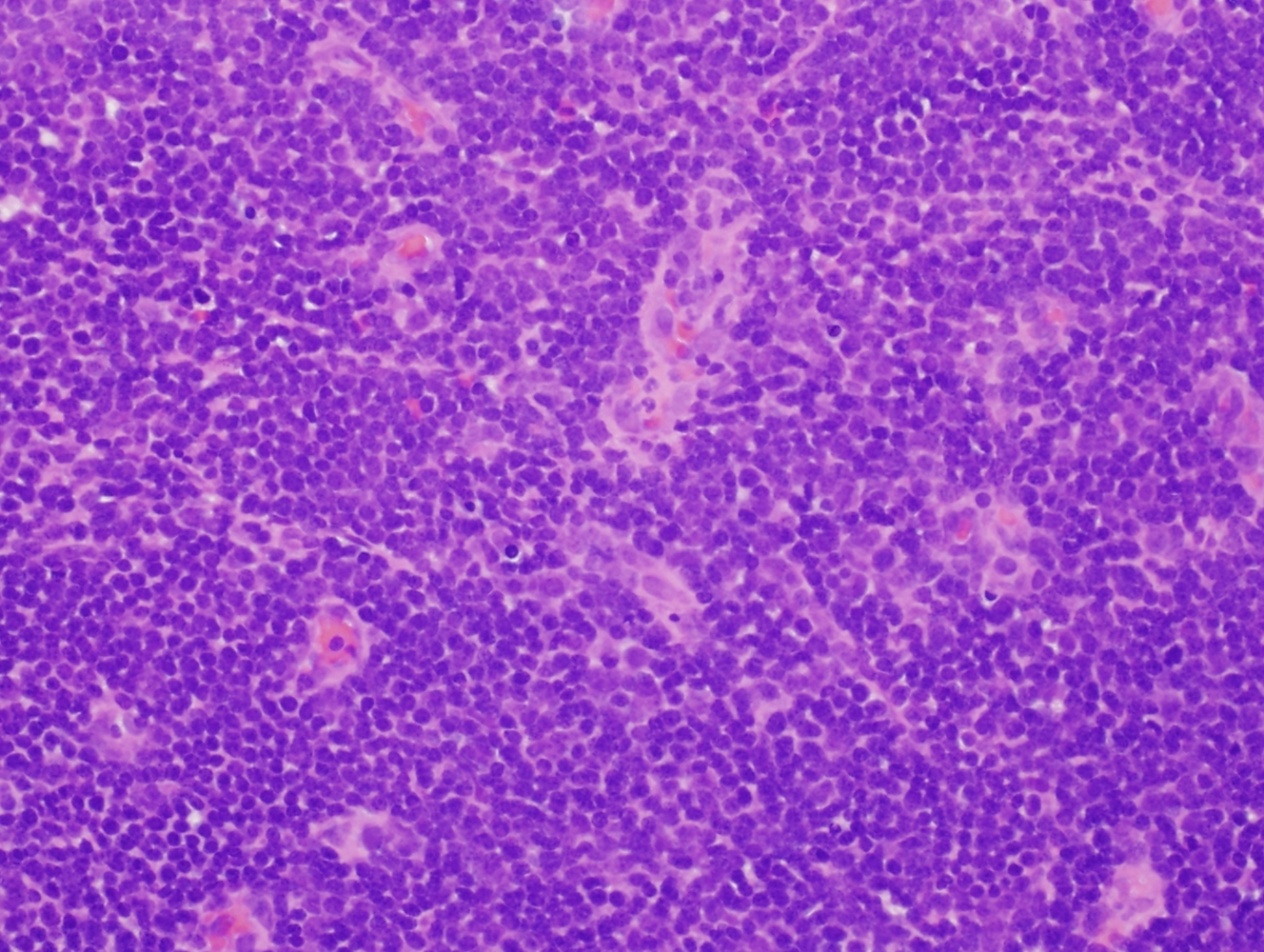 |
| High power view of LN1 demonstrating lymphoblastic morphology of the cells (H&E, 40x). | 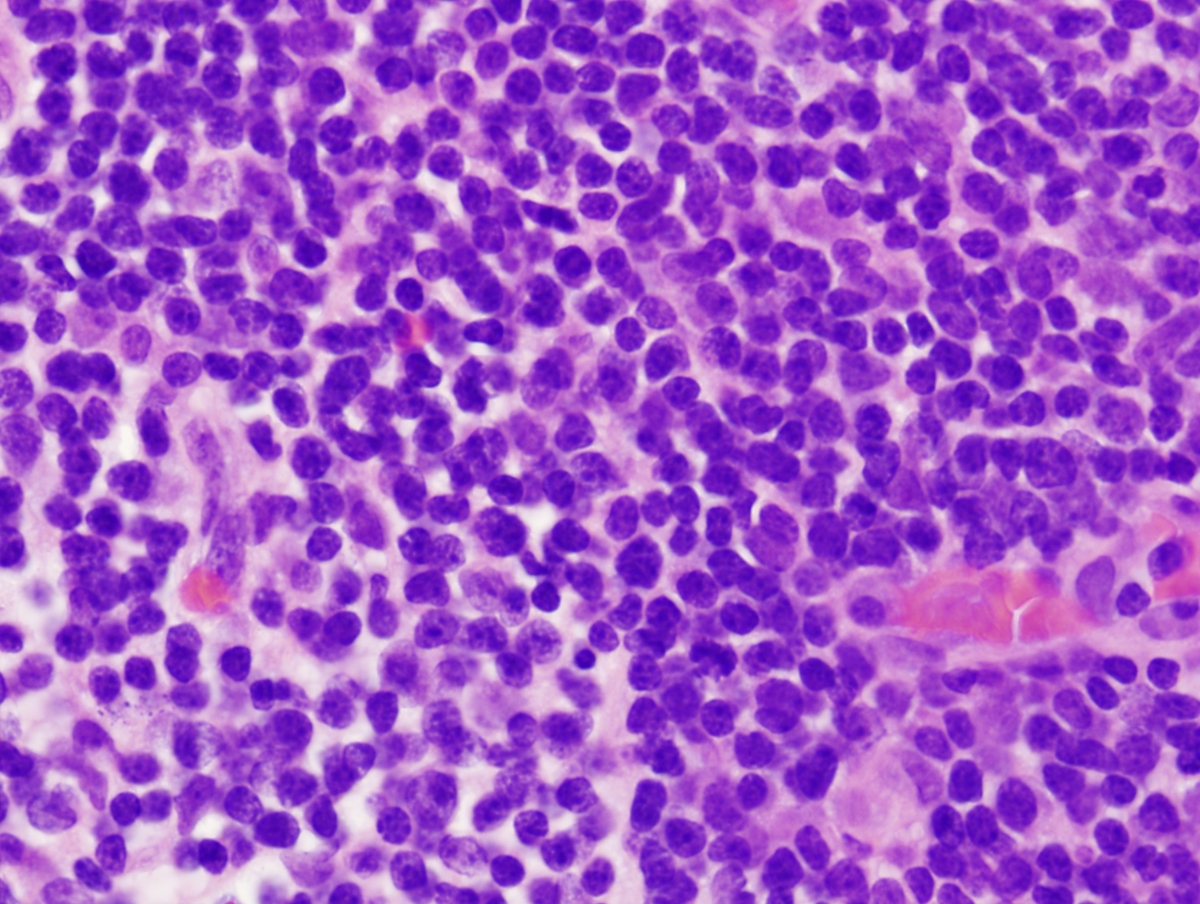 |
| Bone marrow aspirate (BM3) demonstrating profound eosinophilia and myeloid hyperplasia (Wright Giesma, 40x). | 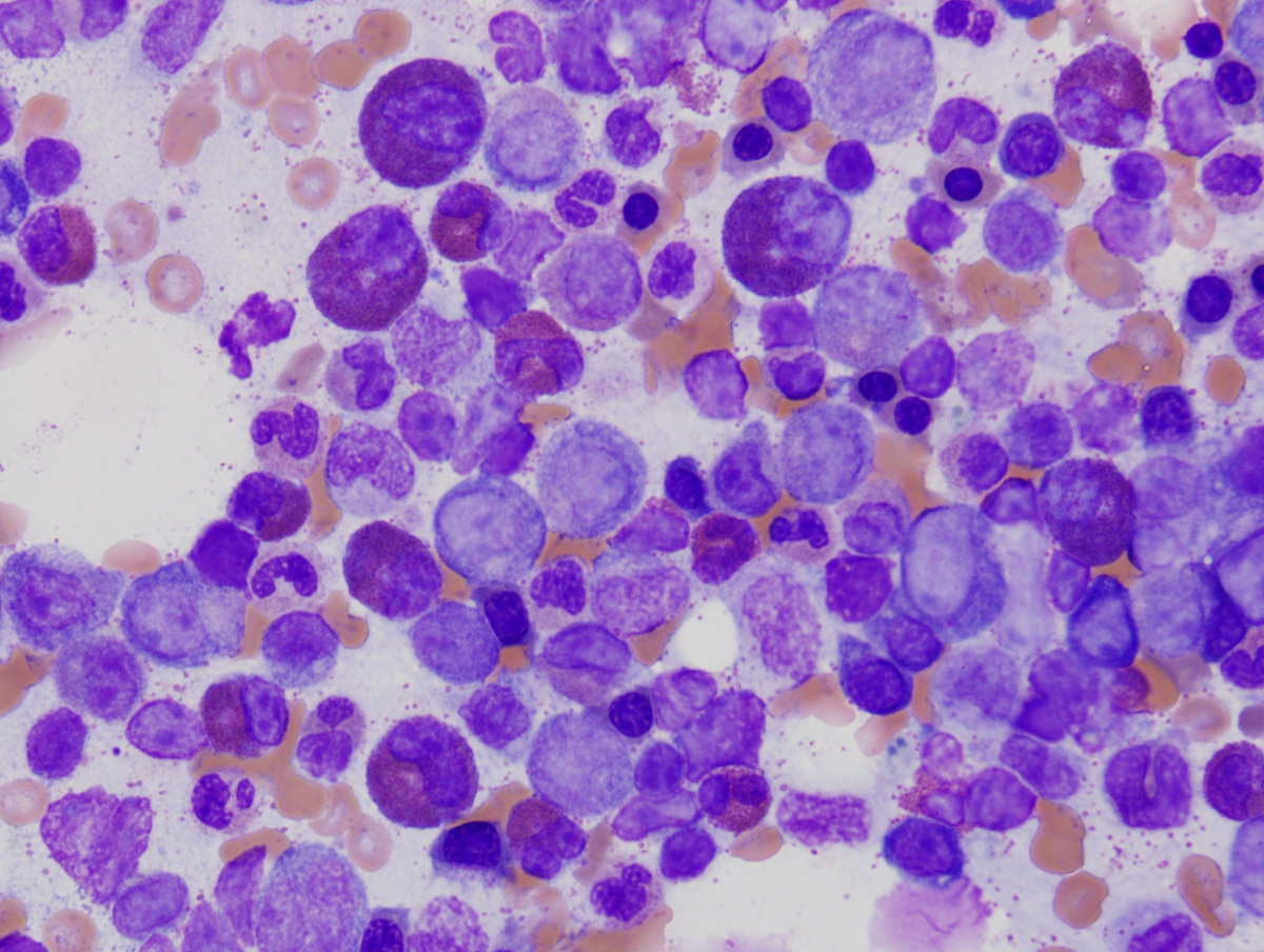 |
| Bone marrow biopsy (BM3) demonstrating eosinophilia and hypercellularity (H&E, 40x). | 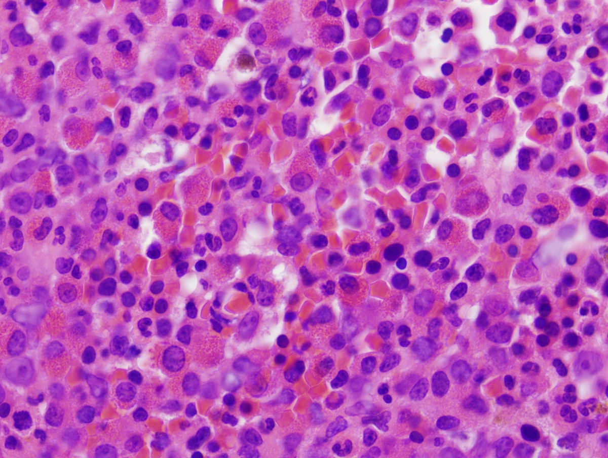 |
| Fluorescent in situ hybridization of LN1 demonstrating PDGFRB translocation. | 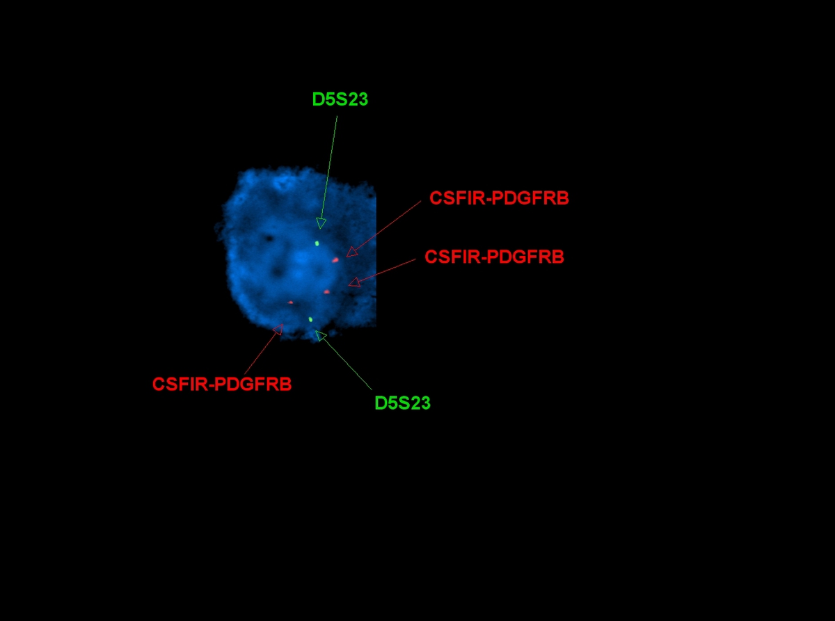 |
| Fluorescent in situ hybridization of BM3 demonstrating PDGFRB translocation. | 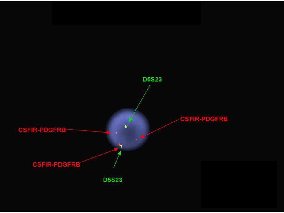 |