Institution: Quest Diagnostics Nichols Institute
Additional authors:Zeqiu J. Han, M.D., Aurelia Meloni-Ehrig, Ph.D., Bong K. Kim, M.D., Christine Bryke, M.D., Dan Jones, M.D. Ph.D.
Session: AML with myelodysplasia-related changes
HISTORY
28 year-old female presented with thrombocytopenia. The provided CBC and differential data was: WBC 4.9 k/uL, RBC 4.58 M/uL, Hb 12.1 g/dL, HCT 37.1%, MCV 81.2 fL, MCH 26.4 pg, MCHC 32.5 g/dL, RDW 21.1%, Platelet 64k/uL. Diff: granulocytes 37.5%, lymphocytes 43.8%, monocytes 18.7%. Absolute granulocyte count 1.80 k/uL, absolute lymphocyte count 2.1K/uL, absolute monocytes count 0.9 k/uL. Secondary causes for thrombocytopenia such as toxic exposure, radiation or autoimmune disorder were ruled out.
DETAILS
Site: bone marrow biopsy and aspirate
Sample processing: The biopsy was fixed with formalin and decalcified according to the standard procedure. Peripheral blood and aspirate smears were stained with Wright-Giemsa stain. Microscopic description: The core biopsy shows hypercellular marrow (80%) with erythroid hyperplasia and relatively decreased myeloid precursors with left shift. Megakaryocytes are present in expected number with predominantly hypolobated/monolobated forms. The aspirate smears contain cellular spicules and show erythroid hyperplasia with left-shift and dyspoietic changes including megaloblastoid changes, nuclear budding, nuclear bridging and nuclear irregularity. Myeloid precursors are left shifted with abnormal nuclear segmentation. The M:E ratio is inverted at 1:2. Frequent small and hypolobated/monolobated megakaryocytes are noted. There is no lymphocytosis or plasmacytosis. A differential count is: blasts 4%, promyelocytes 0%, myelocytes 9%, metamyelocytes 3%, bands/neutrophils 1%, nRBC 63%, eosinophils 4%, monocytes 3%, lymphocytes 12%, plasma cells 1%. The corresponding peripheral blood smear shows normocytic and normochromic red cells with anisocytosis, decreased white cells with dysplastic neutrophils (hypogranulation, monolobated forms). Platelets are reduced in number with occasional giant forms. Blasts are not seen.IMMUNOHISTOCHEMISTRY AND FLOW CYTOMETRY
Immunohistochemistry of CD34 and CD61 are performed on the core biopsy. CD34 stains approximately 5% of immature cells, CD61 highlights the atypical megakaryocytes. Iron stains show the presence of storage iron, ring sideroblasts are not identified.
Flow cytometry identifies approximately 10% myeloblasts, expressing CD34, CD13, CD117, HLA-DR and dim/aberrant CD5. The myeloid forms show immunophenotypic evidence of left-shifted maturation with abnormal CD13/CD38 maturation pattern. A small subset of myeloid precursors also expresses CD117 and HLA-DR. Lymphoid cells are immunophenotypically unremarkable.CYTOGENETIC FINDINGS
Conventional cytogenetic analysis reveals a complex abnormal karyotype with a pericentric inversion of chromosome 3, deletions 5q, 7q, and 13q, and loss of one copy of chromosome 14. The karyotype is:
45, XX, inv(3)(p21q26), del(5)(q13q33), del(7)(q22q34), del(13)(q13q22), -14[16]/46,XX[4]INTERESTING FEATURES
Myelodysplastic syndromes occur mostly in older individuals with a median age of 70 years. It is uncommon in young adult patients, as seen in this case. Although such information is not available, the early age of disease onset of the current case raises the possibility of familial myelodysplastic syndrome/acute leukemia. Familial myelodysplastic syndrome/acute leukemia (MDS/AL) includes two categories: 1. Pure familial MDS/AL. This category includes familial platelet disorder with propensity to myeloid malignancy (FDP/AML), familial AML with mutated CEBPA, familial MDS/AML with GATA2 mutation, familial MDS/AL associated with defective telomere maintenance. The true incidence of these syndromes is unknown, given their relatively recent identification. 2. Syndromes-associated familial MDS/AL, including bone marrow failure syndromes (Diamond-Blackfan anemia, Dyskeratosis Congenita, Shwachman-Diamond syndrome, etc), DNA repair deficiency (Fanconi anemia, Bloom syndrome, etc), and signal transduction aberration (Noonan syndrome, Neurofibromatosis I). Management of these patients may require approach different from that of sporadic MDS patients.
References:1. Churpek J. et al. (2013). Proposal for the clinical detection and management of patients and their family members with familial myelodysplstic syndrome/acute leukemia predisposition syndromes. Leukemia& lymphoma 54(1):28-352. Liew E., et al (2011). Familial myelodysplastic syndromes: A review of the literature. Hematologica 96(10):1536-1542.PROPOSED DIAGNOSIS
Myelodysplastic syndrome (RAEB-1) with complex, poor prognostic chromosome abnormalities.
CONSENSUS DIAGNOSIS
Myelodysplastic syndrome (RAEB-1) with complex karyotype including inv(3)(p21q26)
| Flow cytometry identified abnormal antigen expression on myeloid cells including decreased CD10, increased CD13, abnormal HLA-DR (small subset), and CD117 (small subset) expression. |  |
| Approximately 10% myeloblasts were detected by flow cytometry, expressing CD34, CD13, CD117, MPO, and HLA-DR. | 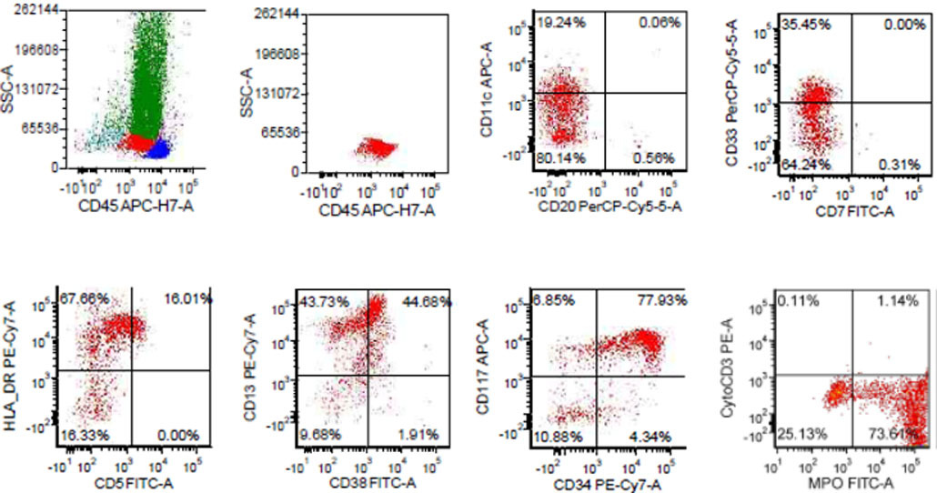 |
| Dysplastic neutrophils on the peripheral blood smear | 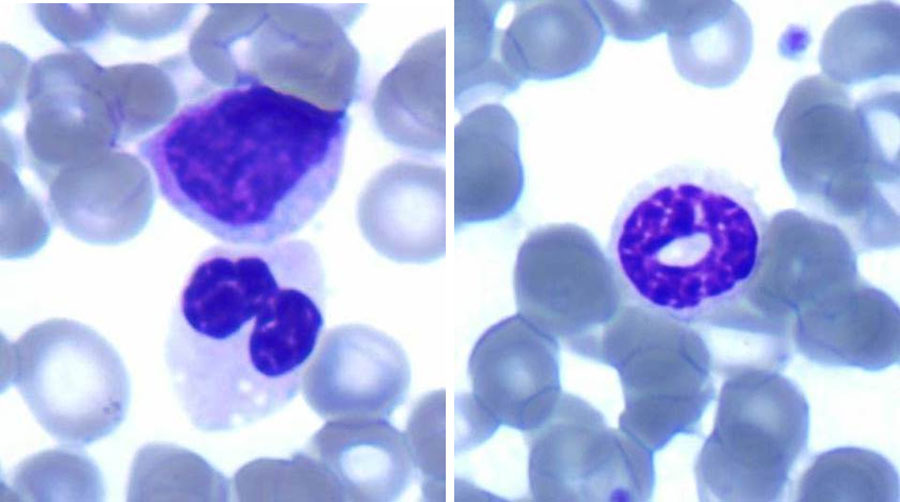 |
| The core biopsy shows hypercellular marrow with erythroid hyperplasia, left-shifted myeloid population and atypical megakaryocytes | 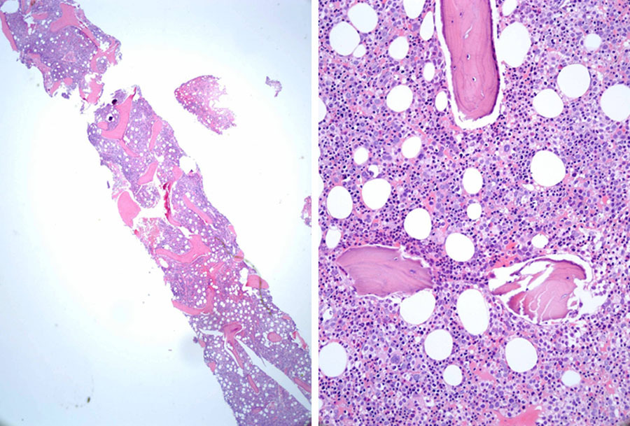 |
| CD34 stains ~5% immature cells, CD61 highlights the atypical megakaryocytes on the core biopsy | 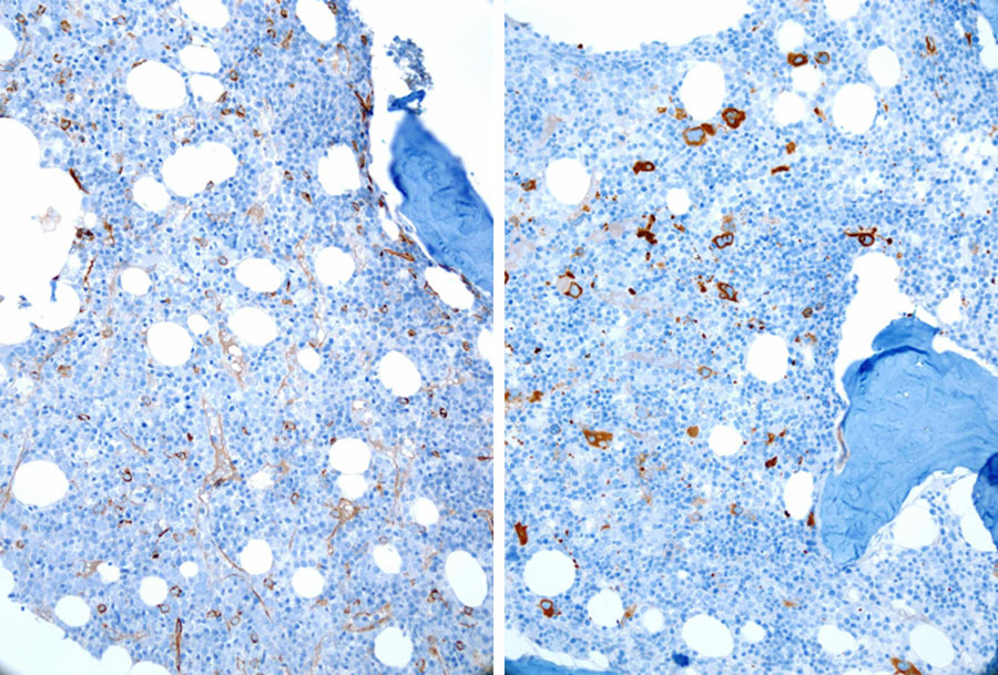 |
| Representative morphologic features from bone marrow aspirate smears (100X). Erythroid precursors show megaloblastoid changes, nuclear budding, nuclear irregularity, and nuclear bridging. Myeloid precursors show dysplastic features including hypogranulation and hyposegmentation. | 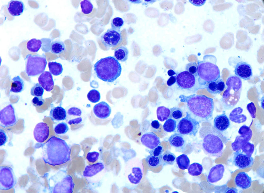 |
| Representative morphologic features from bone marrow aspirate smears (100X). Erythroid precursors show megaloblastoid changes and nuclear budding. | 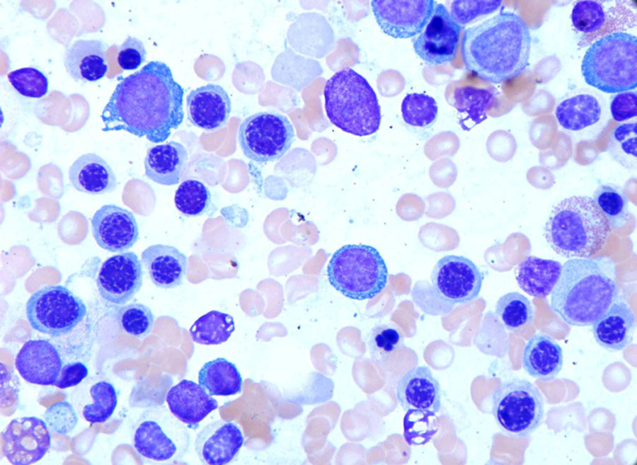 |
| Representative morphologic features from bone marrow aspirate smears (100X). Nuclear bridging is clearly visible in this picture (arrow). | 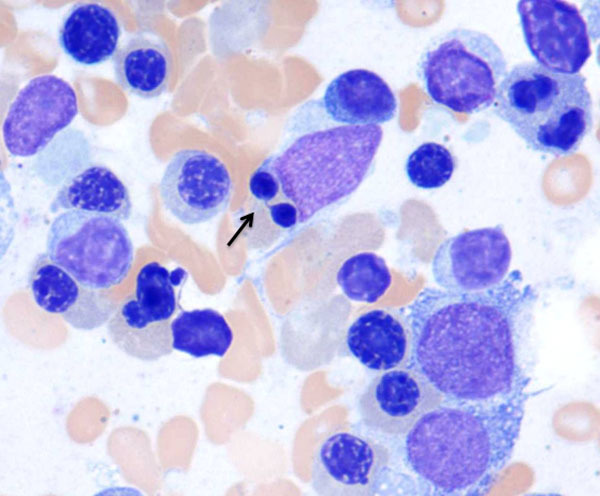 |
| Representative morphologic features from aspirate smears (100X). Myeloid precursors show dysplastic features including hypogranulation and hyposegmentation. |  |
| Representative morphologic features from aspirate smears (100X). Megakaryocytes are small and hypolobated or monolobated. | 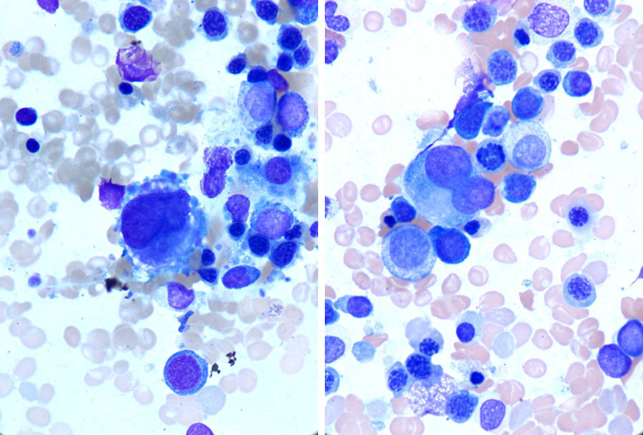 |
| Chromosome analysis revealed inv (3) (p21q26), del (5) (q13q33), del (7) (q22q34), del(13) (q13q22), and -14 in 16/20 metaphase cells | 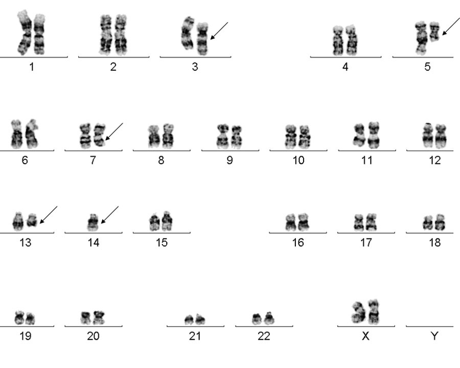 |