Institution: University Health Network
Session: Acute leukemias of ambiguous lineage
HISTORY
80-year-old, previously well male with a 2-week history of epistaxis which resolved with conservative management. He subsequently developed a cough and was being treated with an antibiotic. He began to decline and developed progressive fatigue. He was sent to the ER. The CBC showed a WBC of 129 x 10e9/L with 110 x10e9/L blasts. The patient was started on allopurinol and hydroxyurea.
Past medical history: Prostatectomy for a benign process. Other past surgeries include lipoma removal and cholecystectomy. A bone marrow aspiration was subsequently performed.CBC: Hb 98 g/L, WBC 63.1 x 10e9/L, Platelets 59 x 10e9/L, Neutrophils: 4.42 x 10e9/L, Lymphocytes 3.16 x 10e9/L, Monocytes 0.63 x 10e9/L, Blasts 54.9 x 10e9/L.DETAILS
Aspirate smears were made. The contents were then fixed in B plus fixative and 0.6 ml of hemorrhagic contents were submitted in one block (clot section).
Peripheral blood showed ~30-40% blasts and promonocytes. RBC, platelet and neutrophil morphology were unremarkable.Bone marrow aspirate was markedly hypercellular with decreased trilineage hematopoiesis. Differential: Blasts 70%, neutrophils 4%, lymphocytes 4%, monocytes 19%, plasma cells 1%, erythroblasts 2%.A heterogeneous population of blasts was noted. Some blasts were large with irregular nuclei, multiple nucleoli and abundant vacuolated cytoplasm. Other blasts were smaller in size with frequent nuclear folds and abundant cytoplasm with small vacuoles. Auer rods were not seen.IMMUNOHISTOCHEMISTRY AND FLOW CYTOMETRY
Flow cytometry identified two populations as follows:
Population #1 (blast region) showed blasts present at 17% of all cells in the sample. The blasts expressed CD4/7/11b/13/33/34/56(partial)/64 (partial) and cytoplasmic CD3 (partial). MPO, B and other T cell markers were negative.Population #2 (monocyte region) contained 50% of all cells in the sample. These cells expressed CD4/11b/13/14/33/64. MPO, B and other T cell markers were negative.Cytochemistry: Non-specific esterase <10% positive. Myeloperoxidase <3% positiveImmunohistochemistry: Blasts were positive for lysozyme. A subset were positive for CD3, CD34 and S100 and a smaller subset were positive for myeloperoxidase. Blasts were negative for CD20/123/61, pankeratin, HMB45, melan A, glycophorin C.CYTOGENETIC FINDINGS
Results by G-banded karyotype: Abnormal clonal proliferation with a high hyperdiploid karyotype.
All twenty metaphases analyzed showed trisomies for chromosomes 8,11 and 12, and an extra chromosome X. In addition, five of these abnormal cells had trisomies for chromosomes 10 and 21. No structural abnormalities were identified.INTERESTING FEATURES
Assignment of lineage was challenging by flow cytometry. The cytCD3+ population showed an intensity intermediate between T-lymphocytes and the negative population. Whether this should be considered diagnostic of T-cell lineage was the main question.
The myeloid component showed monocytic differentiation.PROPOSED DIAGNOSIS
Mixed phenotype acute leukemia, T/myeloid, NOS.
CONSENSUS GROUP: ADDITIONAL INFORMATION/STUDIES
Additional immunostaining performed by the conference consensus group:
MPO: Positive in blasts
CD68: Positive in blasts
CONSENSUS DIAGNOSIS
Mixed phenotype acute leukemia, T/myeloid (bilineal) (with hyperdiploidy)
| Peripheral blood | 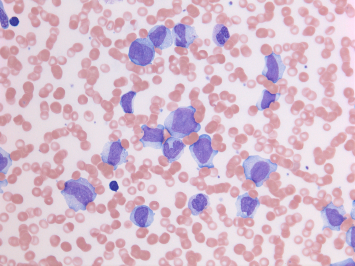 |
| Aspirate | 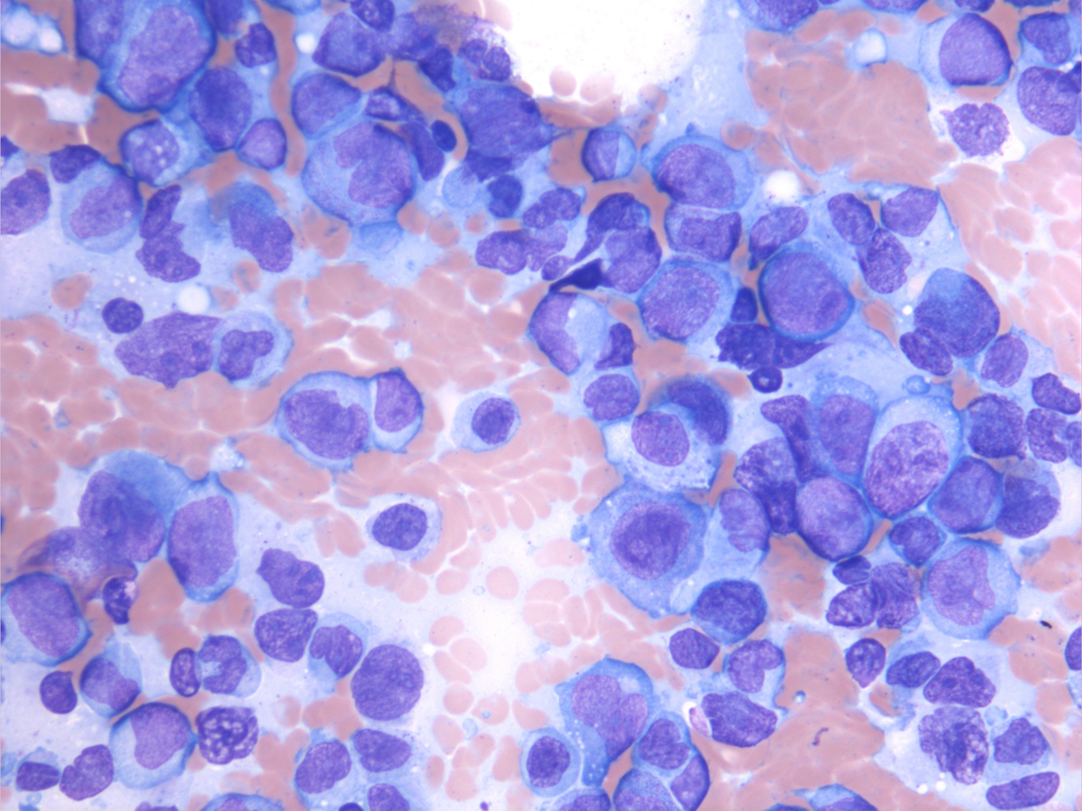 |
| Clot section H&E | 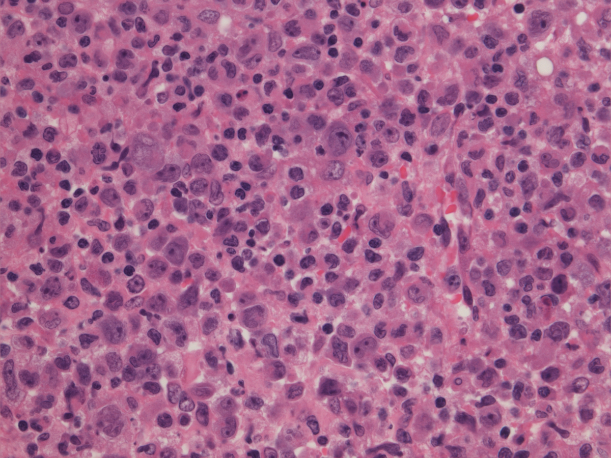 |
| IHC: Lysozyme | 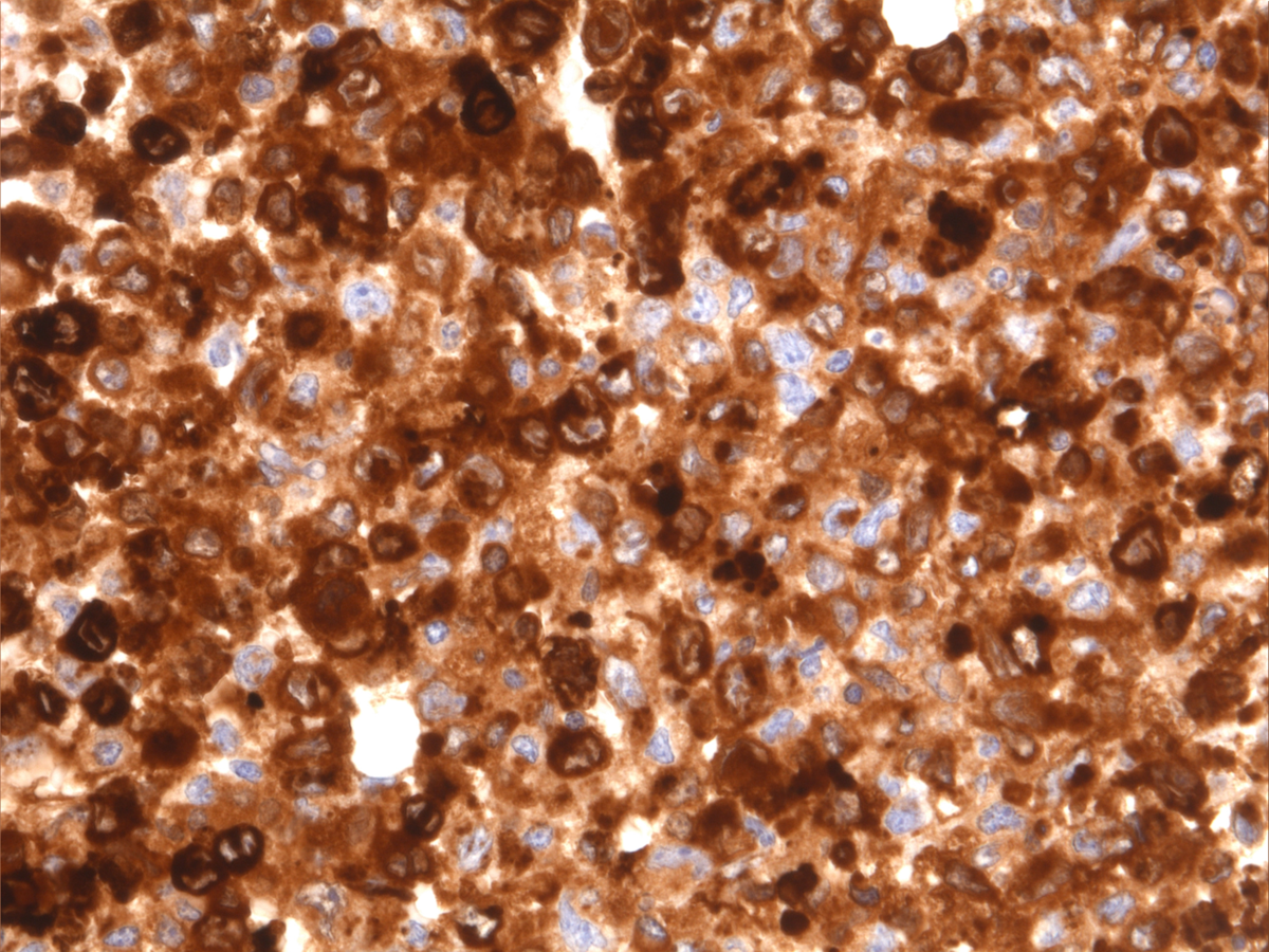 |
| IHC: CD3 | 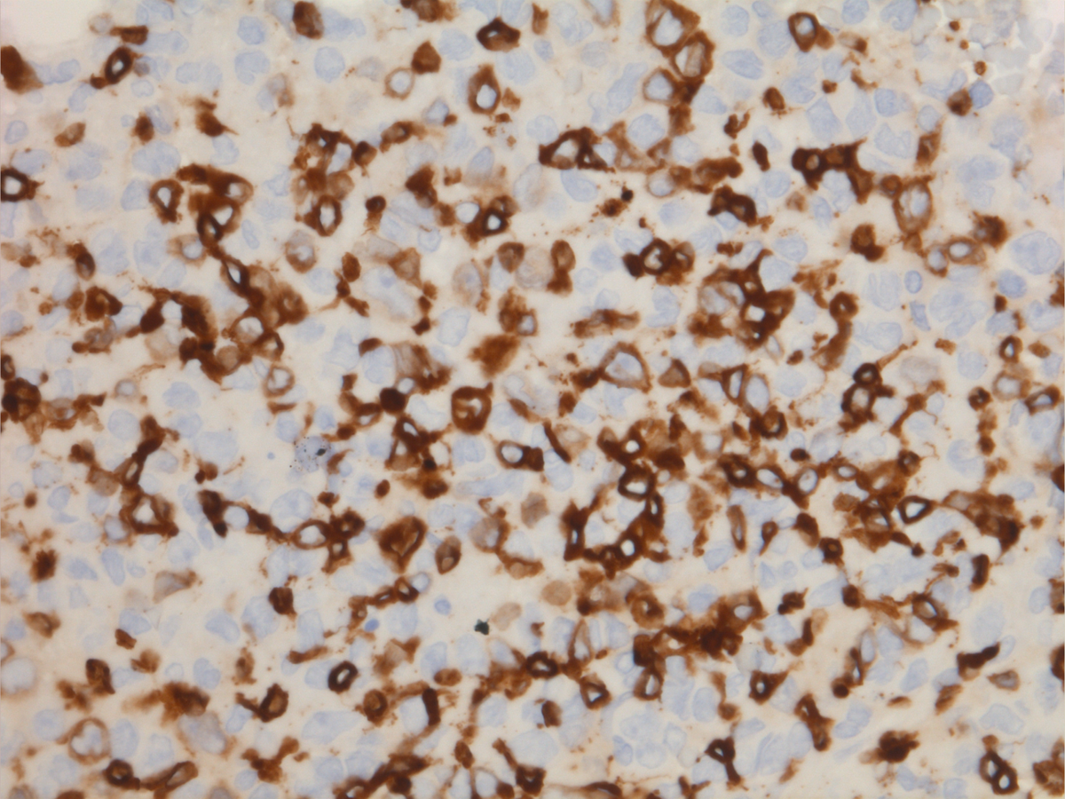 |
| Flow cytometry | 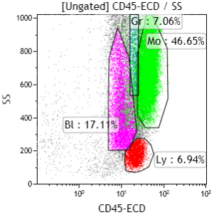 |
| Flow cytometry cytCD3 | 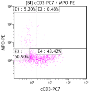 |
| Flow cytometry cytCD3/ungated | 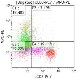 |
| Blasts: intermediate cytCD3 intensity | 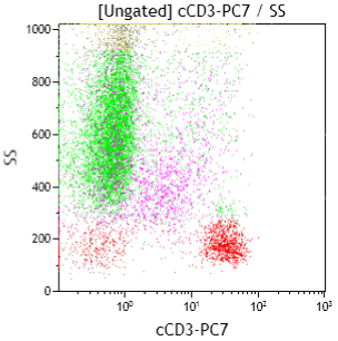 |
| surface CD3 negative | 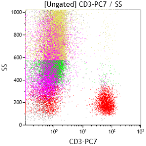 |