Institution: Indiana University, School of Medicine, Department of Pathology
Additional authors:Mehdi Nassiri, MD
Session: AML secondary to myeloproliferative neoplasms and other types of disease progression in MPN
HISTORY
An African American male (born in 1948) was initially present with leukocytosis and splenomegaly in 03/2004. Bone marrow evaluation was performed and diagnosis of chronic myelogenous leukemia, BCR-ABL1 positive, chronic phase was rendered. He was treated initially with imatinib and had only hematologic remission without molecular remission as demonstrated by a positive BCR-ABL1 fusion gene by PCR of peripheral blood. Patient also had persistent anemia and leukopenia since treatment of imatinib. He was treated with Neupogen with improvement. He was switched to dasatinib in 2007 and had a good response. A complete molecular remission was achieved and confirmed by PCR study of peripheral blood in 2010. However, he started to feel dyspneic and fatigue in summer 2011. Laboratory studies showed pancytopenias and increased LDH. Review of peripheral blood smear showed 22% blasts. The clinical concern was CML progression and transformation. Bone marrow biopsy was performed. Cytogenetics, FISH and molecular study for BCR-ABL1 fusion was ordered.
DETAILS
Biopsy in 2004: Marrow biopsy was taken from right posterior iliac crest and measured 0.6 cm. It was submitted in 10% buffered formalin for decalcification. The core biopsy was adequate for evaluation with a cellularity approaching 100%. The myeloid to erythroid ratio was markedly increased. The myeloid elements achieved full maturation with increased eosinophils seen. No evident increased in blasts seen. Rare erythroid elemetns were seen. Megakaryocytes were increased in number with occasional clustering. Small “dwarf” megakaryocytes were present. The aspirate smears were suboptimal for evaluation due to hemodilution and lack of well defined marrow particles. There were increased granulocytes with full maturation. Blasts were not increased. Eosinophils were increased. Rare unremarkable erythroid precursors and megakaryocytes were seen. Bone marrow in 2011: Marrow biopsy was taken from right posterior iliac crest and measures 1.2 cm. It was submitted in 10% buffered formalin for decalcification. The core biopsy was adequate for evaluation with a cellularity of ~90%. The myeloid to erythroid ratio was markedly increased. The myeloid elements showed marked left shift with increased interstitial and para-trabecular aggregates of blasts which showed vesicular nuclear chromatin and prominent nucleolus. Increased eosinophils were also present. Erythroid and megakaryocytic elements were markedly decreased. The aspirate smears were adequate for evaluation. There were markedly increased myeloid elements with left shifted maturation. Blasts were increased (23%) with high n/c ratio; open chromatin and prominent nucleolus. Dysplastic myeloid elements were seen including giant metamyelocytes, abnormal nucleation and abnormal granulations. Eosinophils were increased with immature forms seen. Erythroid precursors were decreased. Megakaryocytes were markedly decreased.
IMMUNOHISTOCHEMISTRY AND FLOW CYTOMETRY
Flow cytometry of marrow in 2004 showed a small population of blasts (~2%) that express CD34, CD117, CD33, CD13 and HLA-DR, consistent with myeloblasts. Immunohistochemical stain of CD34 on biopsy section showed scattered CD34+ blasts (<5% of total cellularity).
Flow cytometry of marrow in 2011 showed increased blasts (~12%) which are positive for CD34, CD117, CD13, CD33, and HLA-DR, consistent with myeloblasts. Immunohistochemical stain of CD34 on biopsy section showed interstitial and para-trabecular aggregates of CD34+ blasts (approaching 20% of total cellularity).
CYTOGENETIC FINDINGS
Cytogenetics of marrow in 2004 showed 46,XY,t(9;22)(q34;q11.2)[20]
Cytogenetics of marrow in 2011 showed 47,XY,+8[18]/48,XY,+8,+10[2].
MOLECULAR FINDINGS
Both FISH and PCR study in 2004 were positive for BCR-ABL1 fusion.
Both FISH and PCR study in 2011 were negative for BCR-ABL1 fusion.
INTERESTING FEATURES
Blastic transformation is one of the unfavorable outcomes of CML which is associated with poor prognosis. For diagnosis of blast transformation, presence of Philadelphia chromosome of BCR-ABL1 fusion is required although additional chromosome abnormalities might co-exist with Philadelphia chromosome in the same clone. Very rarely, a Ph-independent acute myeloid leukemia can evolve with chromosomal abnormalities detected in Ph-negative cells in CML patients as demonstrated by the current cases. The underlying mechanism remains to be determined. It could be due to side effect of CML treatment (imatinib and dasatinib) or genetic damage on Ph-negative cells by BCR-ABL1 associated process. Nevertheless, the occurrence of this phenomenon emphasizes the importance of cytogenetics/molecular study for the appropriate diagnosis/classification.
PROPOSED DIAGNOSIS
Acute myeloid leukemia with trisomy 8 mimicking blast crisis of Ph-positive chronic myeloid leukemia
CONSENSUS DIAGNOSIS
Acute myeloid leukemia with trisomy 8 and t(9;22)(q34;q11.2); BCR-ABL1 negative, in patient with history of chronic myelogenous leukemia, BCR-ABL1 positive
| Figure 1. Bone marrow evaluation in 2004. A. H&E section of marrow biopsy showing hypercellular marrow with maturing trilineage hematopoiesis; B. Bone marrow aspirate smear showing increased maturing myelopoiesis and decreased erythropoiesis; C. IHC of CD34 highlighting rare CD34+ blasts. |  |
| Figure 2. Immunophenotyping by flow cytometry in 2004: ~2% blasts which are positive for CD34, CD117, CD33, CD13, CD38 and HLA-DR. |  |
| Figure 3. Bone marrow evaluation in 2011. A. H&E section of marrow biopsy showing hypercellular marrow with increased blasts; B. Bone marrow aspirate smear showing increased blasts and myeloid left shift; C. IHC of CD34 highlighting increased CD34+ blasts. | 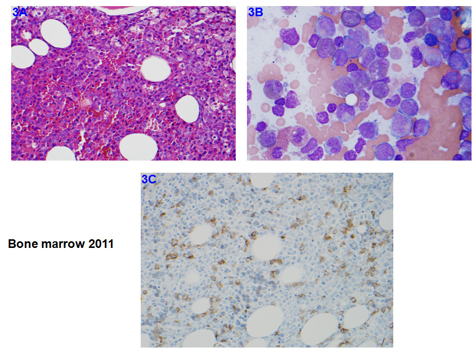 |
| Figure 4. Composite images showing dysmyelopoiesis in marrow aspirate smear in 2011. | 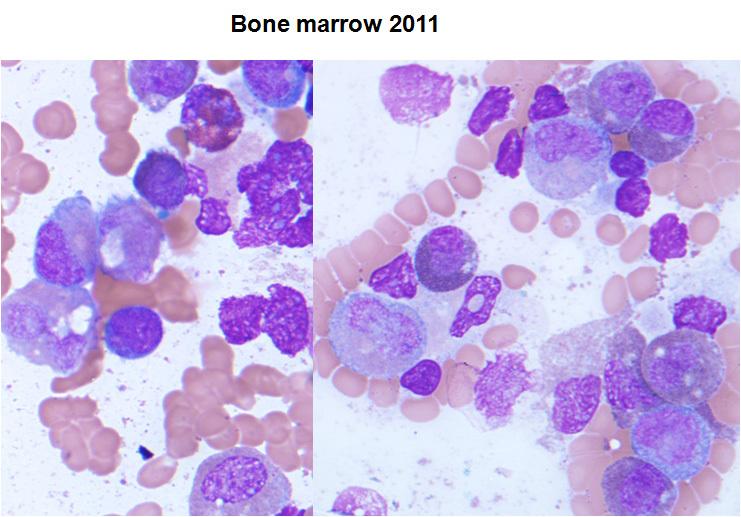 |
| Figure 5. Immunophenotyping by flow cytometry in 2011: ~12% blasts which are positive for CD34, CD117, CD33, CD13, CD38 and HLA-DR. | 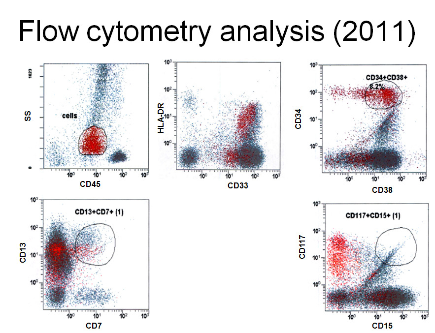 |
| Figure 6. Chromosomal analysis showed abnormal male karyotype: 47, XY,+8[18]/48, XY +8,+10[2]. | ![Figure 6. Chromosomal analysis showed abnormal male karyotype: 47, XY,+8[18]/48, XY +8,+10[2].](cases/14/images/Figure6- Zhou1.jpg) |