Institution: Moffitt Cancer Center
Additional authors:Haipeng Shao
Session: AML with recurrent genetic abnormalities Part I
HISTORY
The patient is a 72-year-old man presented with anemia (hemoglobin of 91 g/L), thrombocytopenia (platelet count of 90 x 109/L) and leukocytosis (white blood cell count of 40 x 109/L) to a local hospital in 6/2012. An outside bone marrow showed hypercellularity, dyspoiesis and “maturation arrest to myelocyte and promyelocyte level without excess blasts”.The bone marrow finding was suspicious for acute promyelocytic leukemia (APL). However, no karyotyping was performed and FISH for PML-RARA was reported negative. He complained of increasing fatigue but denied recurrent infections, unintentional weight loss, fevers, chills and drenching night sweats. After transferred to our institute, a repeated bone marrow biopsy was performed with features similar to the previously described. Conventional cytogenetic study on this marrow failed due to absence of mitosis, while Genome-wide single-nucleotide polymorphism (SNP) array demonstrated some gene abnormalities (see below) associated with 7q22.1 deletion. Additional FISH study with break-apart probe confirmed RARA gene rearrangement. The patient also had multiple cutaneous nodules, which was biopsied with diagnosis of panniculitis, considered to be paraneoplastic.
He received several chemotherapeutic regimens including 5-azacitidine, idarubicin +cytarabine, followed by 1 course of Gemtuzumab Ozogamicin (Myotarg). He had persistent disease despite these treatments. Subsequent bone marrow biopsy showed hypercellularity with excess blasts and dysplasia (details see below). He eventually achieved complete hematologic and molecular/genetic remission after additional therapy with Gemtuzumab and Ozogamicin x course at day 30. The patient has been on Vidaza maintenance therapy and retained in cytogenetic and molecular remission as documented in 1/2013, 7 months after the initial diagnosis.
DETAILS
Biopsy: posterior iliac crest Fixation: 10% neutral buffered formalin The initial bone marrow biopsy performed by the outside contributor was reviewed. The aspirate smear revealed left shifted granulocytic hyperplasia with increased myelocytes (48%) and promyelocytes (16%) (Figure 1). Morphologic dysplasia is noted in a subset of myeloid cells. Auer rods were overtly not seen (Figure 2). The cellularity was nearly 100% with myeloid preponderance and markedly left shifted maturation (Figure 3) admixed with few atypical megakaryocytes and small islands of erythroid precursors. A repeat bone marrow biopsy 11 days later at Moffitt Cancer Center demonstrated similar morphologic features. The bone marrow biopsy post chemotherapy at day 14 detected hypercellularity (95%) with excess myeloblasts (14%). Dysplastic megakaryocytes and granulocytes were identified. Erythroid lineage showed hyperplasia (Figure 4 and 5). The last follow up bone marrow biopsy performed in 1/2013 still exhibited hypercellularity (80%) with myeloid hyperplasia and mild dysplasia (Figure 6)
IMMUNOHISTOCHEMISTRY AND FLOW CYTOMETRY
Flow cytometry on the initial bone marrow aspirate detected a population of immature cells (71% of total events), phenotypically consistent with acute promyecytic leukemia. The cells expressed moderate CD45, partial CD11b, CD117, CD13, CD33, HLA-DR (minor subset) and MPO (strong) but were negative for CD34 (Figure 7).
CYTOGENETIC FINDINGS
Karyotyping showed no mitotic figures on the initial bone marrow aspirate, and FISH studies for PML-RARA fusion, MDS panel and BCR-ABL gene rearrangement were negative. FISH study with break-part RARA probe performed on the peripheral blood sample demonstrated an additional copy of 17q (RARA) in 134/200 cells (67%) (Figure 9). Additional FISH performed on the cell pellet of the second bone marrow aspirate confirmed RARA gene rearrangement. The follow-up karyotyping showed 46,XY,t(11;17)(q23;q21.1)[11]/46,XY[9] (Figure10). Normal male karyotype, 46,XY[20] was detected on the bone marrow samples after initial chemotherapy but FISH study still detected 1.5-2% of cells positive for RARA gene rearrangement consistent with residual disease. Karyotyping and FISH studies were normal after completion of chemotherapy therapy on 10/30/2012 (day 30) and 1/18/2013 (day 80), respectively.
MOLECULAR FINDINGS
NPM1 and JAK2 mutations were not detected in the initial bone marrow aspirate. The whole genome chromosome SNP array (Reveal) analysis was performed on the second bone marrow sample and revealed a 1.66 MB interstitial deletion of 7q22.1->Q22.1(Figure 8), and a 1.78 MB interstitial duplication of 13q31.3->Q31.3
INTERESTING FEATURES
This is an interesting case of t(11;17)(q23;q21) carrying out additional genetic abnormalities detected by SNP array. SNP array disclosed segmental duplication of 13q31.3>Q31.3 in addition to segmental interstitial deletion of 7q22.1>Q22.1. The clinical significance of both genetic aberrations is uncertain. The atypical promyelocytes in APL with t(11;17)(q23;q21) have an unusual morphology with resemblance to myelocytes and no Auer rods. There is always some degree of morphologic dysplasia. The unusual morphology and presence of dysplasia can be misinterpreted as evidence of myelodysplastic syndrome. In addition, t(11;17)(q23;q21) can be detected in usual cases of acute monoblastic leukemia with monocytic differentiation, but the partner gene for RARA on chromosome 17q21 is MLL rather than ZBTB16., The morphology of the atypical promyelocytes and phenotype will resolve the differential diagnosis. t(11;17)(q23;q21) is also reported in de novo MDS without associated with AML or APL. In our patient, hypercellularity with morphologic dysplasia was persistently noted in the 2 bone marrow biopsies after his having achieved complete molecular and cytogenetic remission lasting for 2.5 months, which raised the question of underlying myelodysplastic syndrome. The follow-up period is still too short to determine whether the patient truly has a coexisting myelodysplastic syndrome. The updated information will be followed.
PROPOSED DIAGNOSIS
Acute promyelocytic leukemia with t(11;17)(q23;q12)/ZBTB16-RARA
CONSENSUS DIAGNOSIS
Acute myeloid leukemia with t(11;17)(q23;q12); ZBTB16-RARA
| Figure 1 | 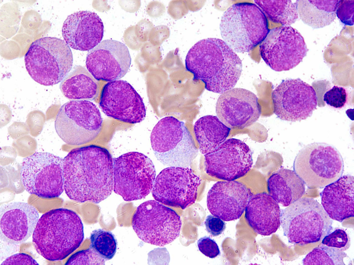 |
| Figure 2 | 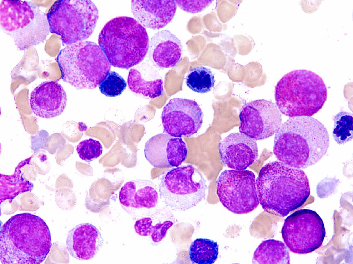 |
| Figure 3 | 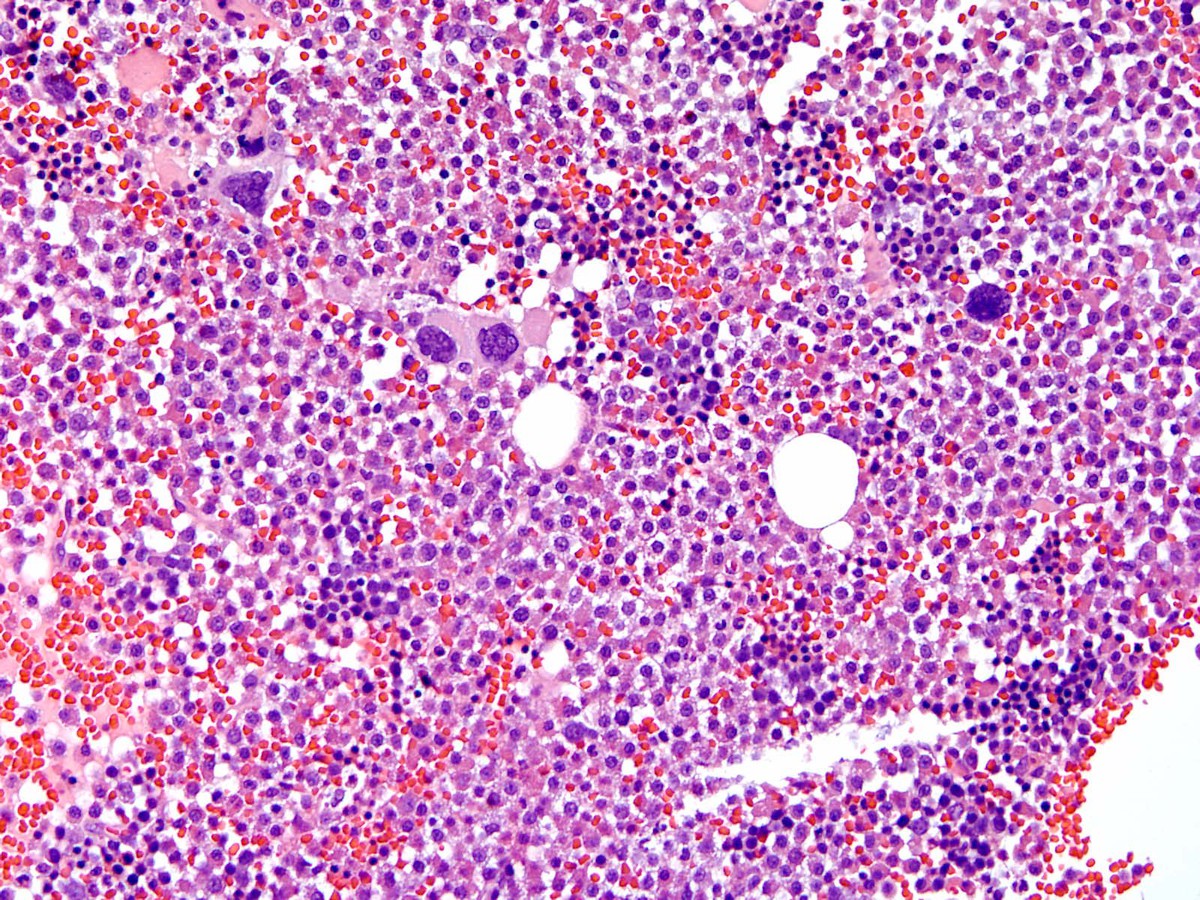 |
| Figure 4 | 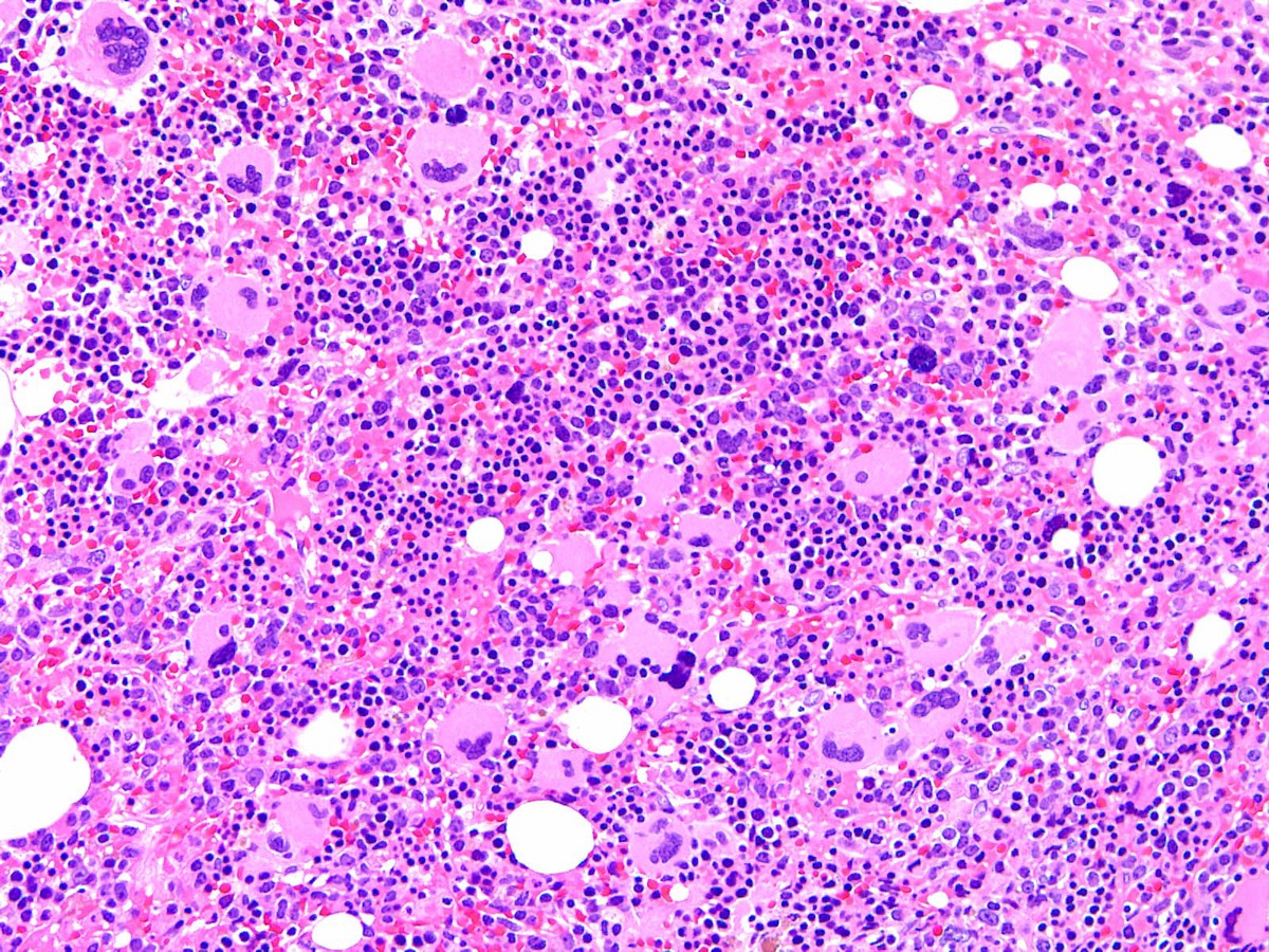 |
| Figure 5 | 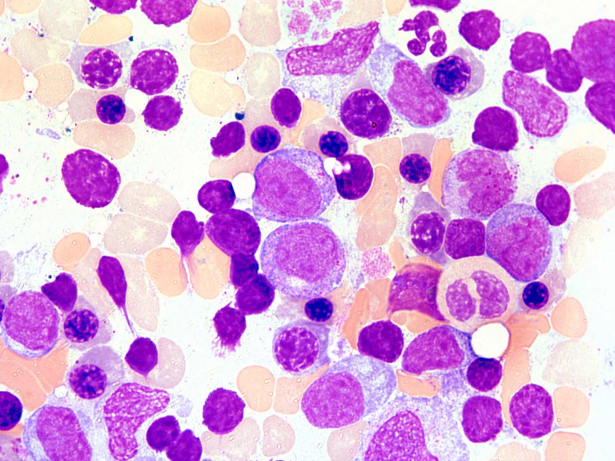 |
| Figure 6 |  |
| Figure 7 | 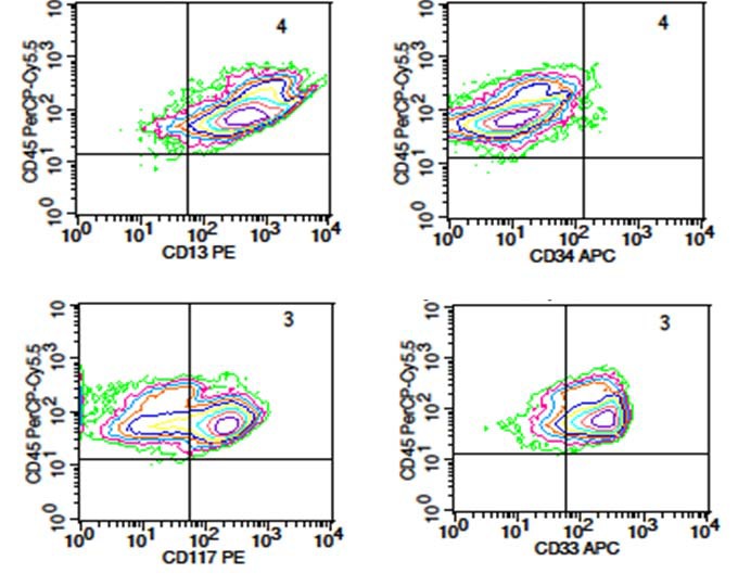 |
| Figure 8 | 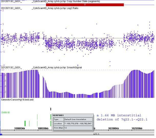 |
| Figure 9 | 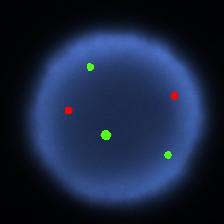 |
| Figure 10 | 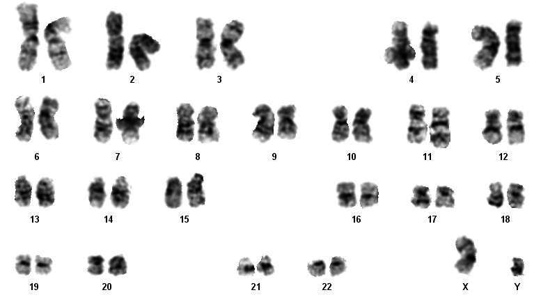 |