Institution: University of Pittsburgh School of Medicine
Additional authors:Miroslav Djokic, MD Malini Sathanoori, PhD, ABMG
Session: B Lymphoblastic Leukemia/Lymphoma
HISTORY
8 year old female with history of B lymphoblastic leukemia diagnosed June 2011:
47~53,XX,+3,+5,i(7)(q10),+8,+8,+14,der(19)t(1;19)(q23;p13),+21,+22[cp20]/46,XX[3]Fluorescence in situ hybridization studies for the TCF3 gene rearrangement are in progress.Bone marrow aspirate obtained on day 7 status post induction therapy demonstrated persistent disease ~27% blasts. 49,XX,i(7)(q10),+8,der(19)t(1;19)(q23;p13),+21,+22[4]/46,XX[16]. Bone marrow aspirates obtained on days 14 and 29 status post induction demonstrated no evidence of residual disease.Relapsed B lymphoblastic leukemia was diagnosed October 2012, with 95% blasts in bone marrow. 46,XX,i(7)(q10),der(19)t(1;19)(q23;p13)[4]/46,XX[16]. Patient was enrolled in COG AALL07P1 (Bortezomib and combination chemotherapy). Bone marrow obtained prior to “Block 2” of induction demonstrated approximately 3% blasts. Submitted bone marrow aspirate was obtained prior to “Block 3” of induction therapy (day 29 status post prior induction).DETAILS
Bone marrow aspirate only.
Aspirate smears demonstrate cellular marrow with trilineage hematopoiesis and increased immature cells. The immature cells include some abnormal appearing cells with irregular nuclear outlines and cytoplasmic vacuoles, representing approximately 4.2% of total cells by manual differential count, as well as some possible hematogones (Images 4 - 7).IMMUNOHISTOCHEMISTRY AND FLOW CYTOMETRY
Flow cytometric immunophenotyping performed with routine 4-color combinations demonstrates CD10 positive B-cells with features consistent with hematogones, but overlapping those seen at diagnosis and at relapse (CD19 positive, CD20 variable, CD10 positive, CD38 positive, CD34 negative, TdT positive) (Images 1 and 2).
Additional flow cytometric studies performed using an 8-color MRD-type combination (CD45 V500, CD20 V450, CD58 FITC, CD123 PE, CD34 Per-Cp Cy5.5, CD19 PE-Cy7, CD10 APC, CD38 APC-H7 ), and analyzed using Infinicyt software (Cytognos) demonstrate two cell populations (Image 3): hematogones (blue events) falling mostly within the outlined reference population generated using 19 control bone marrow samples; an abnormal CD58 positive population (green events) falling outside the reference population and representing 4.4% of total events.CYTOGENETIC FINDINGS
Fluorescence in situ hybridization (FISH) studies confirm the presence of residual disease with one extra signal corresponding to 7q31 in 15 of 230 interphase cells examined (6.5%) and one abnormal metaphase cell confirming the presence of an i(7q) chromosome (Images 8 and 9).
INTERESTING FEATURES
This case highlights difficulties that can be encountered in distinguishing between hematogones and residual B lymphoblastic leukemia and illustrates some of the newer, MRD-type, flow cytometric strategies that can be employed. Identification of a population with an abnormal phenotype required flow cytometric assessment of multiple antigens and was assisted by direct comparison with a composite reference phenotype.
In addition, at diagnosis the case demonstrated an unusual karyotype with the combination of hyperdiploidy and t(1;19)(q23;p13). At relapse the translocation t(1;19) was present but the trisomies were absent. A subset of B lymphoblastic leukemia with hyperdiploidy and a translocation t(1;19)(q23;p13), lacking either the E2A or PBX1 gene rearrangement but having an identical karyotype to B lymphoblastic leukemia with E2A-PBX1(TCF3-PBX1), has been reported. Fluorescence in situ hybridization studies are in progress to further investigate the submitted case.PROPOSED DIAGNOSIS
Residual B Lymphoblastic Leukemia.
CONSENSUS DIAGNOSIS
Residual B-lymphoblastic leukemia
| Image 1 flow cytometric phenotype | 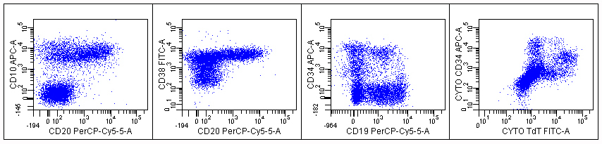 |
| Image 2 flow cytometric phenotype at diagnosis |  |
| Image 3 additional flow cytometric data | 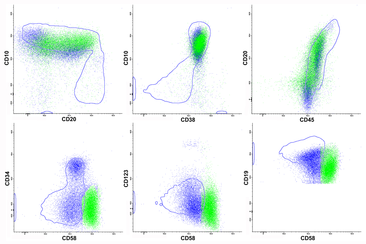 |
| Image 4 aspirate 10x | 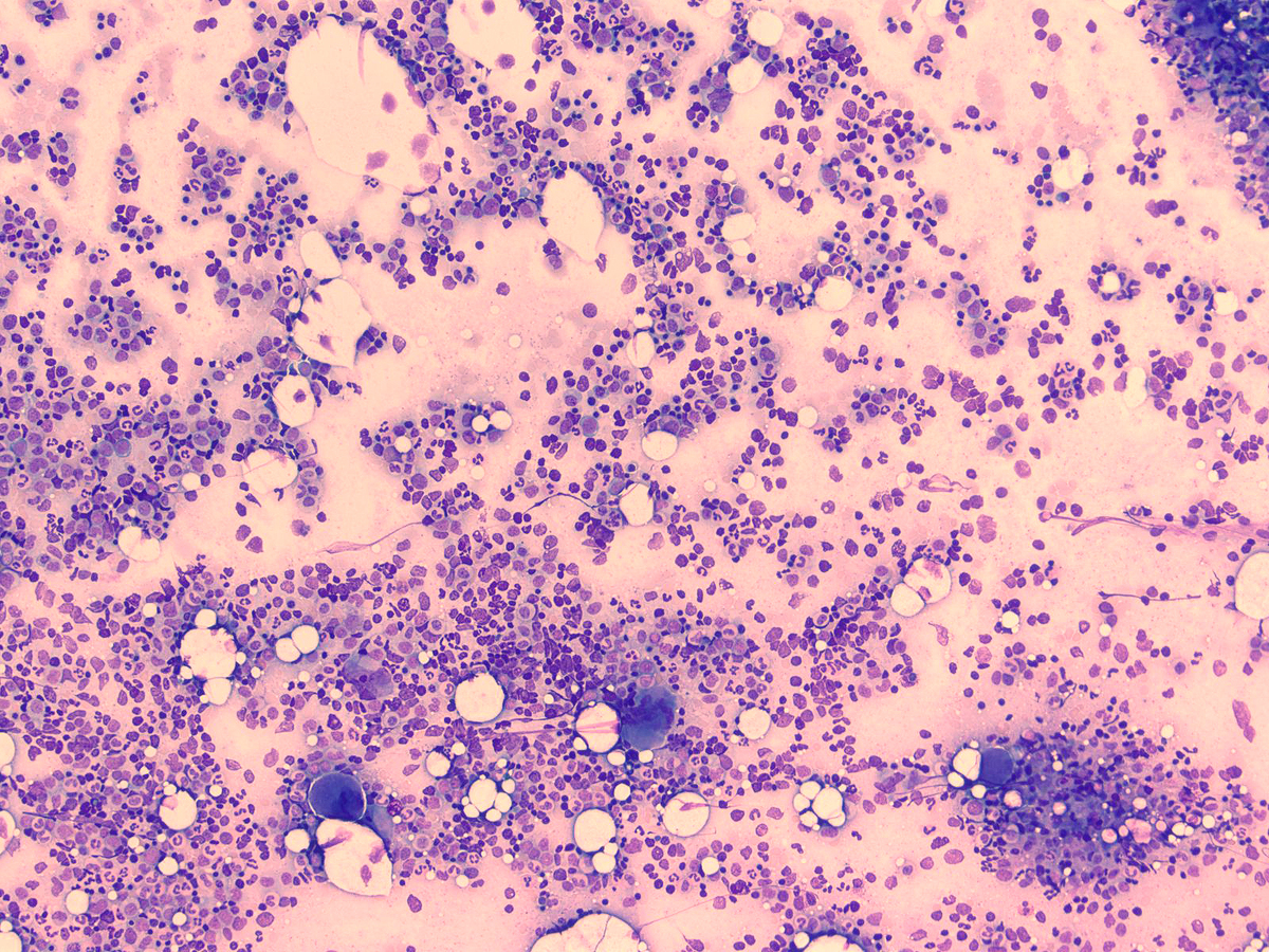 |
| Image 5 aspirate 50x | 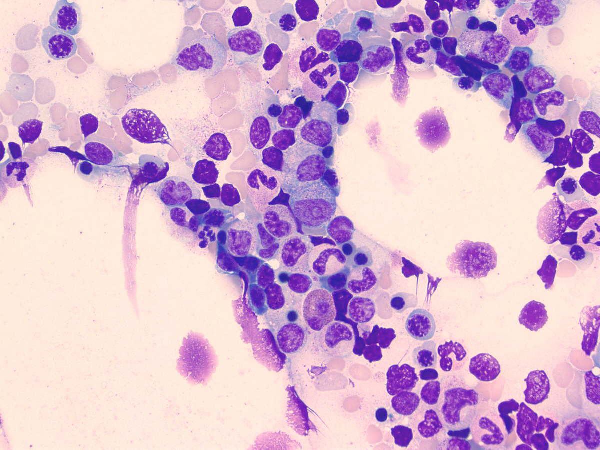 |
| Image 6 aspirate 100x 1 | 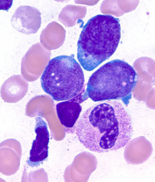 |
| Image 7 aspirate 100x 2 | 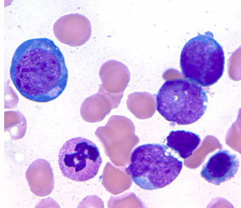 |
| Image 8 interphase FISH |  |
| Image 9 interphase FISH | 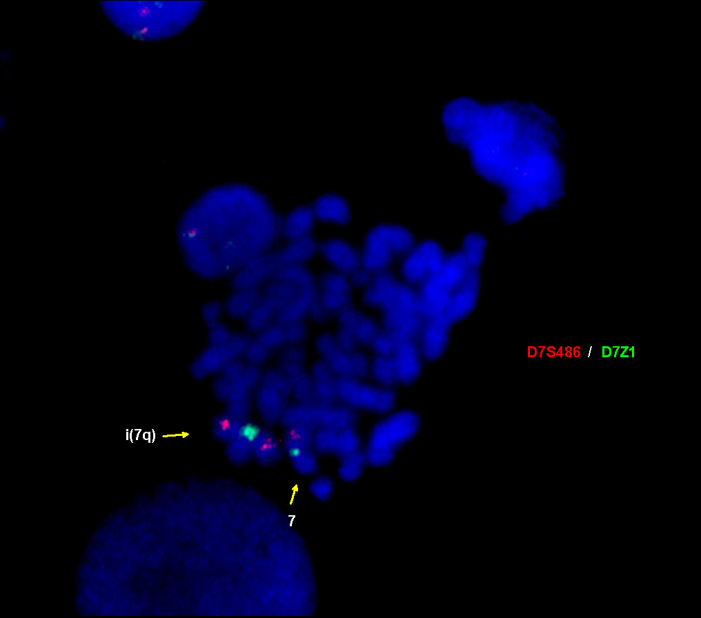 |