Institution: SUNY Upstate Medical University, Syracuse, NY
Additional authors:Katalin Banki MD, Robert E. Hutchison MD, Theresa Haven MT (ASCP), Haider Khadim MD
Session: Therapy-related myeloid neoplasms
HISTORY
23 year old Caucasian female patient presented in July 2010 with marked leukocytosis ( WBC : 118 K/uL) and was found to have circulating blasts (33%) and promonocytes (26%). Bone marrow biopsy revealed 46% blasts expressing myeloid and monocytic markers and a diagnosis of acute myelomonocytic leukemia was made. She was enrolled in CALGB 10523 protocol and underwent induction chemotherapy with 7+3+3 (cytarabine, daunrubicin and etoposide) and showed good response with no evidence of residual disease on Day 14 status post induction. She underwent consolidation therapy with high dose Ara-C and Etoposide in September 2010. She received autologus stem cell transplant with conditioning regimen including Busulfan and Etoposide in January 2011. Her counts recovered steadily following transplant and she continued to be in remission up until March 2012 when she presented with leukocytosis with a WBC count of 23.8 K/uL with 75% circulating blasts. Bone marrow aspirate examination revealed 65% blasts and 29% promyelocytes. Bone marrow biopsy was hypercellular with sheets of blasts and immature myeloid cells. Patient was re-induced and showed good cytoreduction as evaluated on the Day 14 bone marrow study. She was severely neutropenic, developed septicemia, and acute respiratory failure. She died on Day 25 after re induction chemotherapy.
DETAILS
Posterior superior iliac crest: Bone marrow biopsies and aspirate performed in 2010, 2011 and 2012. The bone marrow biopsy was fixed in formalin/B5. The aspirate smears were stained with Wright Giemsa stain.
IMMUNOHISTOCHEMISTRY AND FLOW CYTOMETRY
2010: Blasts were positive for CD33, CD15, CD13, CD11b, CD117 (partial), cMPO, CD64 (partial), and CD4 (partial). CD34 was negative
2012: Blasts were positive for CD33, CD15, CD117, CD 4 (partial) and CD64 (partial and lesser than the study done in 2010). CD34 was negative.
NPM1 immunohistochemical stain: showed abnormal cytoplasmic localization of NPM1 both in 2010 and 2012.
CYTOGENETIC FINDINGS
2010: Normal karyotype, 46 XX
No evidence of t (15; 17) by FISH assy
2012:
46x,t(X;1)(q13,p34),der(15)?(15pter-->15q22::17q22::17q21::15q22-->15q23::17q22-->17qter),der(17)?(17pter-->17q21::15q23-->15qter)[16]
46XX[4]
FISH results: nucish (PMLx2),(RARAx2,(PMLconRARAx1)[10/500]
nucish (PML,RARA)x2[443/500]
FISH study showed a variant signal pattern consistent with a variant 15;17 rearrangement. Combining the data from karyotype and FISH analysis it seems there is a reciprocal tranlocation between 15 and 17 resulting in typical derivative followed by a second rearrangement in which the fusion region from derivative 17 is translocated to the derivative 15 and a portion of the derivative 15 is returned to the 17.
Bone marrow FISH study done status post re-induction Day 17 showed evidence of minimal residual disease (7.6% cells with persistence of translocation 15;17).
MOLECULAR FINDINGS
FLT3 mutational analysis negative both in 2010 and 2012
INTERESTING FEATURES
Few cases of acute promyelocytic leukemia arising as a secondary malignancy following treatment for solid organ tumors has been described in the literature. Acute promyelocytic leukemia occurring as a secondary malignancy following treatment for acute myeloid leukemia is rar and also unusual. We did not come across any similar case in the literature where APL developed following treatment for acute myeloid leukemia. It raises an important issue in terms of classification; should this case be viewed as clonal evolution and relapse of previously diagnosed acute myelomonocytic leukemia now with complex cytogenetics including t(15;17) or a therapy related acute promyelocytic leukemia.
PROPOSED DIAGNOSIS
Therapy – related acute myeloid leukemia, consistent with acute promyelocytic leukemia with t (15;17)(q22;q12);PML-RARA.
CONSENSUS DIAGNOSIS
Therapy-related myeloid neoplasm, acute promyelocytic leukemia with t(15;17)(q22;q12); PML-RARA
| peripheral blood 2010, circulating blasts and promonocytes-1 |  |
| peripheral blood 2010, circulating blasts-2 |  |
| 2010: bone marrow aspirate smear |  |
| 2010 bone marrow aspirate smear: blasts and promonocytes | 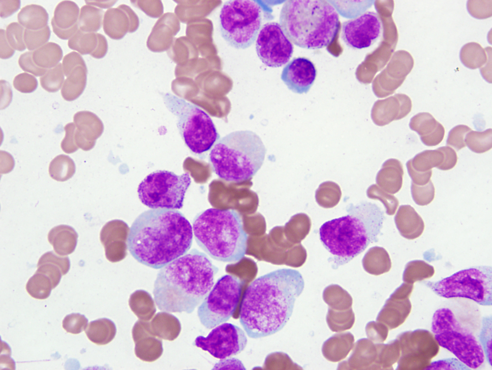 |
| 2010, hypercellular bone marrow biopsy | 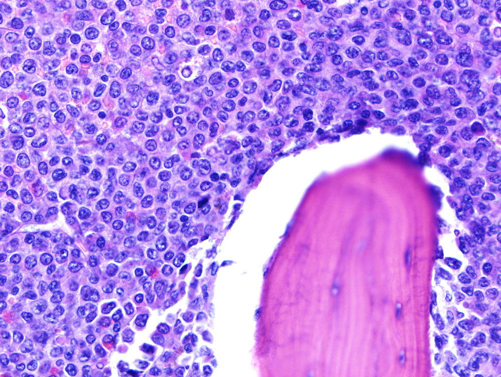 |
| 2011 bone marrow biopsy, patient in remission |  |
| peripheral blood 2012: circulating blasts-1 | 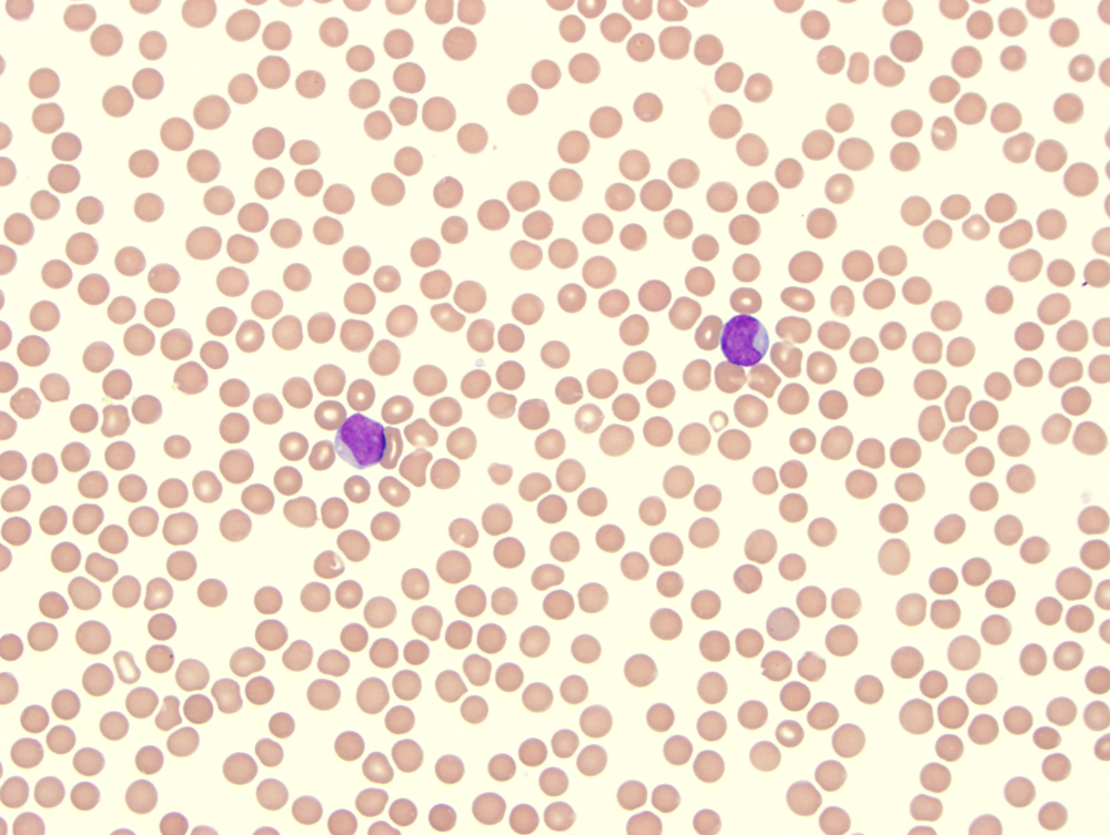 |
| peripheral blood, 2012, circulating blasts-2 |  |
| 2012, bone marrow aspirate smear-1 |  |
| 2012 bone marrow aspirate smear-2 |  |
| bone marrow biopsy, 2012 | 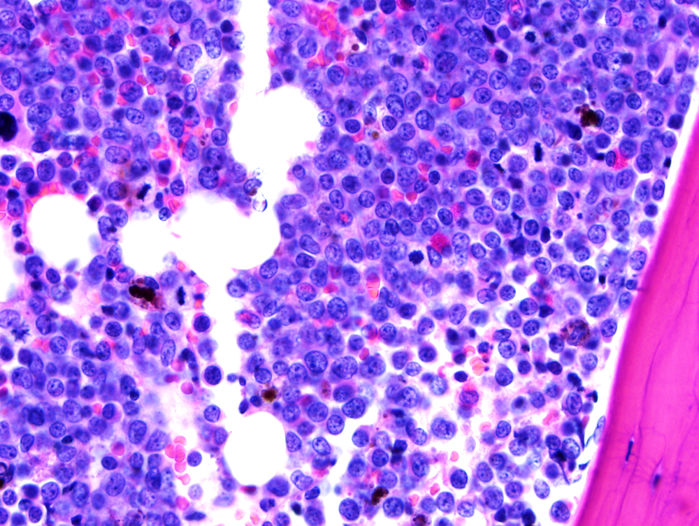 |
| 2010, bone marrow flow cytometry-1 |  |
| 2010 bone marrow flow cytometry-2 |  |
| 2012 bone marrow flow cytometry | 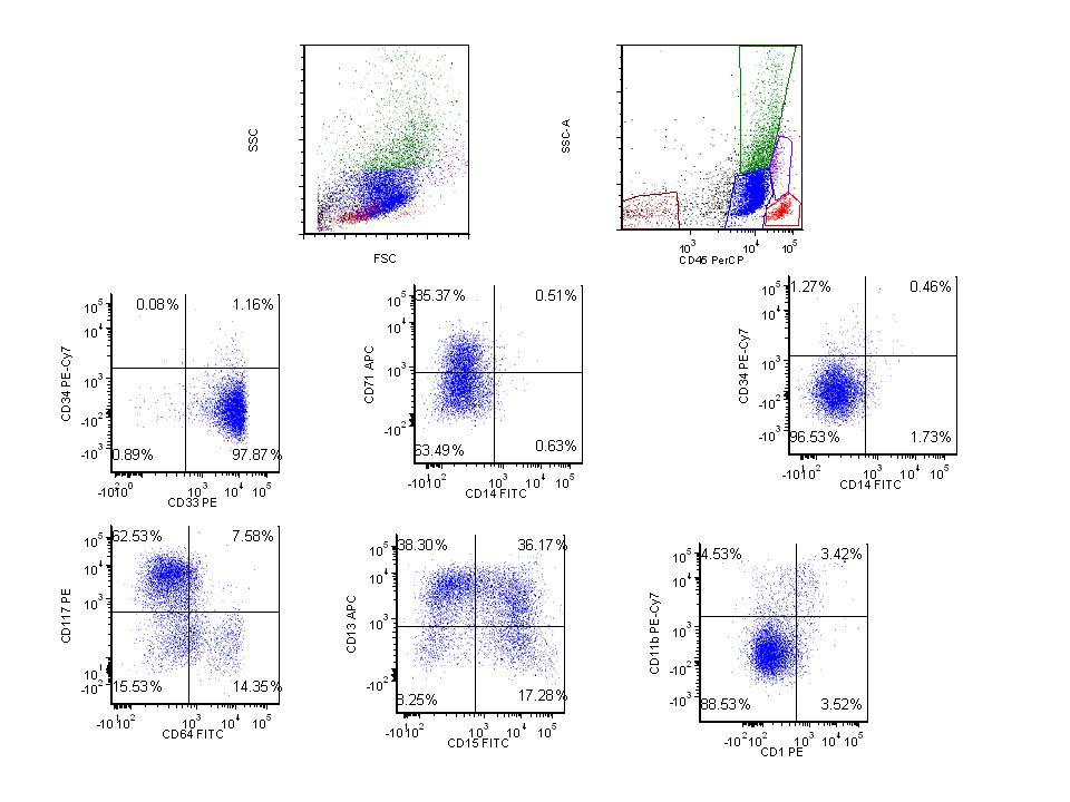 |
| 2010: karyotype | 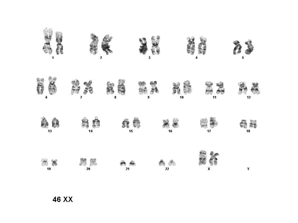 |
| 2010: FISH t( 15;17): Negative study | 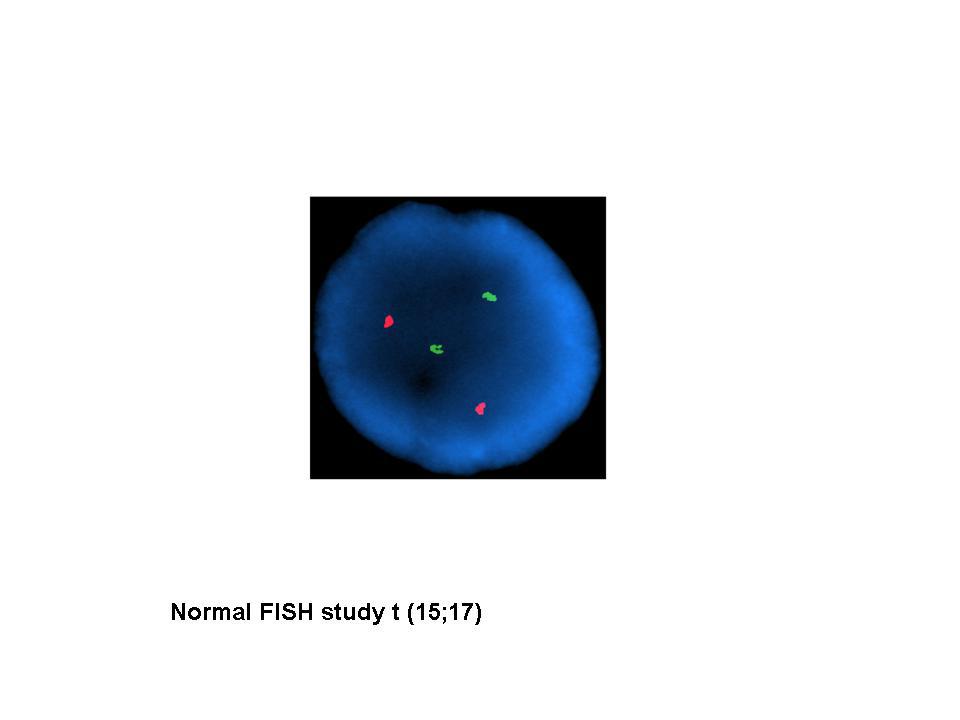 |
| Karyotype: 2012 | 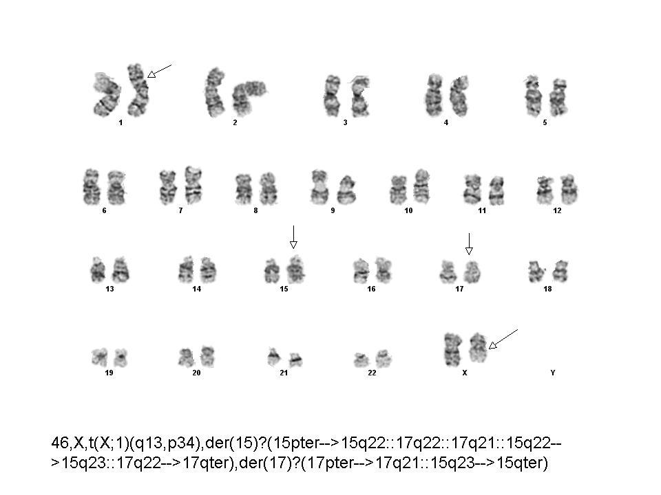 |
| FISH analysis 2012 | 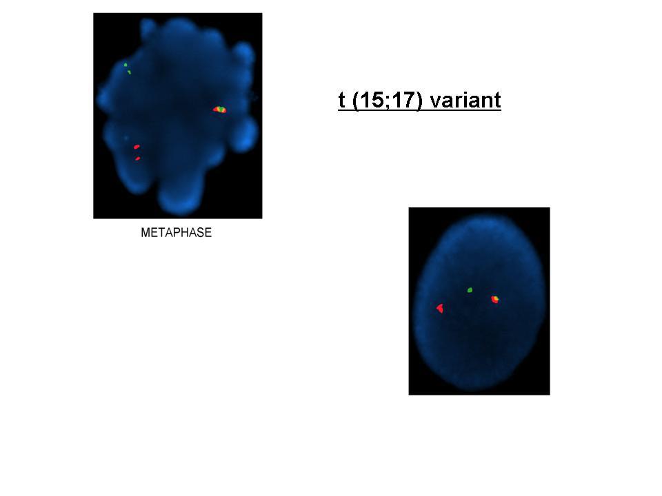 |
| NPM immunohistochemical staining: Abnormal cytoplasmic localization | 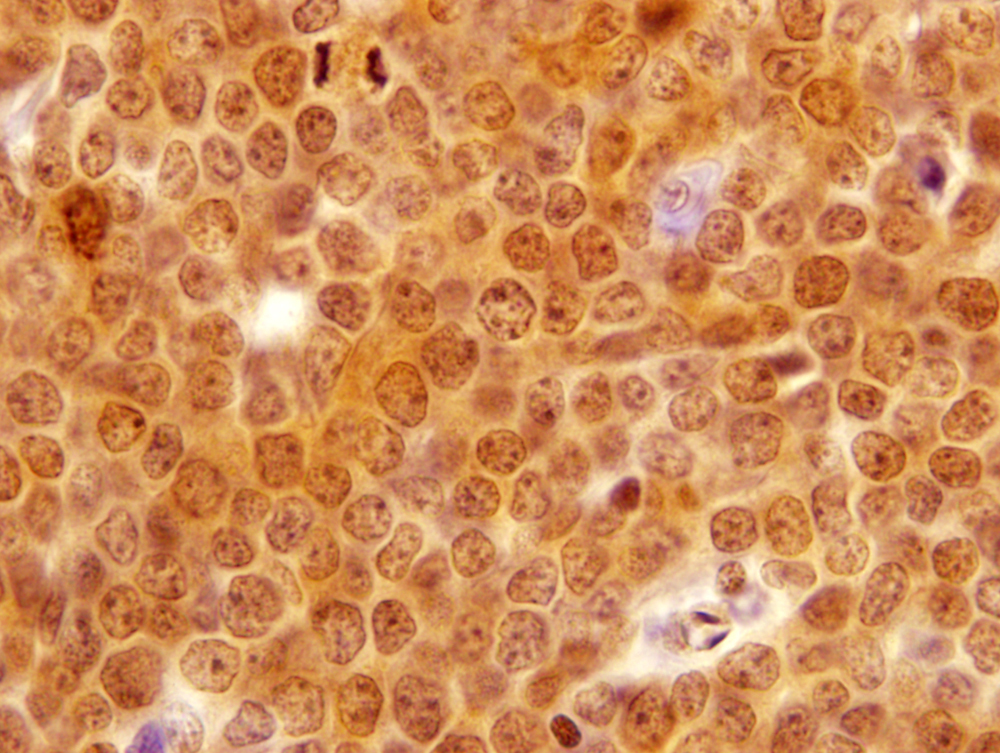 |