Institution: University of Texas Health Science Center at San Antonio
Session: Extramedullary manifestations of myeloid neoplasms
HISTORY
68-year-old female with a several month history of pruritus and a persistent papular skin eruption. The clinical impression was lymphomatoid papulosis versus folliculitis. The patient was said to have a history of "CML". A CBC was not available.
DETAILS
Skin, trunk, multiple biopsies, formalin fixed: Beneath an acanthotic to focally ulcerated epidermis, a moderately dense perivascular and interstitial and periadnexal mononuclear cell infiltrate is present in the superficial and deep dermis. The mononuclear cells are a mixture of small lymphocytes (more prominent in the deeper dermis) and medium-sized to large cells with more dispersed chromatin and occasionally prominent nucleoli. The medium-sized cells have features of monocytes, and some of the larger cells resemble Langerhans cells but lack well defined nuclear grooves. Rare neutrophils are admixed.
IMMUNOHISTOCHEMISTRY AND FLOW CYTOMETRY
The lymphocytes are predominantly CD4+ T-cells with normal antigen expression (CD2+, CD5+, CD7+). The medium to large mononuclear cells (MNC) express CD4, CD33, and CD68. Many MNC express CD1a and CD33. Small clusters of MNC express CD123 and TCL-1. The mononuclear cells lack other T-cell antigens, CD34, CD56, CD117, CD207, TdT, S100, Factor XIIIa, and myeloperoxidase. Staining for CD30, EMA, and ALK-1 is negative. Flow cytometry not performed.
CYTOGENETIC FINDINGS
Not performed.
MOLECULAR FINDINGS
Not performed.
INTERESTING FEATURES
Although the patient was initially reported to have CML, after immunophenotyping was performed the referring dermatologist was questioned and confirmed the patient actually had CMML. CMML in the skin has variable features as described by Vitte F et.al., Am J Surg Pathol 36:1302-1316, 2012, which can make the diagnosis difficult. The histologic and immunophenotypic features in this patient's lesion correspond to mature plasmacytoid dendritic cell proliferations (MPDCPs) of mixed cell type with small clusters of PDC intermixed with lymphocytes and large cells with pale, irregular nuclei similar to large histiocytes or Langerhans cells and corresponding most closely to indeterminate dendritic cells. This case also illustrates the importance of knowing the pathologic spectrum of CMML and having an accurate clinical history.
PROPOSED DIAGNOSIS
Cutaneous chronic myelomonocytic leukemia
CONSENSUS DIAGNOSIS
Cutaneous manifestation of chronic myelomonocytic leukemia with mature plasmacytoid dendritic cell proliferation
| H&E, 4X | 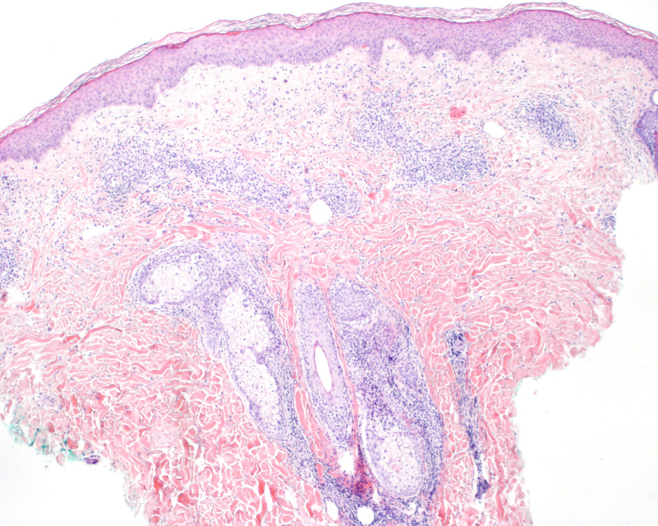 |
| H&E, 12X |  |
| H&E, 4X |  |
| H&E, 75X |  |
| H&E, 75X |  |
| CD3 |  |
| CD4 | 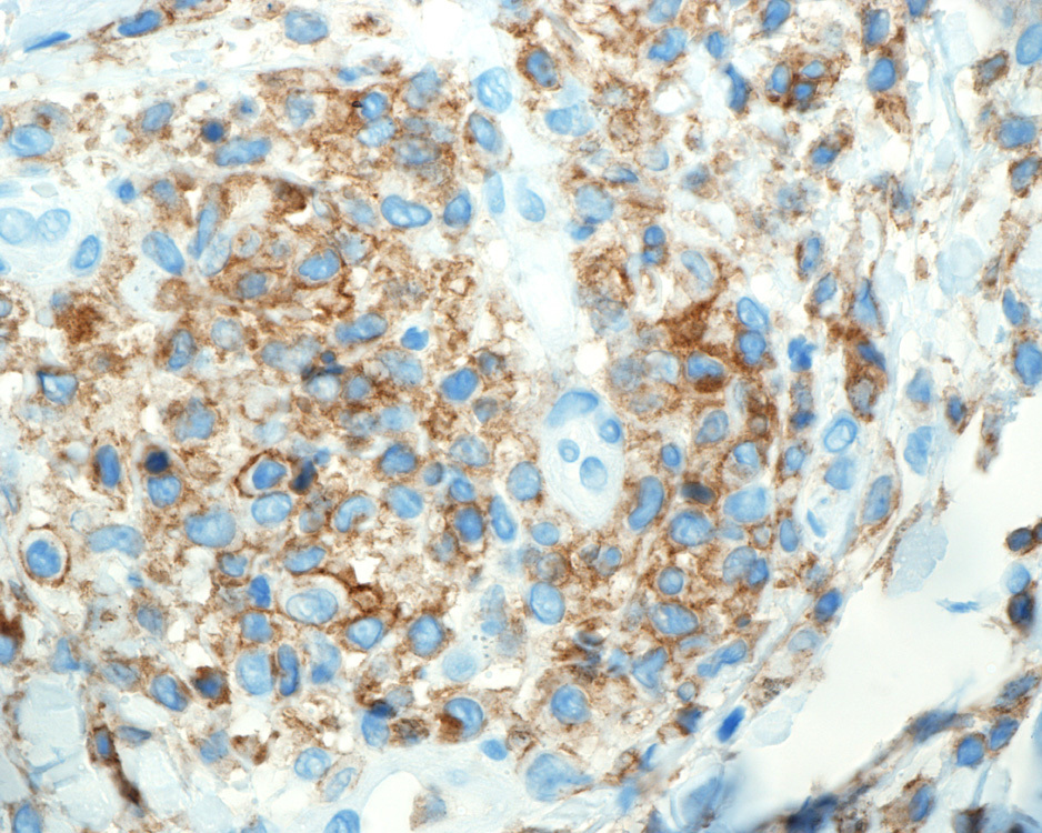 |
| CD8 |  |
| CD68 |  |
| CD33 | 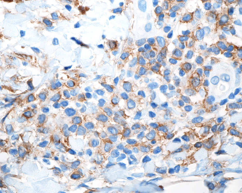 |
| CD117 |  |
| CD1a |  |
| CD207 |  |
| CD123 | 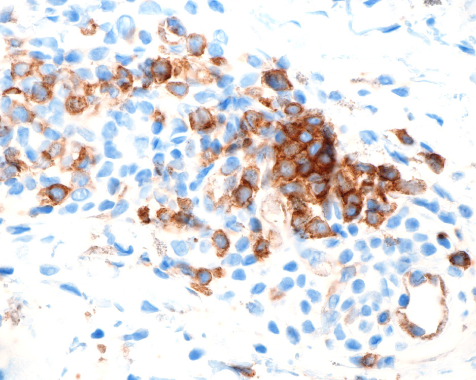 |
| TCL1 |  |
| CD56 | 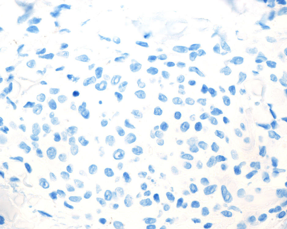 |
| CD34 |  |