Institution: University of Massachusetts Medical School, Department of Pathology
Additional authors:Hongbo Yu, Patricia Miron, Bruce Woda
Session: Therapy-related myeloid neoplasms
HISTORY
A 43 year-old male presented with pancytopenia in December 2009. His bone marrow biopsy revealed left-shifted granulopoiesis with 79% myeloblasts and promyelocytes. Cytogenetic study showed translocation t(15;17) in 20/20 metaphases. Polymerase chain reaction (PCR) test was positive for PML-RARA fusion transcripts. The overall findings were diagnostic of acute promyelocytic leukemia (APL). The patient was treated with all-trans-retinoic acid (ATRA), prednisone and idarubicin, resulting in complete remission. He also received three independent courses of consolidation therapy with ATRA and idarubicin. In May 2010, the patient started maintenance chemotherapy with ATRA, mercaptopurine and methotrexate. He finished two years of maintenance on this regimen. In October 2012, the patient was found to be pancytopenic during a routine follow-up. His bone marrow biopsy showed myelodysplastic syndrome with increased blasts of 10-20%. Cytogenetic study demonstrated t(3;14)(q26.2;q32) and monosomy 7. Fluorescence in situ hybridization (FISH) confirmed the t(3;14) to be an EVI1-IGH rearrangement. The patient was then started treatment with decitabine, and will undergo stem cell transplantation when blast count is improved.
DETAILS
Site: Posterior iliac crest Method of fixation: Formalin Gross: Bone marrow core biopsy Microscopic pathology: Bone marrow biopsy from 12/6/2009 revealed hypercellular marrow (95%) with sheets of myeloblasts and promyelocytes. Erythroid elements and megakaryocytes were markedly decreased. Bone marrow aspirate smears were difficult to evaluate due to artifact. Bone marrow biopsy and aspirate smears from 10/22/2012 showed variably hypocellular marrow, dyserythropoiesis, which is characterized by megaloblastoid maturation and nuclear irregularity, and 10-20% myeloblasts, which is shown by CD34 immunostain on biopsy and by differential count on aspirate smears. Iron stain showed increased storage iron with ring sideroblasts.
IMMUNOHISTOCHEMISTRY AND FLOW CYTOMETRY
12/6/2009
Flow cytometric analysis showed a population of myeloblasts and promyelocytes (81%) with the following immunophenotype: CD34 subset+, CD117 subset+, CD13+, CD33+, myeloperoxidase+, HLA-DR-, CD19-, CD56-, CD3-
10/22/2012
Flow cytometric analysis showed a population of myeloblasts (15%) with the following immunophenotype: CD34+, CD117+, HLA-DR+, CD13+, CD33+
CYTOGENETIC FINDINGS
12/6/2009
Bone marrow karyotype:
46,XY,del(9)(q13q22),t(15;17)(q22;q21)[20].nuc ish(PML,RARA)x3(PML con RARAx2)[94/100]
Interpretation: Out of 20 cells studied from this bone marrow specimen, 20 carried a translocation involving chromosomes 15 and 17 with breakpoints at 15q22 and 17q21. In addition to the t(15;17), all 20 cells demonstrated a deletion of chromosome 9 with breakpoints at 9q13 and 9q22.
10/22/2012
Bone marrow karyotype:
45,XY,t(3;14)(q26.2;q32),-7[13]/46,XY[7].nuc ish(EVI1x2)(5'EVI1 sep 3'EVI1x1)[59/100],(IGH@x2)(5'IGH@ sep 3'IGH@x1)[31/100]ish t(3;14)(5'IGH@+,3'IGH@-;5'IGH@+,3'IGH@-)
Interpretation: Of 20 cells studied, 13 comprised an ABNORMAL clone with a translocation between chromosomes 3 and 14, breakpoints at bands 3q26.2 and 14q32, and monosomy 7. FISH confirmed the t(3;14) to be an EVI1-IGH rearrangement.
MOLECULAR FINDINGS
12/6/2009
PML-RARA hybrid transcripts were detected using PCR.
10/22/2012
PML-RARA hybrid transcripts were not detected using PCR.
INTERESTING FEATURES
Therapy-related myeloid neoplasms (t-MDS/AML) is a relatively infrequent, late, and poor prognostic complication in patients with APL treated with ATRA and anthracycline-based chemotherapy. EVI1 proto-oncogene is located on chromosome 3q26, and has been shown to be involved in pathogenesis of acute myeloid leukemias or myelodysplastic syndromes. Cytogenetic abnormalities involving EVI1 such as t(3;21)(q26;q22) translocation have been reported as a rare recurring translocation in patients with APL relapsing as t-MDS/AML. Here, we described a case of a 46-year-old male with history of APL developed therapy-related myeloid neoplasm (t-MDS) with translocation t(3;14)(q26;q32) and EVI1-IGH rearrangement, which has not been reported previously.
PROPOSED DIAGNOSIS
Therapy-related myeloid neoplasm (t-MDS) with translocation t(3;14)(q26.2;q32) and EVI1-IGH rearrangement
CONSENSUS DIAGNOSIS
Therapy-related myeloid neoplasm, myelodysplastic syndrome with t(3;14)(q26.2;q32); EVI1-IGH
| Bone marrow 12062009 | 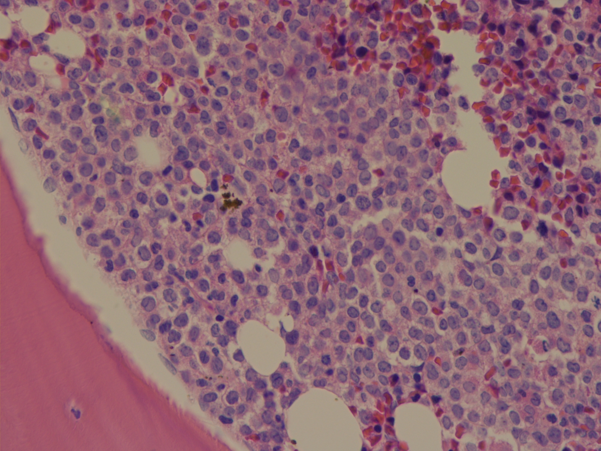 |
| Bone marrow 10222012 | 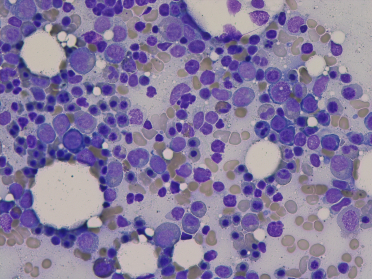 |
| Iron stain on Bone marrow 10222012 | 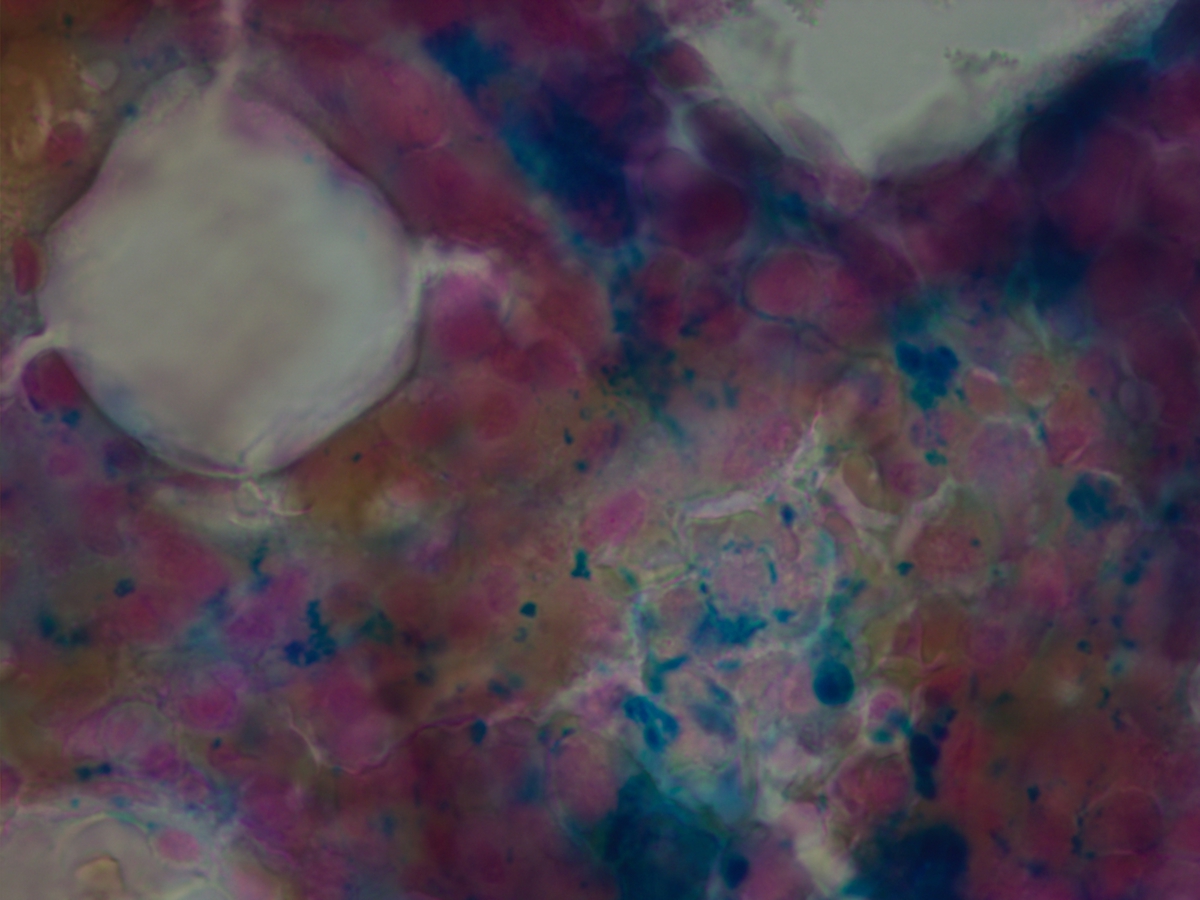 |
| Karyotype | 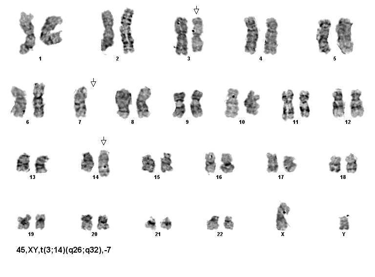 |
| EV1-IGH rearrangement by FISH | 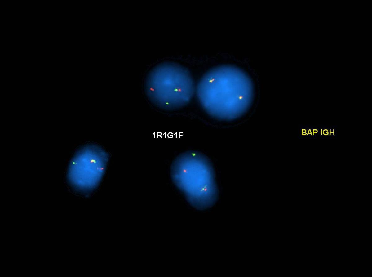 |
| EV1-IGH rearrangement by FISH | 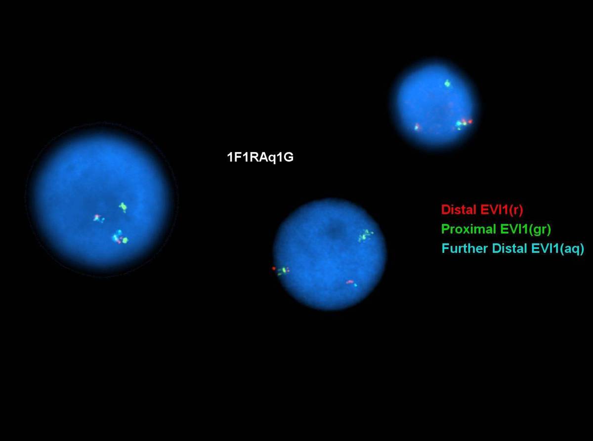 |