Institution: University of Arkansas Medical system
Additional authors:Ginell Post MD, PhD
Session: Erythroleukemia and megakaryoblastic AML and mimics
HISTORY
A 64 –year old male with clavicular fracture, without prior therapy presented to our institution diagnosed with IgG kappa multiple myeloma, stage III-A, in 12/1999. At that time patient had normal karyotype and normal laboratory findings. The patient was treated on Total Therapy 2, randomized to no thalidomide and had single transplantation with melphalan in 2000. Lost appropriate follow up, due to disease progression with normal karyotype in 2001, treated with dexamethasone pulsing through May of 2005. Further disease progression noted in September 2005 with abnormal karyotype and started on single agent velcade, followed by Revlimid until 2008. At that time gene array showed hyperploid molecular subtype, low risk gene array scores in the 70 and 80 gene models. In March 2008 started on regimen with thalidomide, velcade and dexamethasone, but patient noted to have disease progression in 01/2009 with abnormal karyotype of hyperdiploidy subtype. Then patient received VDT- PACE on 01/10/09, showing excellent response with near complete remission status maintained well with VRD (velcade, revlimid and dexamethasone) until April 2010.
Returned in April 2010 with c/o extreme fatigue, at that time laboratory findings significant for pancytopenia and increased M protein levels. Bone marrow examination showed 20% plasmacytosis, dyserythropoiesis with left shifted maturation and hyperdiploid DNA stem line and kappa light chain excess in 52% of cells. Karyotype was abnormal and gene array analysis showed low risk in 70 and high risk in 80 gene model, proliferation subtype. Stopped revlimid therapy in June/2010 due to severe thrombocytopenia, and returned to myeloma clinic due to acute rise in LDH to 2500 and abnormal liver function tests. MRI and PET scan studies revealed hepatosplenomegaly. Assuming disease progression with extramedullary involvement, bone marrow biopsy and liver biopsy were performed.DETAILS
Bone marrow aspirates (wright stain) and right iliac core biopsy (H&E): Almost entire bone marrow biopsy and the aspirate replaced by two separate populations of neoplastic cells. First population is composed of large sized erythroblasts with round nuclei; fine chromatin with prominent nucleoli, deeply basophilic moderate amount of cytoplasm with most of them contains poorly demarcated vacuoles. These cells have nodular and interstitial pattern of infiltration.
Second population is medium sized neoplastic plasma cells with most of them them showing prominent nucleoli with nodular and interstitial pattern of infiltration, comprising about 40% of the cellularity.IMMUNOHISTOCHEMISTRY AND FLOW CYTOMETRY
PAS stain on bone marrow aspirate reveals intense cytoplasmic globular PAS staining in proerythroblasts.
The immunohistochemical stains show that the proerythroblast population (~40%) is positive for CD117, CD4, E cadherin and Glycophorin A (variable, scattered, subset). Also revealed CD138+ve 40% plasma cells that are MUM-1+, CD79a+ and kappa light chain restricted. Liver Biopsy (H&E) : Sinusoidal infiltration of proerythroblast population that are immunohistochemicaly positive for CD117, CD4, and Glycophorin A (variable, scattered, subset), similar to the erythroid population in the bone marrow biopsy. CIg/DNA flow cytometry showed revealed hyperdiploid DNA stem line and kappa light chain excess in 52% of cells.CYTOGENETIC FINDINGS
Bone marrow: Two populations: myeloma and MDS karyotypes
Myeloma karyotype: 52~53,XY,+3,+5,del(6)(q25),+9,+11,+15,+15,+19[cp13]MDS karyotype: 43~45,XY,del(1)(p13p22),?t(1;2)(p32:q33),del(7) (p12p15),add(8)(p22), del(9)(q12q22),add(13) (p11.1),-16, add(16)(q?22),add(19)(p13.1), del(?20)(p11.2), del(?21)(q21)[cp6] Liver biopsy: MDS karyotype 43~46,XY,del(1)(p13p22),?t(1;2)(p32;q33),del(7)(p12p15),add(8)(p22),del(9)(q12q22),add(13)(p11.1),-16,add(16)(q?22),add(19)(p13.1), del(?20)(p11.2),del(?21)(q21),inc[cp12]/46,XY[cp2]INTERESTING FEATURES
The usual course of therapy related myeloid neoplasm in MM patients is progressive cytopenias, dyspoietic morphologic features and MDS chromosomal abnormalities, that mostly observed by peripheral blood and bone evaluation.
This case is unusual with sudden onset of therapy related myeloid neoplasm in the form of pure erythroid leukemia with extensive extramedullary involvement revealing similar MDS complex karyotypes in the bone marrow and liver. Diagnosis of this patient’s acute myeloid leukemia (erythroid leukemia) complicated by difficulty in morphological differentiation of high grade plasma cells and pronormoblasts on bone marrow aspirates.PROPOSED DIAGNOSIS
BM biopsy: Plasma cell neoplasm with concurrent therapy related myeloid neoplasm (t-AML)
Liver Biopsy: Therapy related myeloid neoplasm (t-AML; erythroid leukemia)CONSENSUS GROUP: ADDITIONAL INFORMATION/STUDIES
Iron stain showed increased storage iron with no ring sideroblasts. Patient died shortly after liver biopsy due to bleeding.
CONSENSUS DIAGNOSIS
Therapy-related AML, with features of pure erythroid leukemia
| Bone marrow aspirate - High grade plasma cells intermingled with pronormoblasts |  |
| Bone marrow aspirate: PAS+ve cytoplasmic vacules in dyspoietic pronormoblasts | 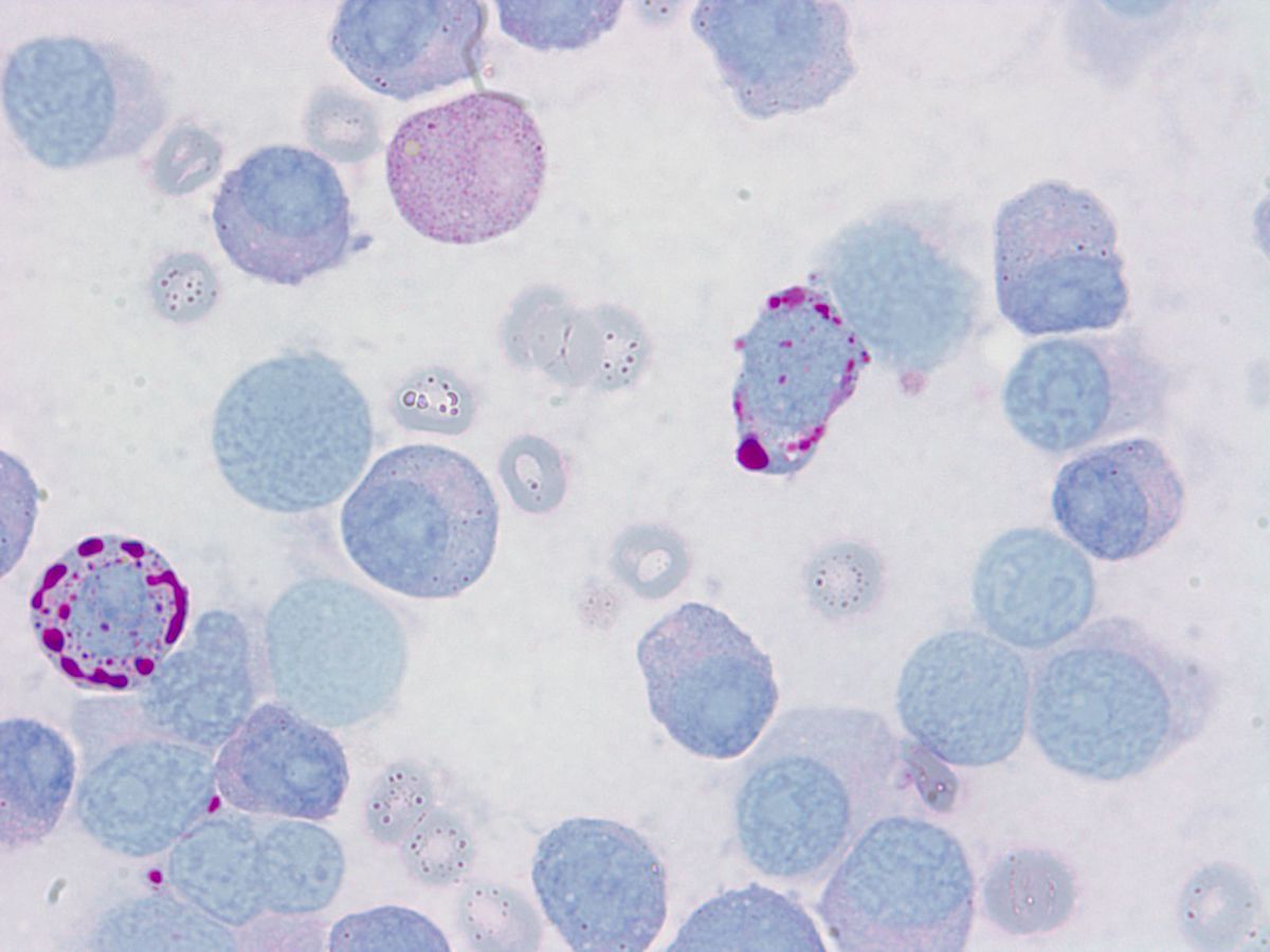 |
| Bone marrow Biopsy: sheets of pronormoblasts | 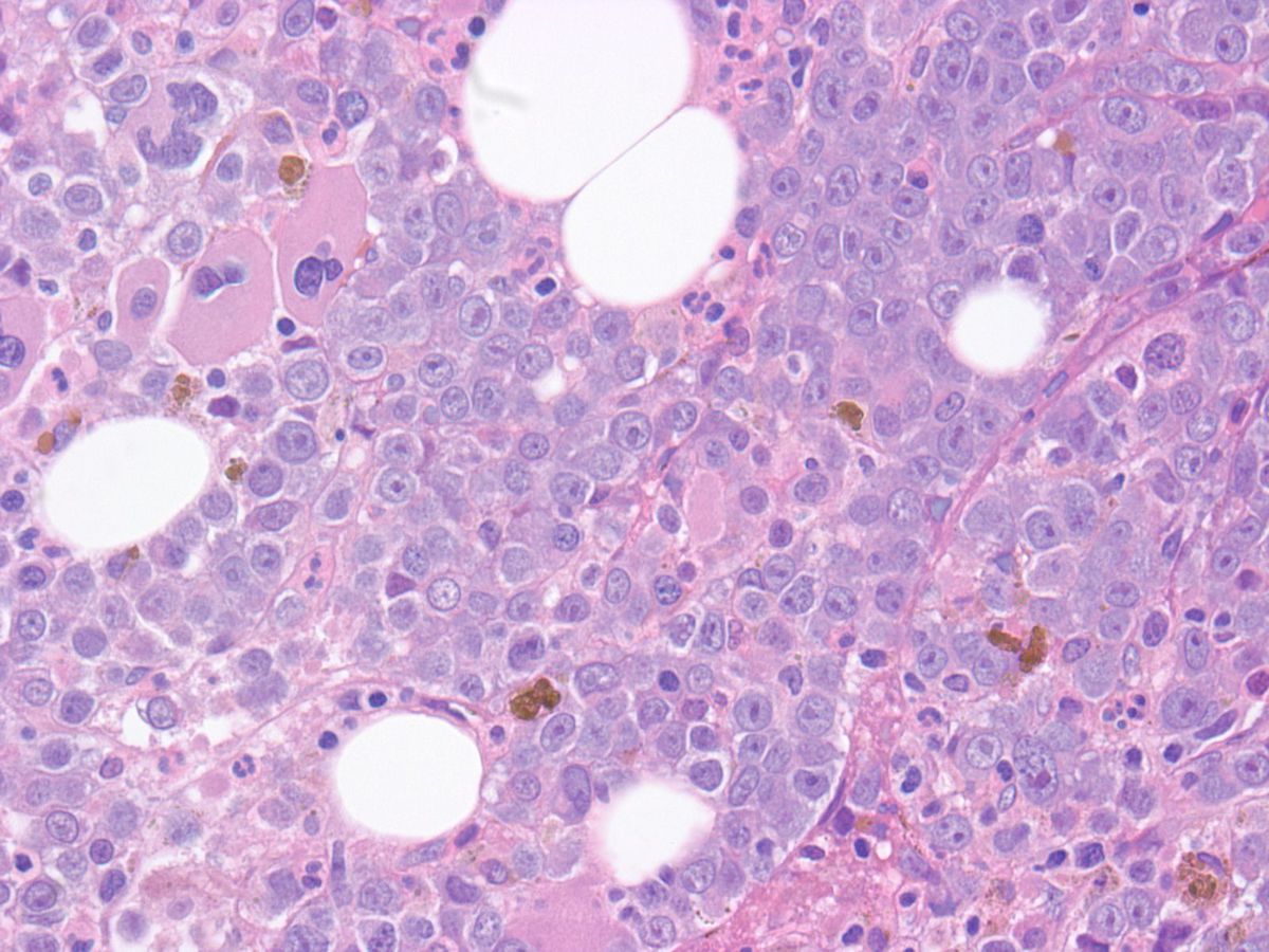 |
| Bone marrow biopsy: Sheets of plasma cells | 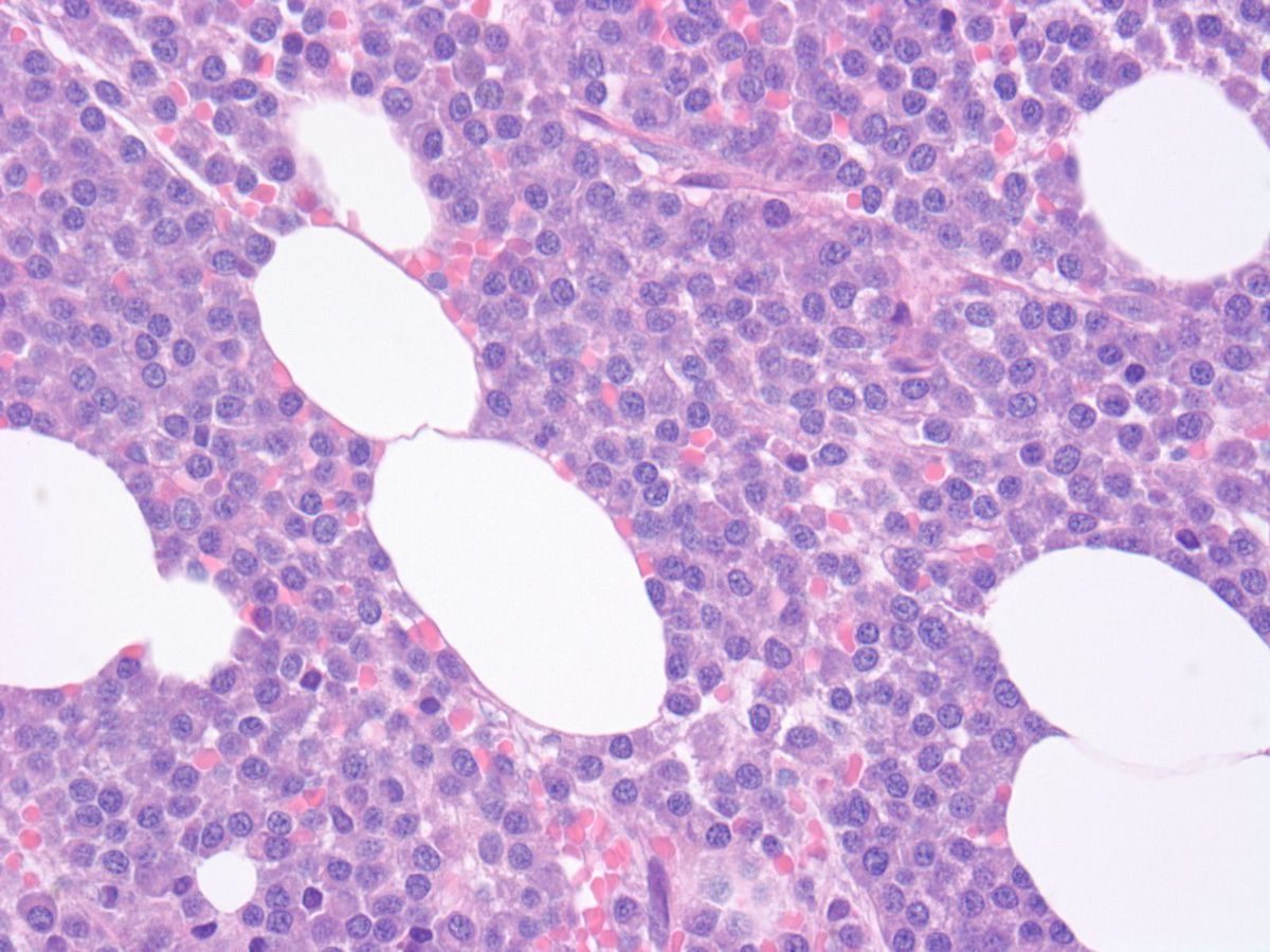 |
| Bone marrow biopsy: Glycophorin A - Variably positive erythroids |  |
| Bone marrow biopsy: CD138 positive intermingled plasma cells. |  |
| Liver: Sinusoidal infiltration of proerythroblast population | 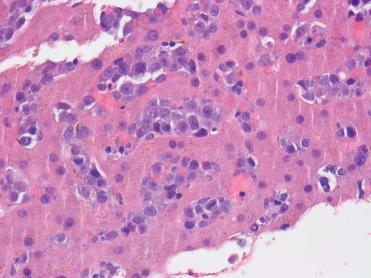 |
| Liver: Glycophorin positive erythroblasts | 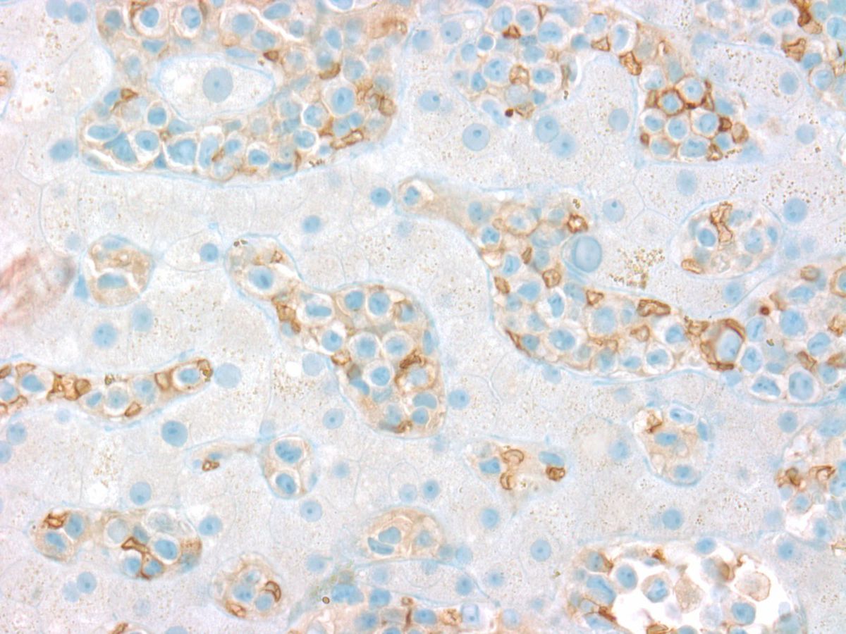 |