Institution: Vanderbilt UMC, Indiana University SOM
Additional authors:Claudio Mosse, Heath Jones, David Head
Session: Erythroleukemia and megakaryoblastic AML and mimics
HISTORY
An 18 month male infant with Down Syndrome and a history of neonatal transient abnormal myelopoiesis with spontaneous regression (by age 1 month) presents now with a hemoglobin of 2.9 gm%, platelet count of 25,000/ul, and WBC count of 16,000/ul with circulating blasts. The child's history has been unremarkable in the intervening months.
After diagnosis of the current illness, the patient was treated on COG protocol AAML 0431 (treatment of AML or MDS in Down Syndrome), entered remission, and remains in remission (at 8 months).DETAILS
A posterior iliac crest aspirate/biopsy was obtained. The aspirate (W-G stain) is cellular but aparticulate. Megakaryocytes are abundant with small size, hypolobate nuclei, and unusual central cytoplasmic inclusions. Myeloid elements are decreased with no atypia. Erythroid elements have irregular nuclear contours and megaloblastoid change. Blasts are increased (51%) with variable amounts of light to dark blue cytoplasm. Cytoplasmic surface blebs are not prominent. The biopsy (B Plus fixative, H&E) is hypercellular (>95%). Blasts are increased. Megakaryocytes are abundant with small size, hypolobate nuclei, with some having peripherally displaced nuclei surrounding a central cytoplasmic mass. Myeloid and erythroid elements are decreased. A reticulin stain demonstrates fine reticulin fibrosis.
IMMUNOHISTOCHEMISTRY AND FLOW CYTOMETRY
Immunohistochemical stains show partial staining of immature cells with CD61 and glycophorin A, minimal staining with E- cadherin, and staining of numerous dysplastic small megakaryocytes with CD61.
With flow cytometry, gating on blasts (48% of total cells) with CD45/side scatter, immature cells have the following immunophenotype: CD19(negative), CD33(moderate), CD45(dim), CD13(partial dim), CD34(partial moderate), CD61(negative), CD117(moderate), HLA-DR(negative), CD11b(negative), CD14(negative), CD15(negative), CD16(negative), CD64(negative), CD2(negative), CD4(moderate), CD7(bright), CD56(negative), and CD36 (60%+).CYTOGENETIC FINDINGS
Karyotype: Trisomy 21 (18/20 metaphases)
FISH: All normal, except +211. nuc ish 8q22(RUNX1T1x2),21q22(RUNX1x3)[180/200]2. nuc ish 15q22-24(PMLx2),17q21(RARAx2)[200]3. nuc ish 16q22(CBFBx2)[200]4. nuc ish 11q23(MLLx2)[20]5. nuc ish 5p15.2(D5S23,D5S721x2),5q31(EGR1x2)[200]6. nuc ish 7cen(D7Z1x2),7q31(D7S486x2)[200]7. nuc ish 8cen(D8Z2x2)[200]8. nuc ish 20q12(D20S108x2)[200]MOLECULAR FINDINGS
NPM1 MUTATION Not Detected
FLT3 ITD Not DetectedINTERESTING FEATURES
This patient is a typical Down syndrome (DS) patient with acute myeloid leukemia (AML). We have reviewed morphologic samples and immunophenotyping from 162 patients with DS and MDS or AML on this COG protocol (AML 0431). Dysplasia in both MDS and AML patients is restricted to megakaryocytes and erythroids. Erythroid dysplasia is predominantly megaloblastoid change, with nuclear dysplasia in a minority of patients. Megakaryocytes are typically hyperplastic, with dysplasia characterized by small size and hypolobate, often multiple, nuclei,with unique features of a large granular eosinophilic central mass in frequent megakaryocytes, displacing nuclei peripherally, and when nuclei are multiple forming a ring-like wreath of nuclei around the mass. Granulocytic dysplasia is rarely present, and if present minimal. The leukemia most typically shows megakaryoblastic (M) differentiation, but some cases show either erythroblastic (E) or mixed M/E differentiation. Only rare cases show granulocytic differentiation, and appear to represent standard non-DS AML. These findings appear to correlate perfectly with the involvement of GATA1 mutation in DS MDS and AML. These patients differ markedly from MDS and MDS-related AML in other clinical settings in chemotherapy response as well as morphology, suggesting that they are not an informative model for the latter diseases. Finally, most of these cases were diagnosed by submitting institutions using WHO nomenclature (Myeloid leukemia associated with DS). While such lumping may be sufficient for current therapeutic strategies, it is an insufficient description of disease pathology and may ultimately restrict progress in diagnosis and treatment of this disease set.
PROPOSED DIAGNOSIS
Acute megakaryoblastic leukemia in a patient with Down Syndrome
CONSENSUS DIAGNOSIS
Myeloid proliferation related to Down syndrome, consistent with acute myeloid leukemia with megakaryoblastic and erythroid differentiation
| Bone marrow aspirate, W-G stain, low power showing packed marrow |  |
| Higher power showing blasts with basophilic cytoplasm, occasional surface blebs, and megaloblastoid erythroid changes |  |
| Peripheral smear, W-G stain, showing blasts with variably abundant blue cytoplasm, and erythroids with megaloblastoid change |  |
| Bone marrow biopsy, H&E stain, showing dysplastic megakaryocytes and blasts. Note two dysplastic megakaryocytes (upper right and lower central) with eosinophilic cytoplasmic mass displacing nucleus. |  |
| Note similar dysplastic megakaryocytes with central masses, center of photomicrograph and lower right |  |
| Similar dysplastic megakaryocyte, upper right, with 2 masses without visible attached cytoplasm or nuclei, lower right | 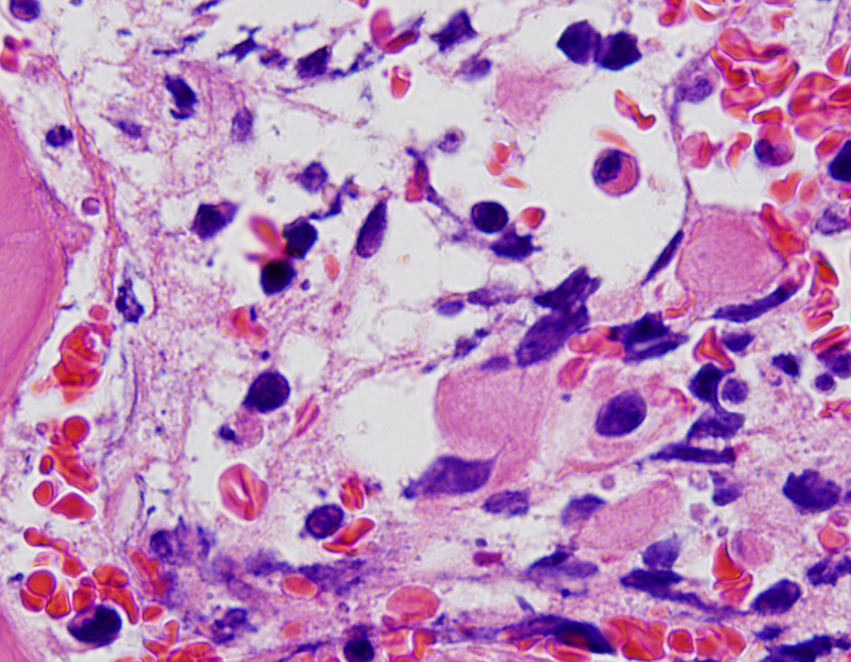 |
| Bone marrow aspirate, W-G stain, showing similar megakaryocyte with this preparation. |  |