Institution: University of New Mexico
Additional authors:Alexei G. Bakhirev
Session: T Lymphoblastic Leukemia/Lymphoma
HISTORY
A 64-year-old man presented with generalized symptoms including fatigue and weight loss and an enlarged left clavicular lymph node. Imaging study revealed diffuse mediastinal and retroperitoneal lymphadenopathy. His past history included a twelve year history of chronic myelogenous leukemia (CML) treated with imatinib. Quantitative PCR for t(9;22) BCR-ABL fusion had been performed only once, in August of 2011, and showed a BCR-ABL/ABL ratio of 0.92.
Laboratory work-up revealed a white blood cell count of 33.5 x 10^6 cells/L (51% neutrophils, 1% lymphocytes, 11% monocytes, 0% eosinophils, 2% basophils, 5% metamyelocytes, 20% myelocytes, 7% promyelocytes, and 2% blasts); hemoglobin of 9.7 g/dL and platelet count of 486 x 10^6 /L.DETAILS
Excisional biopsy of the clavicular lymph node was performed and portions of fresh specimen were sent for flow cytometric and cytogenetic analysis. Remaining material was fixed in neutral formalin and AZF for morphologic examination.
The lymph node architecture is effaced by uniform small to intermediate lymphoid cell proliferation with focal areas of “starry sky” appearance with histiocytes and areas of necrosis. Mitotic figures are increased.IMMUNOHISTOCHEMISTRY AND FLOW CYTOMETRY
Flow cytometry on the lymph node shows 90% of total events to be abnormal T-cells expressing TdT, CD1a, cytoplasmic CD3, CD2, CD4, CD5, CD7, CD8, and CD45. The cells are negative for surface CD3, CD10, CD20, CD23, CD117, and CD34.
CYTOGENETIC FINDINGS
Lymph node:
1. FISH, dual color dual fusion BCR/ABL1 probe: 17% of cells with typical BCR-ABL1 fusion and 55.5% with variant BCR-ABL1 fusion pattern with three fusion signals2. Karyotype:49~50,XY,del(4)(q31),+8,t(9;22)(q34;q11.2),+19,+der(22),t(9;22)(q34;q11.2)MOLECULAR FINDINGS
BCR-ABL transcripts were detected in a concurrent peripheral blood specimen, with a BCR-ABL/ABL ratio of 1.027.
INTERESTING FEATURES
Lymphoblastic transformation accounts for 20-30% of blast crises in CML, with the majority being of B-cell lineage. This case demonstrates a rather rare and previously disputed T-cell lymphoblastic transformation of CML in lymph nodes with bone marrow (performed one week later) and peripheral blood showing essentially a chronic phase CML, all despite imatinib therapy. Cytogenetic abnormalities in the T lymphoblasts were Philadelphia chromosome and additional abnormalities, including an extra Philadelphia chromosome, trisomy 8, trisomy 19 and deletion of the long arm of chromosome 4 (4q-). Additionally, bone marrow FISH study revealed typical and atypical BCR/ABL1 fusion patterns similar to the lymph node. This case also highlights the importance of regular monitoring of molecular disease status in patients with CML being treated with tyrosine kinase inhibitors, as failure to achieve a decrease in BCR-ABL transcripts or identification of increasing transcript levels may indicate disease recurrence, treatment non-compliance, or imatinib resistance.
PROPOSED DIAGNOSIS
T lymphoblastic lymphoma blast phase of chronic myelogenous leukemia.
CONSENSUS DIAGNOSIS
T-acute lymphoblastic phase of chronic myelogenous leukemia
| High power view of H&E stained lymph node section showing uniform small cells with numerous mitoses |  |
| Flow cytometric analysis showing aberrant population expressing TdT, CD1a, cytoplasmic CD3, CD2, CD4, CD5, CD7, and CD8 (red). |  |
| FISH with BCR/ABL dual fusion probe showing atypical pattern with three fusion signals | 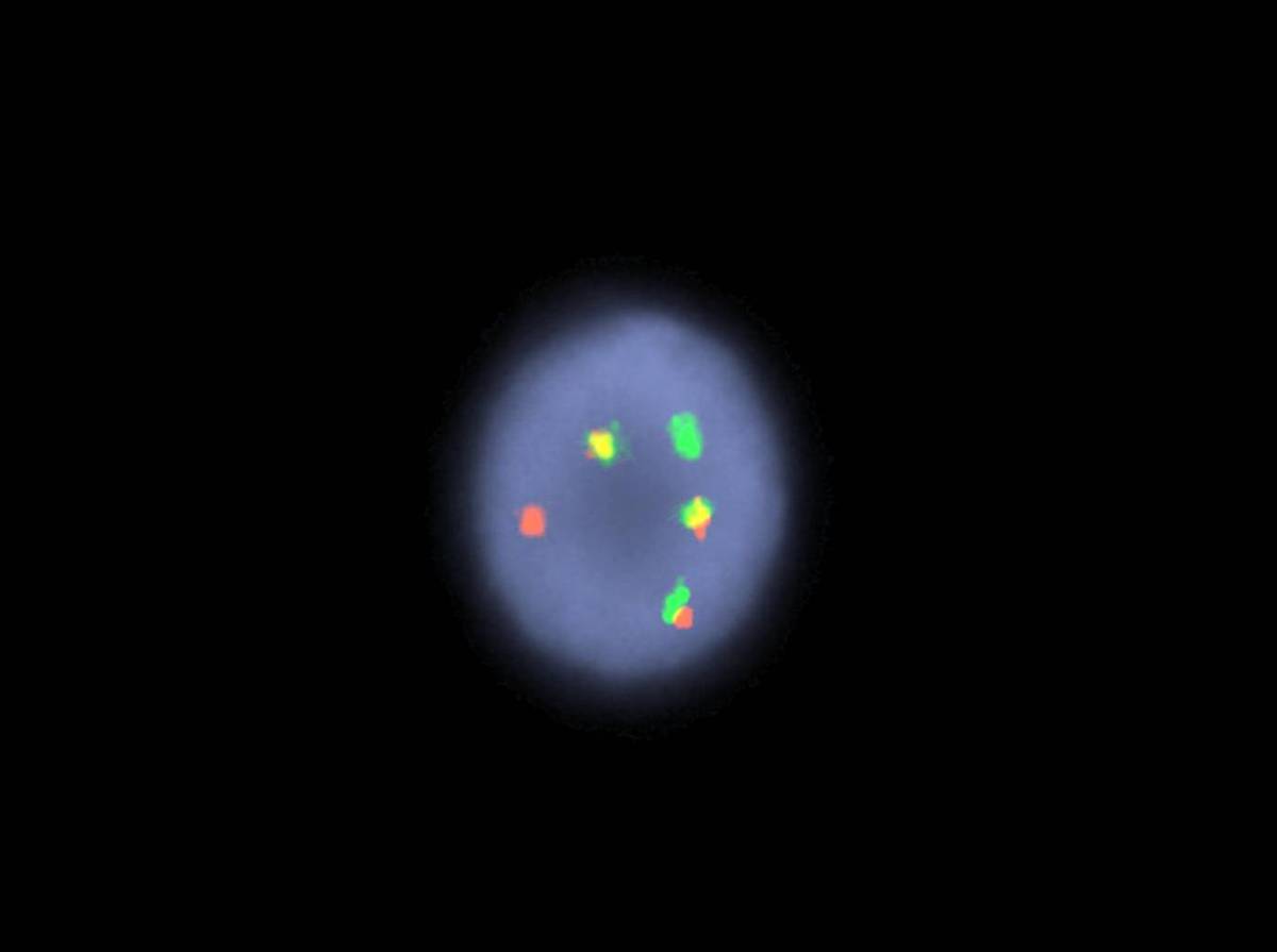 |
| Cytogenetic analysis of lymph node showing complex karyotype with translocation (9;22)(Philadelphia), an additional Philadelphia chromosome, trisomy 8, trisomy 19 and deletion of the long arm of chromosome 4 (4q-) | 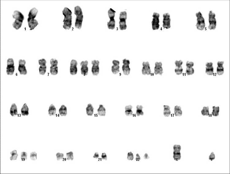 |