Institution: Wake Forest School of Medicine
Session: Erythroleukemia and megakaryoblastic AML and mimics
HISTORY
A 40 year old man with no significant past medical history presented to his physician with dyspnea on exertion, dizziness, and fatigue. A CBC revealed pancytopenia (WBC 1.3K µL, hemoglobin 7.0 g/dL, platelets 6000 µL, ANC 400). A bone marrow biopsy was performed and was reported to have 50% blasts. He was transferred to our institution and the bone marrow biopsy was repeated.
DETAILS
The bone marrow biopsy was fixed in B+. Examination of the bone marrow core biopsy and clot sections revealed a hypercellular marrow (100% cellular) with marked erythroid hyperplasia and megaloblastic morphology. The aspirates had marked erythroid hyperplasia (67% of all cells). The erythroid precursors were present in all stages of maturation. There was marked atypia including irregular nuclei, multinucleation, megaloblastic changes, and cytoplasmic-nuclear dyssynchrony. Mature megakaryocytes were markedly decreased numbers. Myeloid blasts were <1% of all cells. There were numerous large nuclei that had been stripped of their cytoplasm. Myeloid elements were too few to adequately assess for dysplasia.
IMMUNOHISTOCHEMISTRY AND FLOW CYTOMETRY
CD71 immunohistochemistry confirmed the erythroid hyperplasia. Myeloperoxidase highlighted rare positive cells. There was no increase in blasts by CD34. CD117 had very faint expression in some of the erythroid precursors. CD42b highlighted numerous cells.
By flow cytometry, erythroid precursors comprised approximately 60% of all cells analyzed and expressed CD71 and CD36. There was dim expression of CD235a and no co-expression of CD117. CD34 positive blasts were not increased (<1%).
CYTOGENETIC FINDINGS
58,XY,+1,+1,+2,+3,+6,+8,+9,+12,+15,+17,+19,idic(19)(p13.3),+21
MOLECULAR FINDINGS
None performed.
INTERESTING FEATURES
Based on the aspirate smear differential count, this case did not meet the criteria for acute erythroid leukemia. However, the predominance of abnormal erythroid precursors (likely more than 80% of the marrow cells in the core biopsy) favors the diagnosis of acute erythroid leukemia over a myelodysplastic syndrome. The presence of an abnormal megakaryocyte component prompts to speculate that this neoplasm is derived from a common erythroid-megakaryocytic precursor. Although not a formal part of the WHO classification, there is a basis for such leukemias.
PROPOSED DIAGNOSIS
Acute erythroid leukemia, "pure erythroleukemia" subtype, with megakaryocytic component.
CONSENSUS GROUP: ADDITIONAL INFORMATION/STUDIES
Iron stain showed absent storage iron. The patient received induction chemotherapy, and achieved complete response, alive at 9 months of follow-up.
CONSENSUS DIAGNOSIS
Acute erythroid leukemia (pure erythroid leukemia)
| Bone marrow core biopsy 400x |  |
| Bone marrow core biopsy 600x | 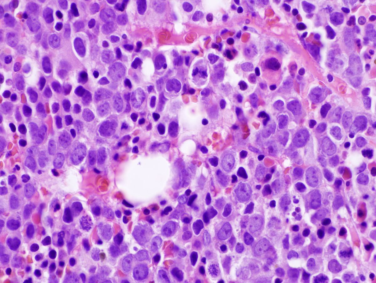 |
| Bone marrow core biopsy CD71 400x | 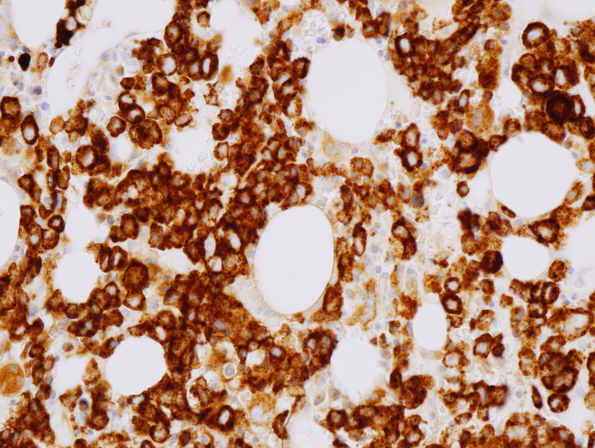 |
| Bone marrow core biopsy CD34 400x | 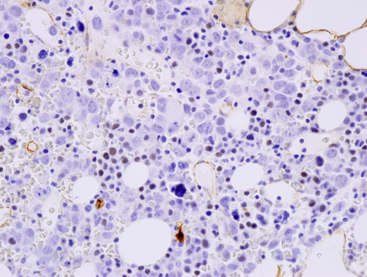 |
| Bone marrow core biopsy CD42b 400x |  |
| Bone marrow core biopsy CD117 400x |  |
| Bone marrow aspirate. Atypical erythroid precursors. 1000x |  |
| Bone marrow aspirate. Atypical erythroid precursors. 1000x | 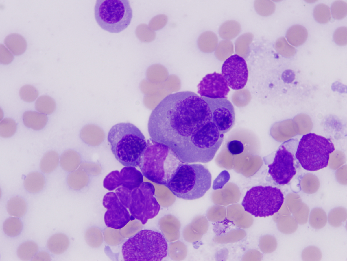 |
| Additional figure 1: Flow | 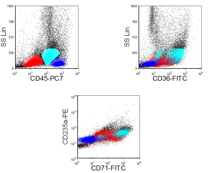 |