Institution: Wake Forest School of Medicine
Session: B Lymphoblastic Leukemia/Lymphoma
HISTORY
A 65-year-old asymptomatic man treated for B-lymphoblastic leukemia with normal cytogenetics presented to his oncologists for an end of therapy bone marrow biopsy. The patient’s original lymphoblasts expressed CD19, CD20, CD34, CD10 (bright), and terminal deoxynucleotidyl transferase (TdT) without expression of myeloid or T-cell antigens. He received standard therapy with induction, consolidation, and maintenance over two years. Chemotherapy included cyclophosphamide, daunorubicin, vincristine, l-asparaginase, dexamethasone, cytarabine, methotrexate, and 6-mercaptopurine. The end of therapy marrow showed 52% atypical lymphocytes that was worrisome for relapse.
DETAILS
The bone marrow core biopsy was fixed in B5 and stained with hematoxylin and eosin. The marrow core biopsy and clot sections were 40-50% cellular with an infiltrate of lymphoid cells. Multilineage hematopoiesis was preserved. Examination of the smears revealed 52% lymphocytes, but no overt increase in blasts. Background hematopoiesis was unremarkable.
IMMUNOHISTOCHEMISTRY AND FLOW CYTOMETRY
By immunohistochemistry, the majority of the lymphocytes expressed CD20 and CD10. TdT was expressed in a minority of the lymphocytes. CD34 positive cells were not increased. Flow cytometry revealed that the atypical lymphocytes were hematogones with a normal spectrum of B-cell maturation by CD20, CD10, and CD34 and not recurrent leukemia. There was no light chain restricted B-cell population.
CYTOGENETIC FINDINGS
46, XY
MOLECULAR FINDINGS
Not performed.
INTERESTING FEATURES
Hematogones (B-cell precursors) decrease with age, with adults having 1-4% hematogones. Above 5% hematogones has arbitrarily been assigned as ‘increased’. Hematogone hyperplasia has been seen in adults and children status post treatment for B-lymphoblastic leukemia and stem cell transplant. Some of these cases have persisted for over 1 year. Increased numbers of hematogones may complicate diagnosis since the morphologic features can be very similar to leukemic lymphoblasts. Although unusual, hematogone hyperplasia must be considered in the differential diagnosis in patient’s status post treatment for B-lymphoblastic leukemia. A bone marrow performed 11 months later also showed hematogone hyperplasia (40% hematogones). The patient continues to remain in remission after 4 years from end of therapy.
PROPOSED DIAGNOSIS
B-cell precursor (hematogone) hyperplasia.
CONSENSUS DIAGNOSIS
Bone marrow with prominent regenerating B-cell population (hematogones)
| Leukemic blasts are from the initial diagnostic marrow (right). Immature lymphoid cells seen in follow-up bone marrow biopsies (left) . |  |
| Clot section. | 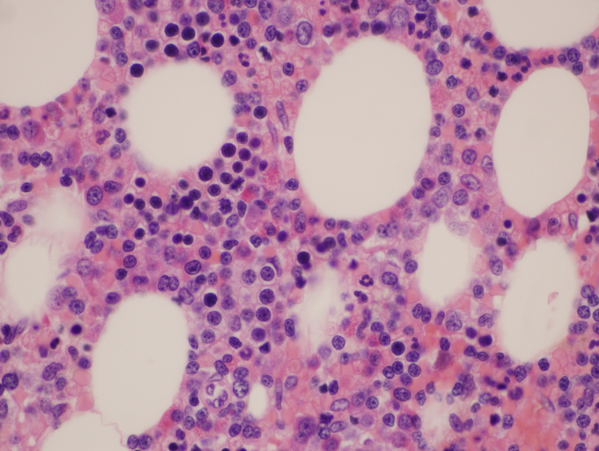 |
| CD10 | 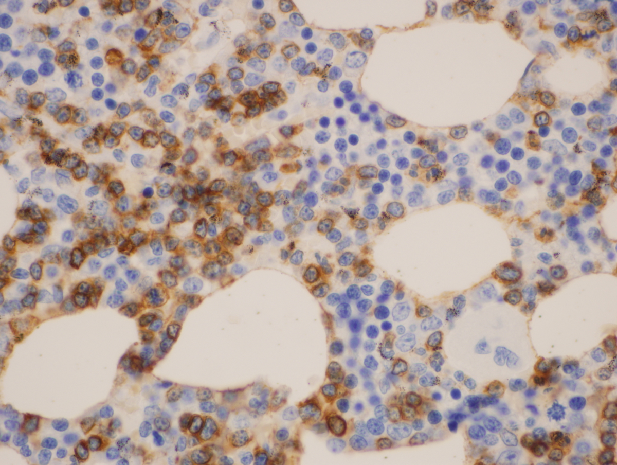 |
| CD20 | 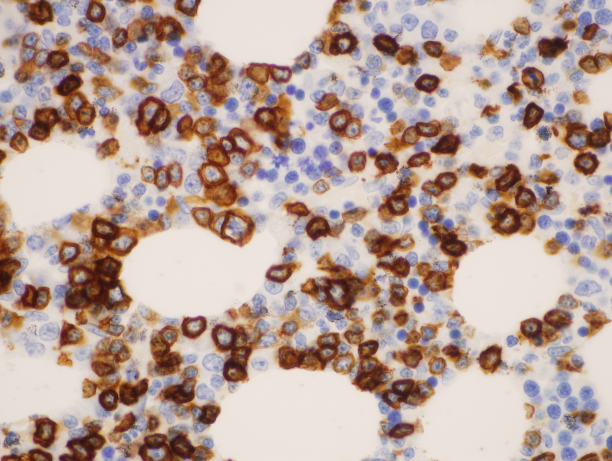 |
| TdT | 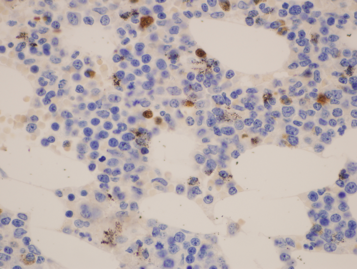 |
| CD34 | 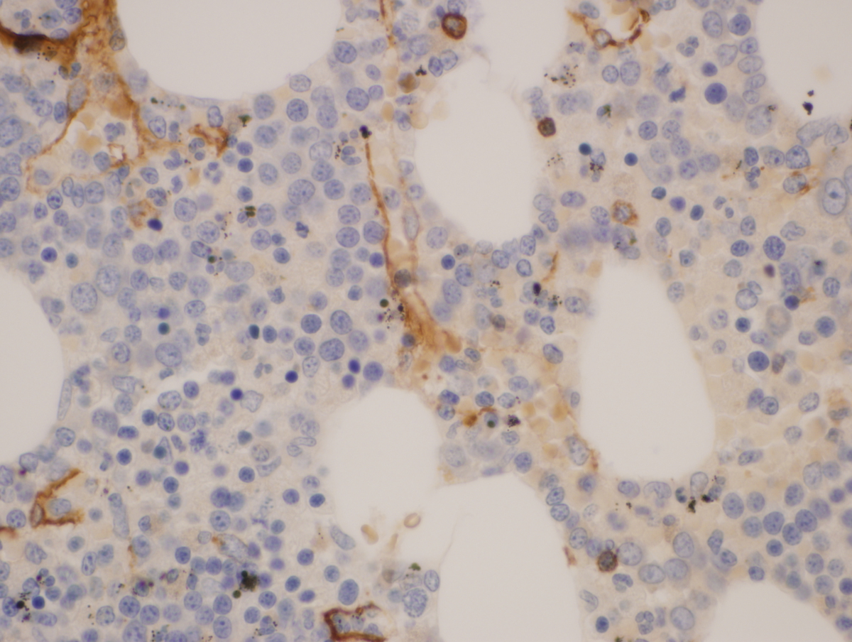 |
| Flow cytometry at initial presentation. |  |
| Flow cytometry after treatment showing hematogone hyperplasia. |  |