Institution: University of Pittsburgh Medical Sciences
Additional authors:Lydia C. Contis
Session: AML with recurrent genetic mutations Part II
HISTORY
The patient is an 84-year old man with past medical history of chronic renal failure, anemia, thrombocytopenia (HIT).
DETAILS
Multiple bone marrow aspirate smears were submitted.
Gross: Bone marrow biopsy, is received in B+ fixative and consists of a single core of tan, cancellous bone measuring 1.0 x 0.2 cm. The specimen is completely submitted to histology for decalcification in block A.Microscopic findings: Hypercellular marrow with increased blasts. Blasts are intermediate in size, with moderate amount of cytoplasm with prominent auer rods (some thicker and resembling crystalline inclusions), prominent granules including some resembling pseudo-chidiak higashi granules. Erythrophagocytosis is seen in some blasts. Maturing myeloid elements with an ocassional auer rod are also seen. (Images 1, 2 and 3). The crystals stained for sudan black and were mostly negative for myeloperoxidase (Image 4).IMMUNOHISTOCHEMISTRY AND FLOW CYTOMETRY
Flow cytometric immunophenotypic studies performed on the bone marrow demonstrate a population of immature myeloid cells with the following phenotype (Image 5):
Dim CD45+, CD34(-), CD13/33+, CD14(-), partial CD36+, partial CD64+, partial CD117+, HLA-DR mostly (-), CD15+, CD33+, CD13+CD11b+ and bright myeloperoxidase positive. These cells comprise approximately 30-43% of total analyzed cells. Few T-cells, few B-cells and granulocytes are also present. Some immature granulocytes appear to demonstrate reduced expression of CD13.CYTOGENETIC FINDINGS
FISH studies for PML/RARA and AML1 (RUNX1)
Classical cytogenetics demonstrated: Abnormal male bone marrow chromosome analysis with appears to be missing the Y chromosome. In addition, each cell has more than 30 copies of double minutes (45,X,-Y,>30dmin[20]) and FISH demonstrated amplification of C-MYC gene in 180/213 cells analysed (84.5%). (Images 6,7)MOLECULAR FINDINGS
Not done
INTERESTING FEATURES
A number of blasts contain multiple Auer rods, cytoplasmic crystals, and pseudo-Chediak-Higashi type granules with double minutes on cytogenetic studies. Many features of this acute myeloid leukemia are like AML-M2, some features are also like AML-M3 (multiple auer rods, lack of HLA-DR on flow cytometry).
The presence of double minutes has been reported in 1-10% of patients of AML especially in APL-like morphology with pseudo-chediak higashi like granules and prominent auer rods. C-MYC amplification is seen in several but not all cases and has been associated with disease aggressiveness and resistance to chemotherapy. Normal karyotype with many double minutes has been shown to respond better to chemotherapy as compared to fewer double minutes and complex karyotype.PROPOSED DIAGNOSIS
Acute myeloid leukemia, NOS
CONSENSUS DIAGNOSIS
Acute myeloid leukemia, NOS, with MYC amplification
| Image 1: Pseudochediak higashi granules | 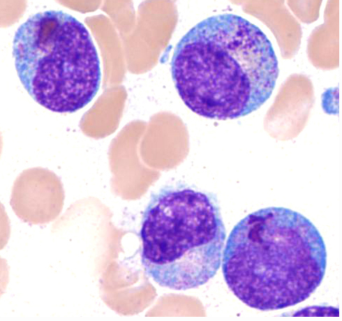 |
| Image 2: Multiple auer rods | 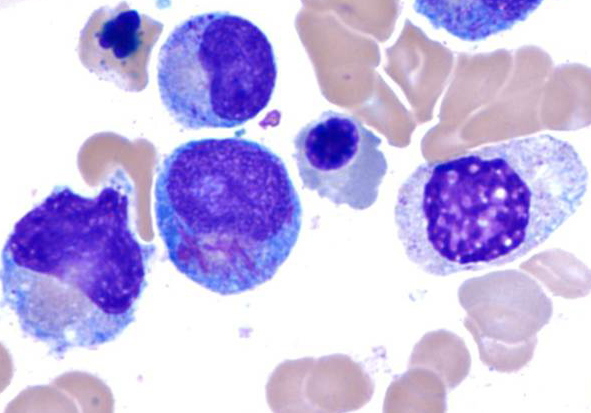 |
| Image 3: Crystalline inclusions | 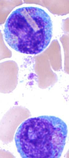 |
| Image 4: Cytochemical stains, myeloperoxidase and Sudan black | 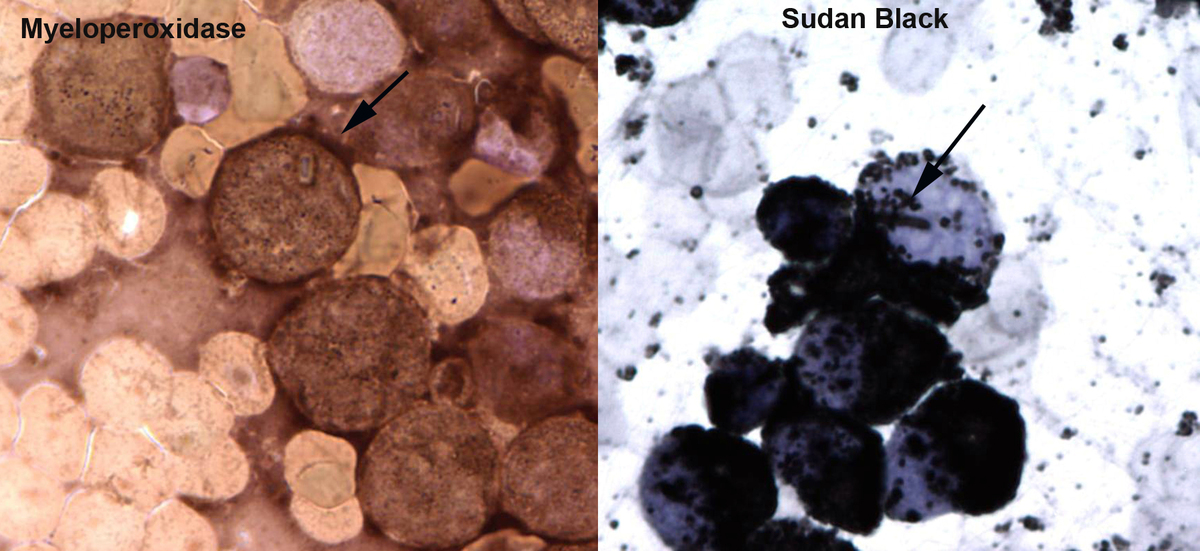 |
| Image 5: Flow cytometry | 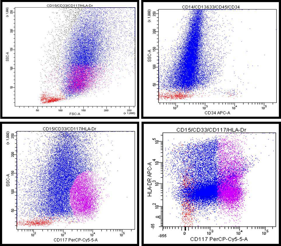 |
| Image 6: Karyotype, many double minutes | 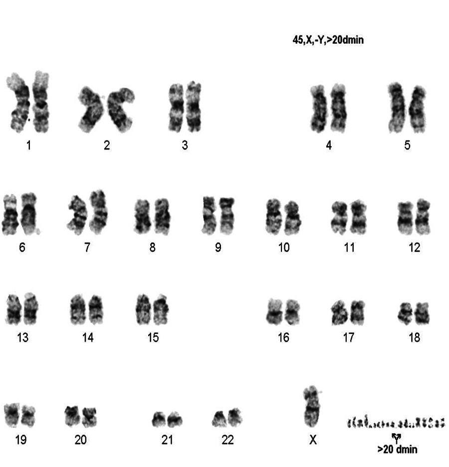 |
| FISH for MYC showing amplification | 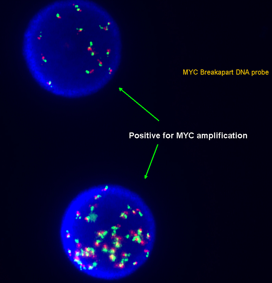 |