Institution: The Methodist Hospital
Additional authors:A. Zieske, M.D.
Session: Myeloid and lymphoid neoplasms with eosinophilia and abnormalities of PDGFRA, PDGFRB, or FGFR1
HISTORY
39 year old female presents to an outside institution with a paraspinal mass. Subsequent biopsy of the paraspinal mass performed at the referring institution showed T-lymphoblastic leukemia/lymphoma (T-ALL). The patient subsequently came to The Methodist Hospital for treatment. In house review of the of slides from the paraspinal mass confirmed T-ALL, and the slides were returned to the referring institution. A subsequent in house staging bone marrow biopsy was performed. The findings from this staging bone marrow biopsy are discussed in this case report.
DETAILS
1. Paraspinal mass biopsy. This specimen was obtained at an outside institution, and fixed in formalin. Our institution reviewed the slides from the referring institution.
2. Peripheral blood, bone marrow biopsy (iliac crest), touch preparation, clot. The clot was fixed in formalin. The bone marrow core biopsy was decalcified and fixed in formalin.
IMMUNOHISTOCHEMISTRY AND FLOW CYTOMETRY
1. Paraspinal mass biopsy:
Submitted immunostains were reviewed. The lymphoblastic lymphoma cells are positive for CD45, CD34, CD99, CD117, CD43, CD7, CD3(partial), and CD4. The lymphoblastic lymphoma cells are negative for CD1a, CD2, CD5, CD8, CD10, CD20, CD79a, CD30, ALK-1, TdT, TIA, MPO, and cytokeratin.
2. Bone marrow core biopsy:
Immunostains performed with adequate controls were interpreted as follows:
CD3: Scattered small T-cells
CD20: Scattered sall B cells
MPO: highlights myeloid hyperplasia
E-Cadherin: positive in erythroid precursors
CD34: Positive in approximately 1-2% blasts
CD99: Negative
Tryptase, CD117, and CD25: A population of scattered tryptase positive mast cells are present. The mast cells appear to co-express CD117 and CD25 in a small subset of cells.
Flow cytometry (bone marrow):
-Partial expression of CD56 on granulocytes
-No clonal B cells or aberrant T cell antigen expression identified
-No increase in immunophenotypic myeloblasts
-No abnormal mast cell immunophenotypes
CYTOGENETIC FINDINGS
1. Paraspinal mass:
FISH studies showed the following results:
Nuc ish (5’FIP1L1x2,3’FIP1L1x1, PDGFRAx2)(FIP1L1 con PDGFRAx1)[8/202]/5’FIP1L1x2,3’FIP1L1x1,PDGFRAx1)[109/202]/(5’FIP1L1,3’FIP1L1,PDGFRA)x1[55/202]
4% of cells showed a CHIC2 deletion and fusion of FIP1L1 and PDGFRA. 54% of cells showed deletion of CHIC2 and PDGFRA from the 4q12 region. 27 % of cells showed loss of one copy of CHIC2, FIP1L1, and PDGFRA
2. Bone marrow:
FISH studies:
Nuc ish (5’FIP1L1x2, 3’FIP1L1x1, PDGFRAx1)(FIP1L1 con PDGFRA x1)[130/205}/(5’FIP1L1x2, 3’FIP1L1x1, PDGFRAx2)(FIP1L1 con PDGFRA x1)
A deletion of CHIC2 and fusion of FIP1L1 and PDGFRA was identified in 9% of cells. A deletion of CHIC2 and PDGFRA from 4q12 region was identified in 63% of cells.
FISH analysis for BCR/ABL: Absence of BCR/ABL fusion
Conventional cytogenetics: Normal female karyotype
MOLECULAR FINDINGS
INTERESTING FEATURES
This patient was referred to The Methodist Hospital for treatment of a previously diagnosed T-ALL in a paraspinal mass. Subsequent staging bone marrow showed myeloid hyperplasia with numerous eosinophils, some with atypical morphology. FISH studies confirmed a FIP1L1-PDGFRA fusion. The same FIP1L1-PDFRA fusion was later confirmed by FISH in the paraspinal mass specimen. These findings collectively support the diagnosis of Myeloproliferative neoplasm with eosinophilia and T Lymphoblastic lymphoma associated with PDGFRA rearrangement.
PROPOSED DIAGNOSIS
Myeloproliferative neoplasm with eosinophilia and T Lymphoblastic lymphoma associated with PDGFRA rearrangement.
CONSENSUS DIAGNOSIS
Myeloid and lymphoid neoplasm with FIP1L1-PDGFRA rearrangement, presenting with T lymphoblastic lymphoma and myeloproliferative neoplasm with eosinophilia
| Low power view of paraspinal mass biopsy. Sections show lymphoblast infiltration that are intermediate in size with fine chromatin, irregular nuclear contours, and scant cytoplasm. Numerous tingible body macrophages are seen. | 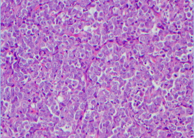 |
| High power view of paraspinal mass biopsy. Sections show lymphoblast infiltration that are intermediate in size with fine chromatin, irregular nuclear contours, and scant cytoplasm. Numerous tingible body macrophages are seen. | 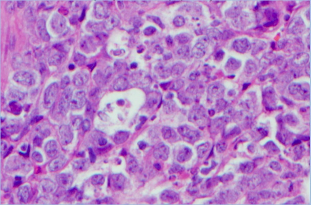 |
| Peripheral blood smear shows leukocytosis with absolute neutrophilia and absolute eosinophilia. The granulocytes are left shifted, including rare blasts, and show reactive changes, including toxic granulation and vacuolization. The eosinophils show reactive or atypical features, including hyperlobation, polarization of granules, degranulation, and cytoplasmic vacuolization |  |
| Peripheral blood smear shows leukocytosis with absolute neutrophilia and absolute eosinophilia. The granulocytes are left shifted, including rare blasts, and show reactive changes, including toxic granulation and vacuolization. The eosinophils show reactive or atypical features, including hyperlobation, polarization of granules, degranulation, and cytoplasmic vacuolization | 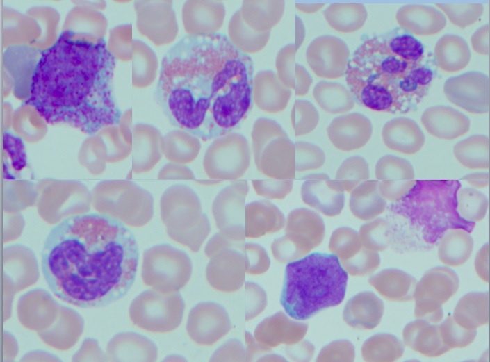 |
| Bone marrow core biopsy shows myeloid hyperplasia with a left shift in granulocyte maturation and increased eosinophils |  |
| Bone marrow core biopsy shows myeloid hyperplasia with a left shift in granulocyte maturation and increased eosinophils |  |
| Bone marrow core biopsy shows myeloid hyperplasia with a left shift in granulocyte maturation and increased eosinophils |  |
| Bone marrow core biopsy shows myeloid hyperplasia with a left shift in granulocyte maturation and increased eosinophils |  |
| IHC for MPO shows myeloid hyperplasia. IHC for CD34 highlights rare scattered blats (1-2% of bone marrow cellularity). A reticulin stain highlights mild reticulin fibrosis | 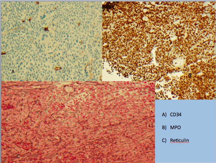 |
| Immature eosinophilic precursors with occasional basophilic granules |  |
| Immature eosinophilic precursors with occasional basophilic granules |  |