Institution: UT M.D. Anderson Cancer Center
Additional authors:Carlos E. Bueso-Ramos, M.D., Ph.D., Cameron Yin, M.D., Ph.D., L. Jeffrey Medeiros, M.D.
Session: AML with recurrent genetic mutations Part II
HISTORY
A 65-year-old man presented with dizziness and fatigue for the previous 3-4 days. He had a history of bladder cancer (carcinoma in situ) post resection and eight courses of BCG instillation eight years ago. His past medical history also includes type 2 diabetes, hypertension and chronic cystitis. He does not drink alcohol and is a never smoker. He has no known exposure to radiation or benzene.
Lab: peripheral blasts 60%, WBC 59.9, Hgb 12.4, Hct 36.8, platelets 109, absolute neutrophil count 8.39Physical exam: scattered bruising on arms, 3cm x 1.5cm erythematous lesion with irregular borders above left scapulaDETAILS
Bone marrow biopsy, aspirate smears, clot section and touch preparation obtained. Bone marrow biopsy specimen was decalcified in 10% formic acid and formalin fixed.
Bone marrow differential counts are:Total cells counted 100Blasts 63 % H (0-5)Progranulocytes 2 % (2-8)Myelocytes 11 % (5-20)Metamyelocytes 0 % L (13-32)Granulocytes 6 % L (7-30)Eosinophils 0 % (0-4)Lymphocytes 6 % (3-17)Plasma cells 0 % (0-2)Monocytes 10 % H (0-5)Reticulum Cells 0 % (0-2)Pronormoblasts 0 % L (1-8)Normoblasts 2 % L (7-32)M:E Ratio 9.5 H (3-4)Microscopic examination revealed significantly increased immature cells, showing irregular and often folded nuclei, dispersed chromatin, distinct to prominent nucleoli and small amounts of cytoplasm. Special stains on aspirate:Myeloperoxidase: Positive in approximately 70% of blastsButyrate esterase: Frequently positiveIMMUNOHISTOCHEMISTRY AND FLOW CYTOMETRY
Flow cytometric analysis of the bone marrow aspirate demonstrates a population of blasts, positive for CD45, CD34 (small subset), CD117, CD13, CD33, CD38, CD64 (subset), CD15 (subset) and MPO. The blasts are negative for CD10, HLA-DR, CD41, TdT, and B- and T-cell markers.
CYTOGENETIC FINDINGS
Diploid male karyotype 46,XY[20]
Negative for CBFB gene rearrangement by FISHMOLECULAR FINDINGS
Diagnostic bone marrow specimen:
Positive for NPM1 mutation.Negative for FLT3 mutations. Relapse bone marrow specimen 9 months later:Positive for NPM1 mutation.Positive for FLT3-ITD mutation.INTERESTING FEATURES
This patient was initially diagnosed AML with mutated NPM1 and wild-type FLT3, which indicated a good prognosis. He responded well to therapy and went into complete remission. However, 9 months later, the patient replased with FLT3-ITD mutation. He stoped repond to therapy and died in 2 months.
PROPOSED DIAGNOSIS
Acute myeloid leukemia with monocytic differentiation
CONSENSUS DIAGNOSIS
Acute myeloid leukemia with mutated NPM1
| GeneScan results of FLT3 mutation status change in an acute myeloid leukemia patient | 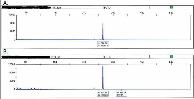 |
| Additional figure 1 | 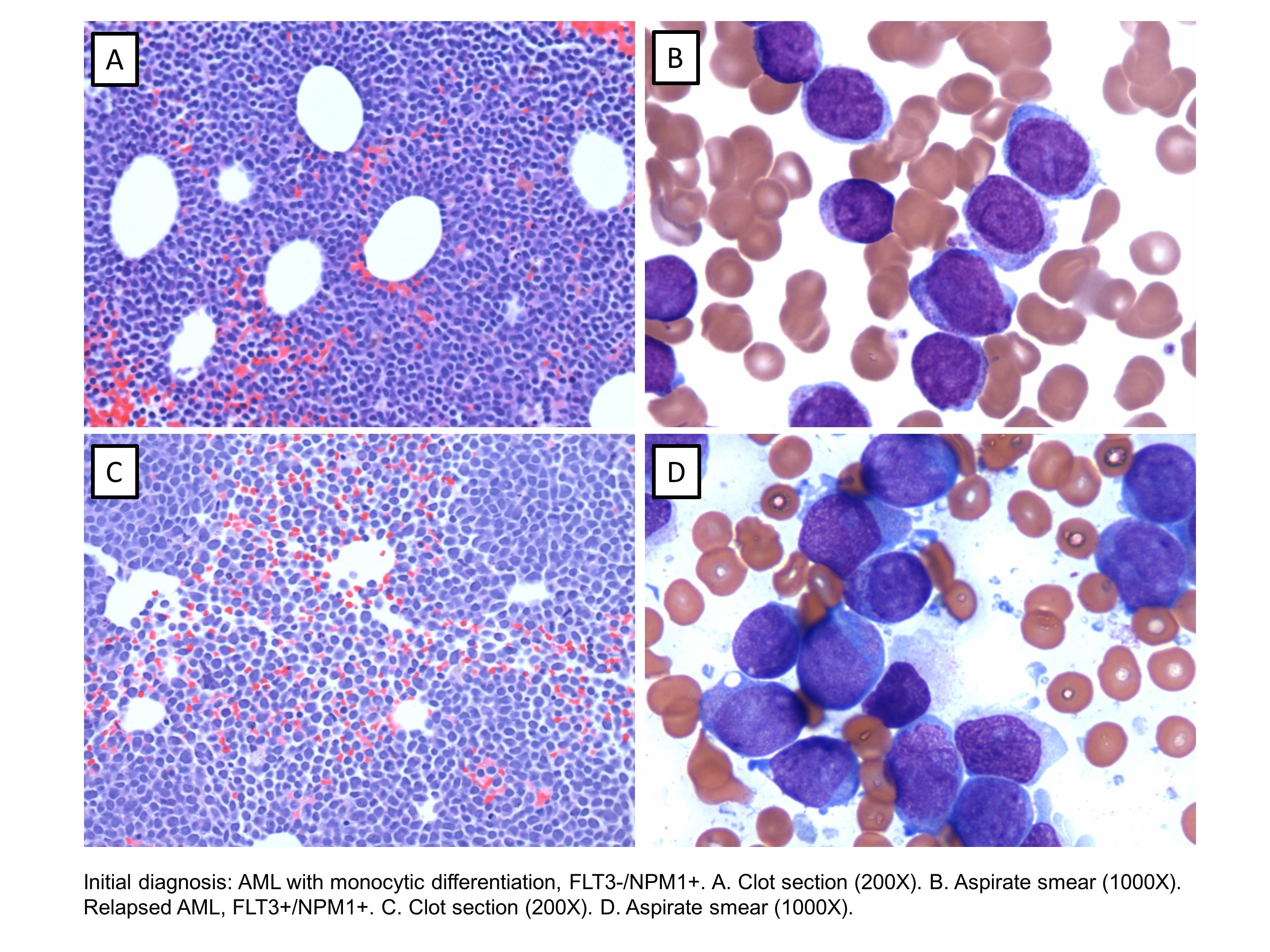 |
| Additional figure 2 | 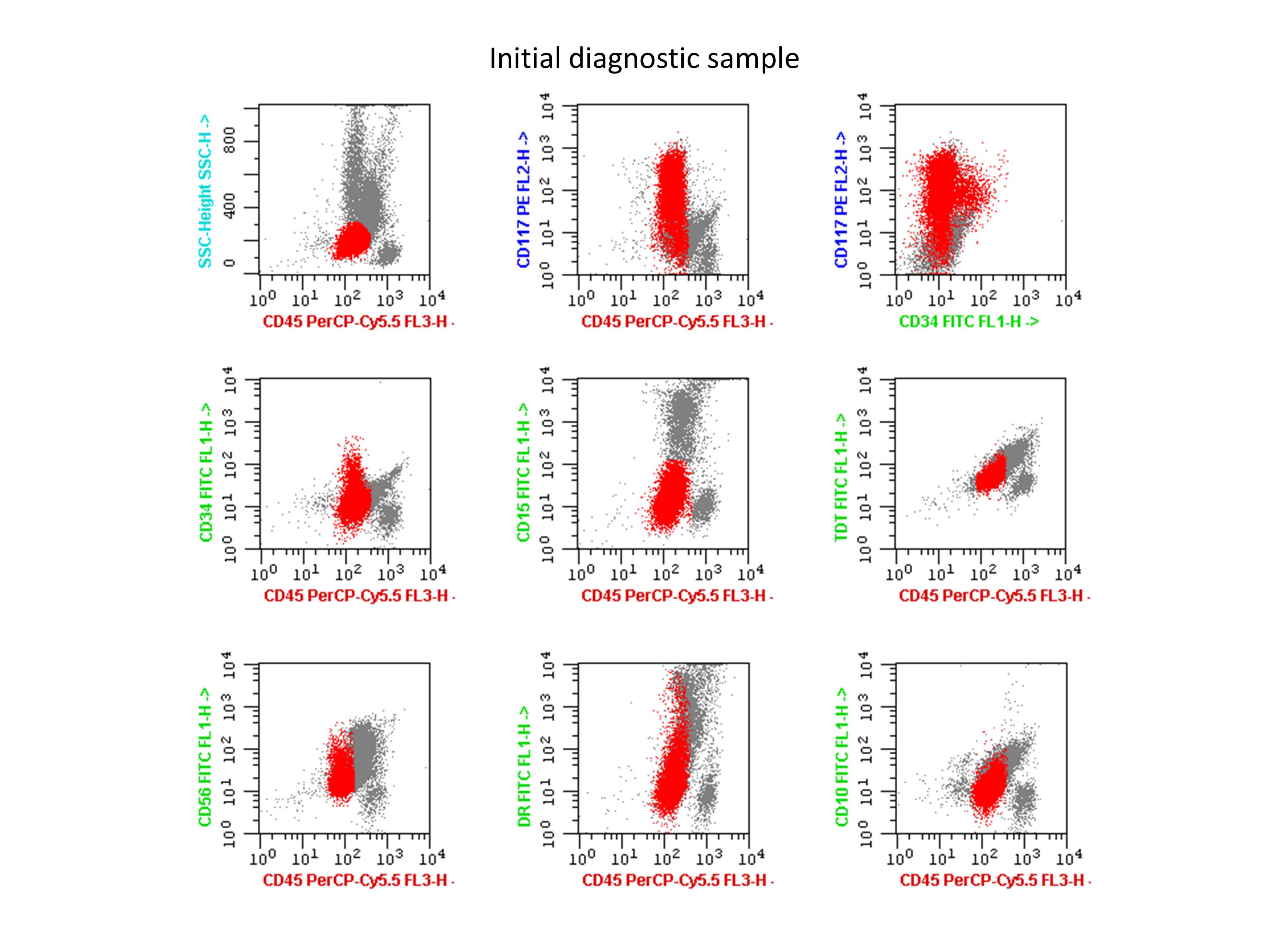 |
| Additional figure 3 | 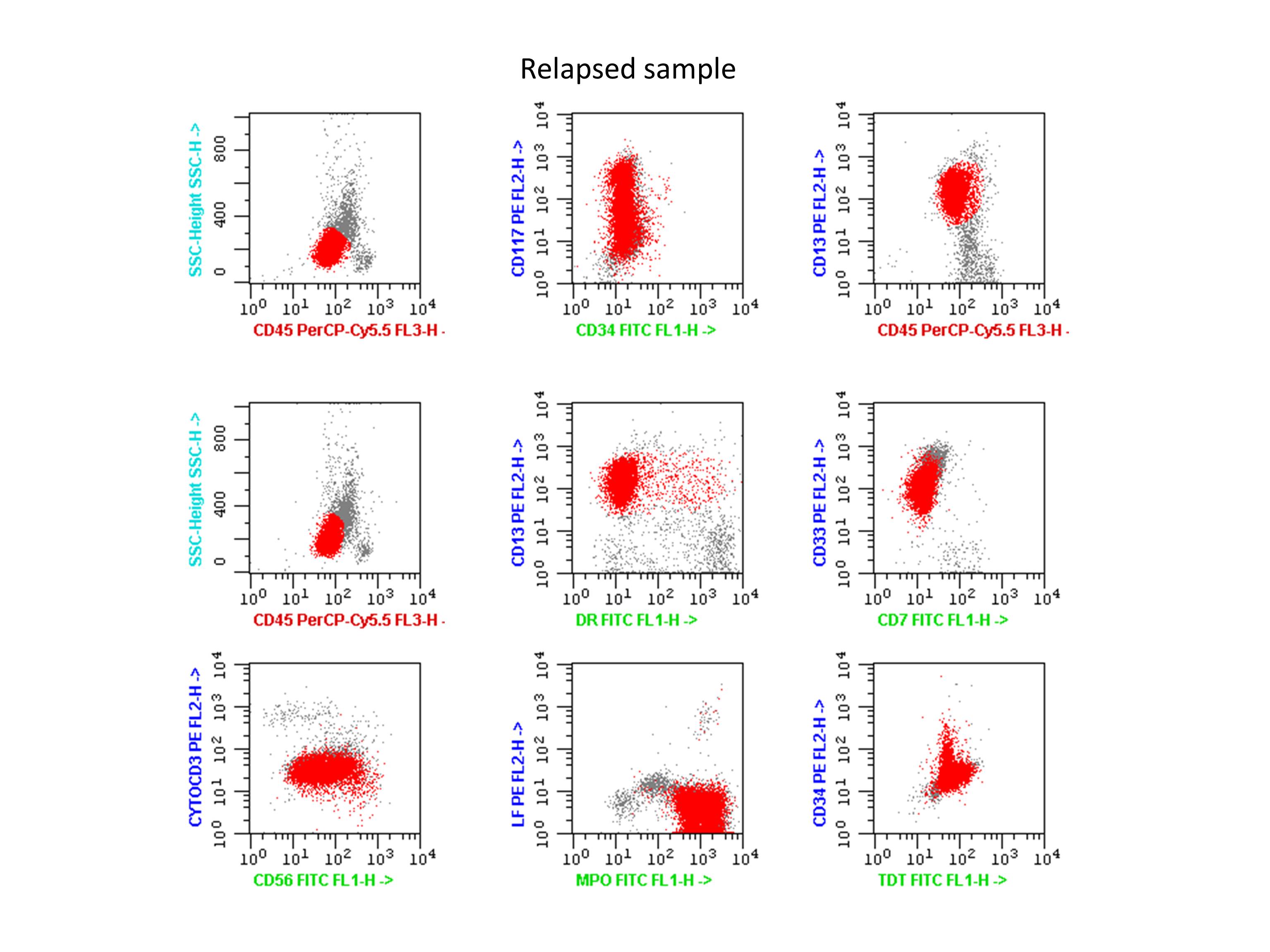 |