Institution: AP-HP; Université Paris Descartes
Additional authors:Agnès Le Tourneau, Amélie Trinquand, Anne Vincent-Salomon, Vahid Asnafi, Hélène Pacquement, Nicolas Boissel, Elizabeth MacIntyre
Session: Extramedullary manifestations of myeloid neoplasms
HISTORY
15 year-old male with mediastinal tumor, 15x9x15 cm, in the anterior and superior area with left susclavicular infiltration. Dyspnea.
Anemia and neutrophilia. No blasts on the blood film.
Clinical summary:
Initial mediastinal mass (13.5 cm ) with left susclavicular adenopathy and mediastinal adenopathies. (June 2012)
•Induction using a protocol AML type (ALFA 07/02)
•No response on PET
•Induction according to lymphoblastic Lymphoma Protocol (FRALLE2000T and HyperCVAD of GRAALL protocol) (Vincristine/Cyclophosphamide/Asparaginase/Corticotherapy)
•Progression
•Aracytin high dose with antiCD33 (Gemtuzumab ozogamicin)
•Venoocclusive disease.
•Numerous chemotherapeutic lines until March 2013 with CT and MTX high dose
•Stabilisation of the mediastinal mass
•Mediastinal Irradiation 24 Gy in May followed by intensification and allograft.
DETAILS
Surgical biopsy of the susclavicular and mediastinal mass. Formalin fixation
Cell Blood Count at diagnosis:
RBC : 4.14x10^12 /L
WBC : 12.9x10^9 /L (80/0/0/12/8)
Plt : 480x10^9 /L
Hb : 11.4 g/dl
Hct : 33.2%
Marrow smears : Presence of 11% blasts with few granules ; by flow : 1% of blast with the same phenotype as the mediastinal mass
IMMUNOHISTOCHEMISTRY AND FLOW CYTOMETRY
IHC of mass : tumor cells express CD45, CD34, CD99, CD56.
No expression of CD1a and TdT.
Tumor cells do not express CD68/KP1, CD20, CDa
IHC of bone marrow: only rare, scattered CD34 positive cells.
Flow :
Cells express CD34, CD117, CD13 (partially), CD33, HLADR, CD56 (bright)
Partial CD7 expression without cytoplasmic or membranous CD3; no CD4, CD2, CD5 expression. No expression of B cell markers not TdT.
CYTOGENETIC FINDINGS
Karyotype normal (with 11% of bone marrow infiltration)
MOLECULAR FINDINGS
Presence of FLT3 ITD
No mutation of NPM1
No CALM-AF10 rearrangement
Clonal rearrangement of TCR Delta, TCR gamma, IgH DJ and IgH VDJ.
INTERESTING FEATURES
Unusual clinical presentation of a phenotypically myeloid tumor
Presence of clonal rearrangement of IgH VDJ and TCR gamma rearrangement without phenotypical lymphoid differentiation.
Unusual absence of expression of CD68/KP1 in myeloid sarcoma
Importance of flow cytometry to identify minimal myeloid marker expression
PROPOSED DIAGNOSIS
Myeloid sarcoma/acute myeloid leukemia with minimal differentiation
CONSENSUS GROUP: ADDITIONAL INFORMATION/STUDIES
Additional immunostains performed by the conference consensus panel:
MPO: Negative
CD117: Subset dimly positive
CD33: Positive in subset
CD43: Strongly positive
TCL1: A subset of cells in nodules are positive
CONSENSUS DIAGNOSIS
Myeloid sarcoma with mutated FLT3 ITD involving mediastinum and supraclavicular region, with 11% blasts in bone marrow (at time of diagnosis)
| HE |  |
| Giemsa |  |
| CD3 |  |
| CD5 | 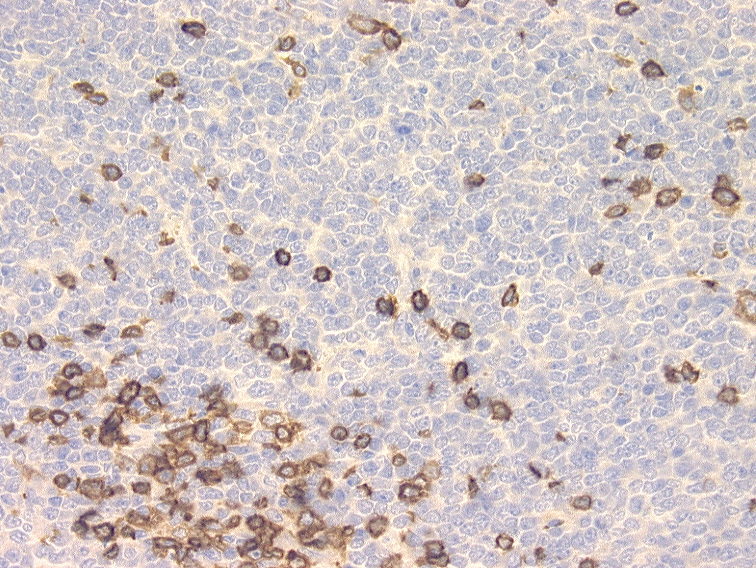 |
| CD34 | 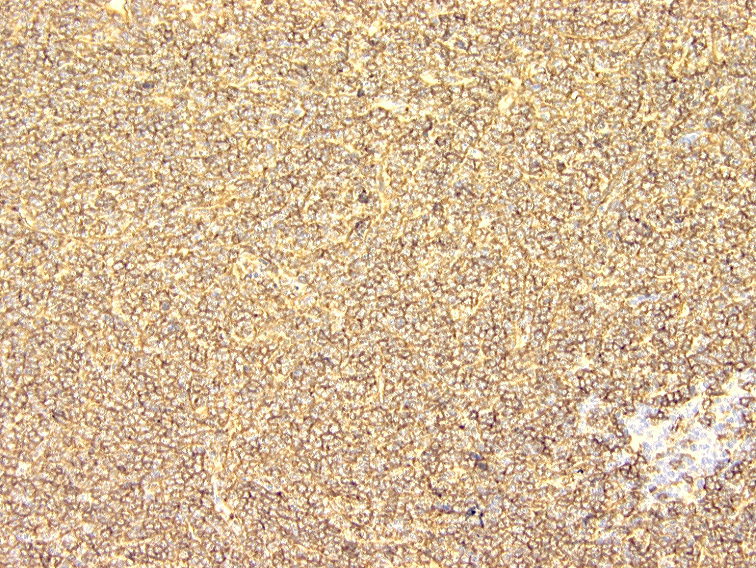 |
| MPO |  |
| Flow of the tumor | 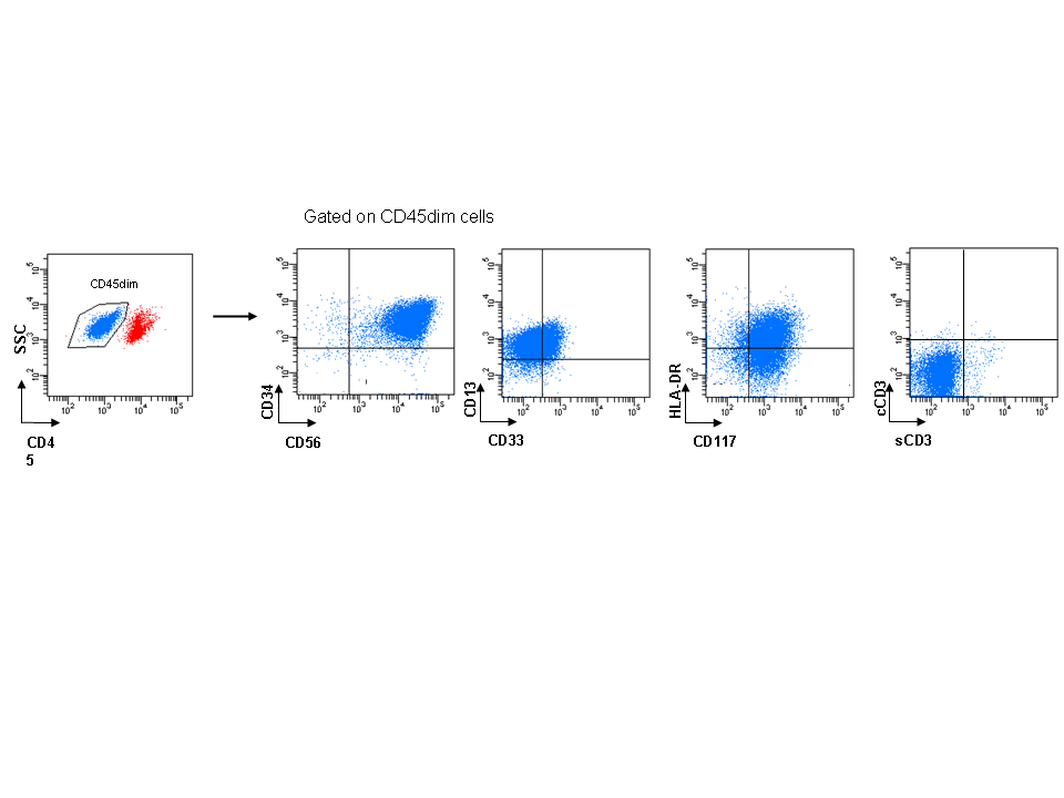 |
| TCR rearrangement | 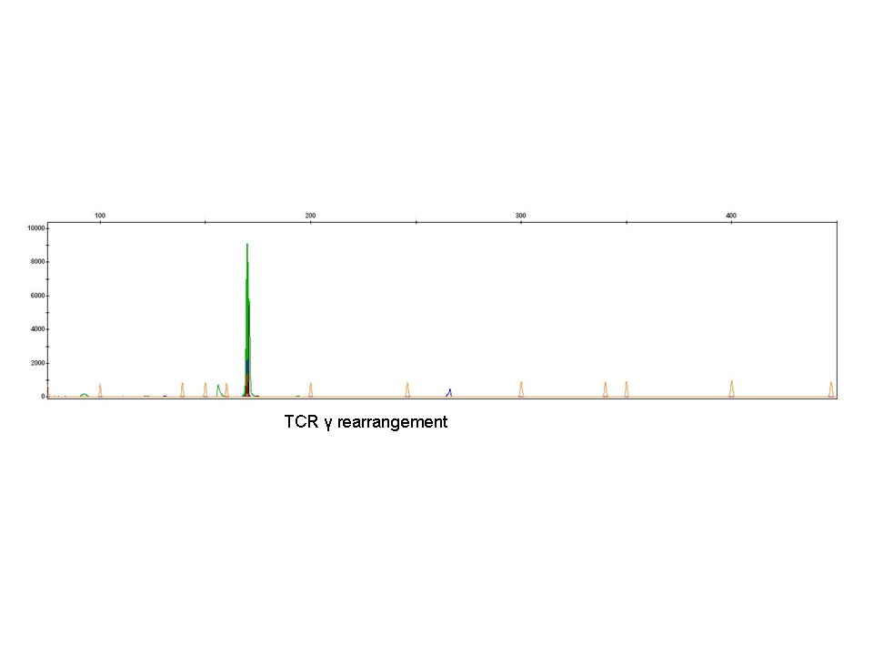 |
| Flow of the tumor | 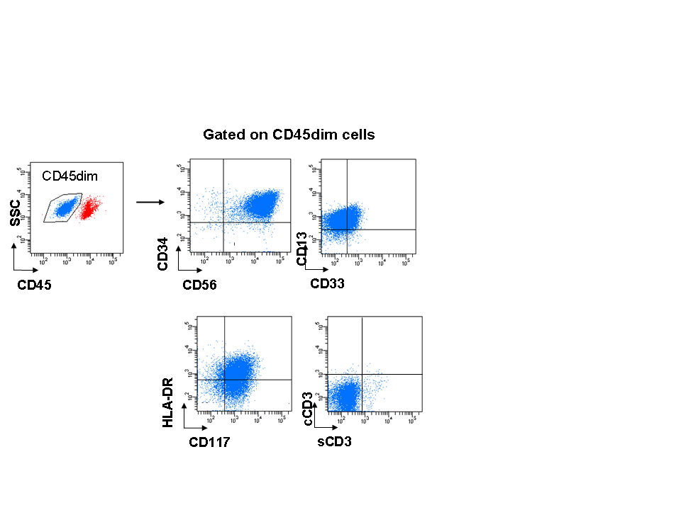 |
| Additional figure 1: TCL1 IHC, 100x | 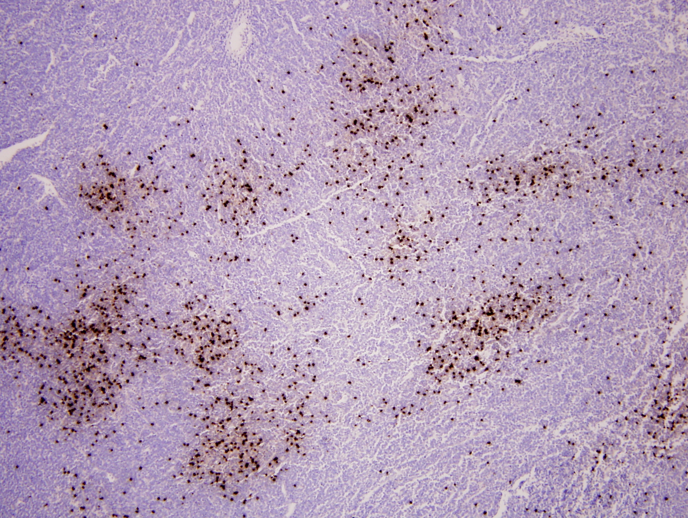 |
| Additional figure 2: TCL1 IHC, 400x | 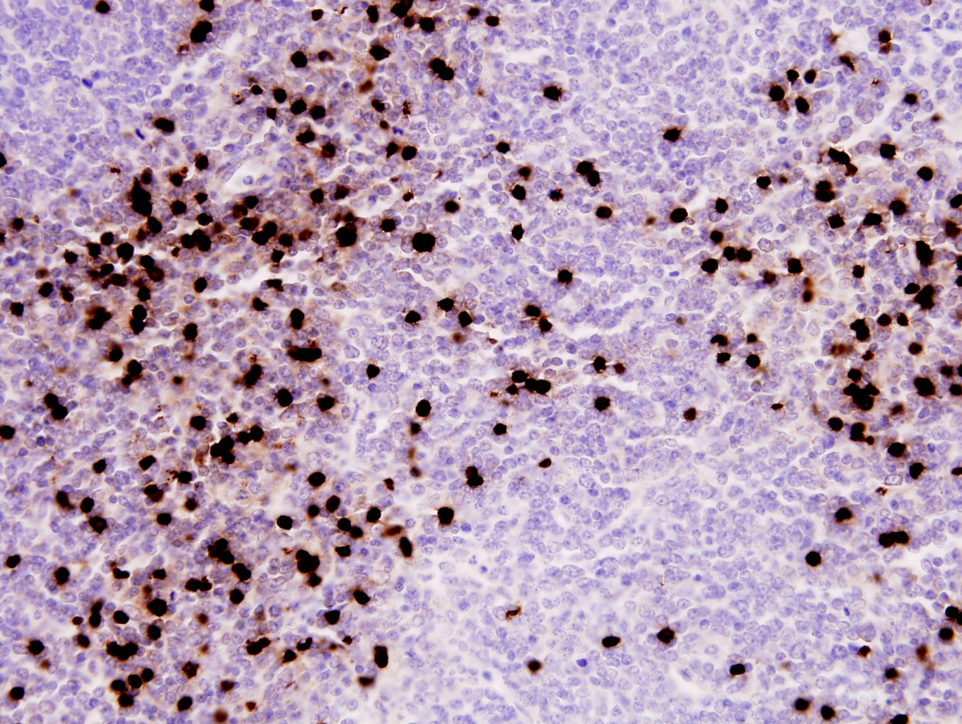 |
| Additional figure 3: CD34 IHC (Bone marrow) | 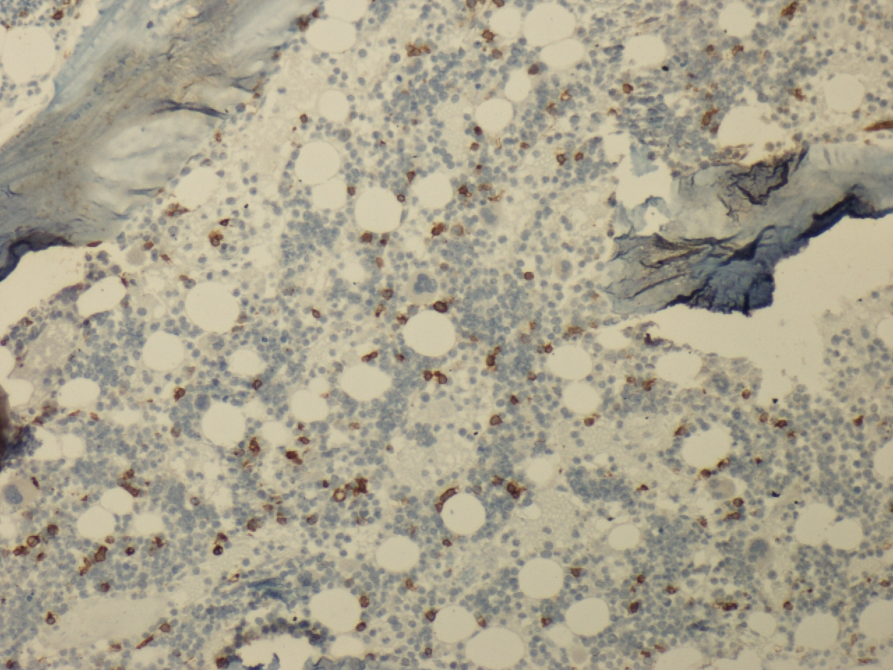 |
| Additional figure 4: CD34 IHC (Bone marrow) |  |
| Additional figure 5: Flow |  |
| Additional figure 6: Flow | 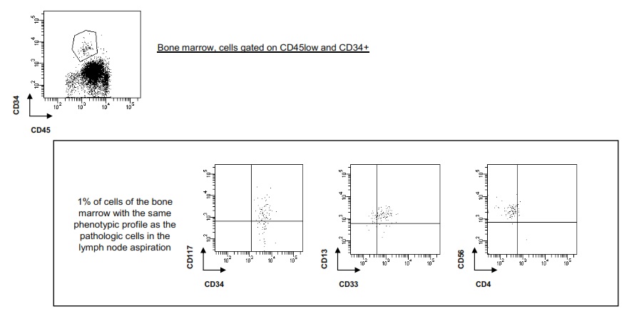 |