Institution: University of Vermont / Fletcher Allen Health Care
Session: Acute leukemias of ambiguous lineage
HISTORY
A 58-year-old man with a history of paraplegia and multiple chronic medical issues presented with a four-week history of fatigue, weight loss, and fevers (39°C). Given his history of chronic urinary tract infections, antibiotics were given empirically; his symptoms did not respond to two courses of therapy. He presented to the emergency department, where a complete blood count revealed pancytopenia (Hb 5.2 g/dl; WBC 1.10 K/μl; plts 18 K/μl). Review of a peripheral smear revealed the presence of circulating blasts, and bone marrow biopsy was performed. As a conclusive diagnosis was not reached based on this study, and physical examination had revealed inguinal lymphadenopathy, a lymph node biopsy was performed. Upon establishment of the diagnosis, the patient elected to pursue palliative care.
DETAILS
Bone marrow biopsy (left posterior iliac crest; formalin fixed tissue): Hypercellular with diffuse infiltration by blasts featuring irregular nuclei, variably discernible nucleoli, and only small amounts of cytoplasm. Lymph node, left inguinal, biopsy (most blocks formalin fixed; a minority fixed in B+): The lymph node measured 3.8 cm in greatest dimension and nodular. Microscopically, the lymph node architecture was effaced by an abnormal infiltrate comprised predominantly of cells intermediate in size with irregular nuclei, fine chromatin, inconspicuous nucleoli, and scant cytoplasm. The abnormal infiltrate was vaguely nodular in areas, and mitotic activity was readily identified.
IMMUNOHISTOCHEMISTRY AND FLOW CYTOMETRY
Bone marrow: Flow cytometry showed a population of abnormal cells expressing dim CD33, dim CD7, dim CD4, CD38, HLA-DR, partial CD11c, and CD11b; AML was favored.
Immunohistochemistry showed the abnormal cells to be positive for CD68 but negative for CD34, myeloperoxidase, CD117, CD3, CD20, and lysozyme.
Lymph node: Flow cytometry showed a population of abnormal cells expressing dim CD33, CD7, dim CD4, dim partial CD5, dim CD11b, CD38, and HLA-DR; the findings were interpreted as representing involvement by a hematopoietic disorder of ambiguous lineage.
Immunohistochemistry highlighted two immunophenotypically distinct blast populations. Blasts forming nodules expressed CD4, CD5, CD7, and CD99, while blasts at the periphery of the nodules expressed CD7, lysozyme, CD33, TdT, and CD99.
CYTOGENETIC FINDINGS
Lymph node: 46,XY,der(12)t(11;12)(q21;p11.2)[6]/45,XY,dic(12;18)(p11.2;p11.2)[5]/46,XY[9].nuc ish(TELx1)[152/200]/(MLLx3)[75/200]
MOLECULAR FINDINGS
None performed.
INTERESTING FEATURES
The diagnosis of mixed phenotype acute leukemia, T/myeloid, was not clear on the basis of bone marrow findings but was reached after examination of an enlarged lymph node which featured a nodular pattern of infiltration by the blasts. The two blast populations were distinguished on the basis of their immunohistochemical staining profiles.
PROPOSED DIAGNOSIS
Mixed phenotype acute leukemia, T/myeloid, NOS
CONSENSUS GROUP: ADDITIONAL INFORMATION/STUDIES
Additional immunostaining performed on lymph node sections by the conference consensus group:
CD3: Positive in blasts
CD1a: Negative
A subset of blasts at the periphery of the lymph node express CD11c and MPO.
CONSENSUS DIAGNOSIS
Mixed phenotype acute leukemia, T/myeloid, NOS
| Blast in peripheral blood | 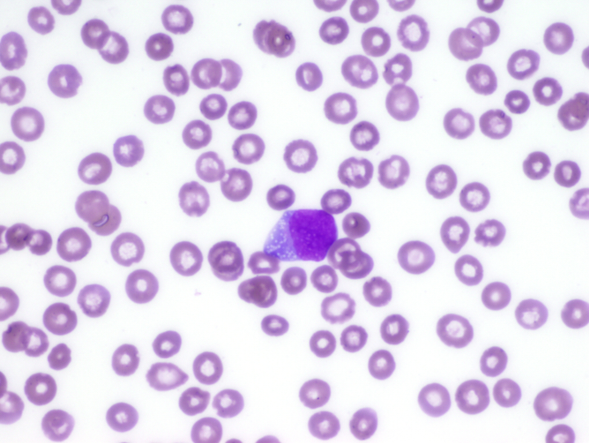 |
| High magnification view of bone marrow biopsy | 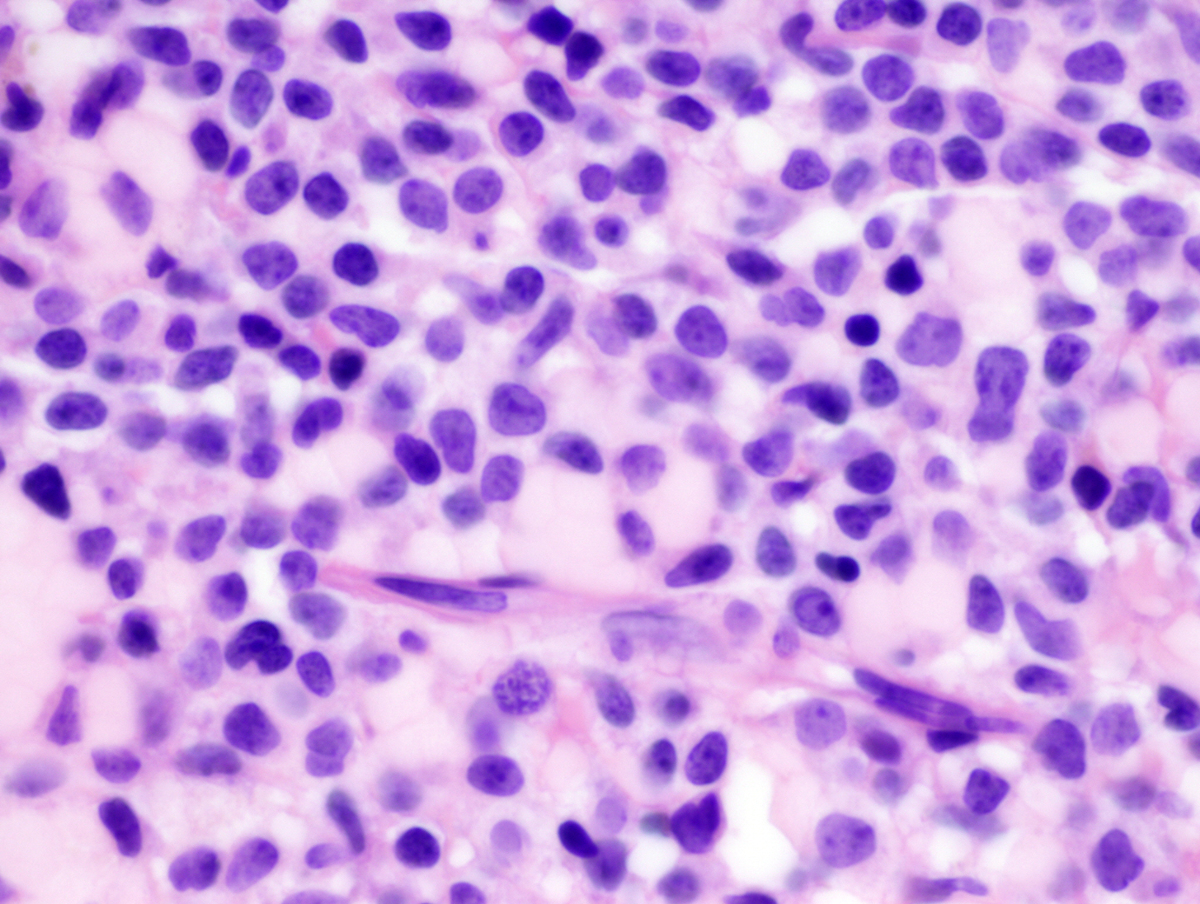 |
| Additional figure 1: CD11c IHC, 100x | 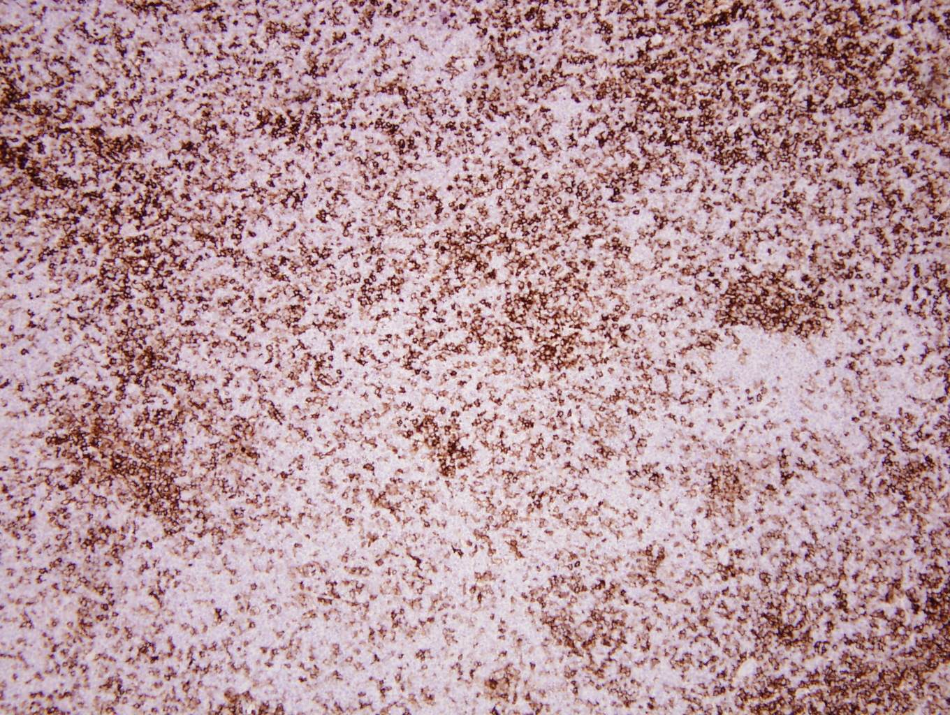 |
| Additional figure 2: CD11c IHC, 400x |  |
| Additional figure 3: MPO IHC, 100x | 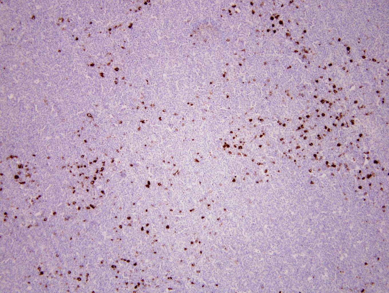 |
| Additional figure 4: MPO IHC, 400x |  |
| Additional figure 5: CD1a IHC, 200x | 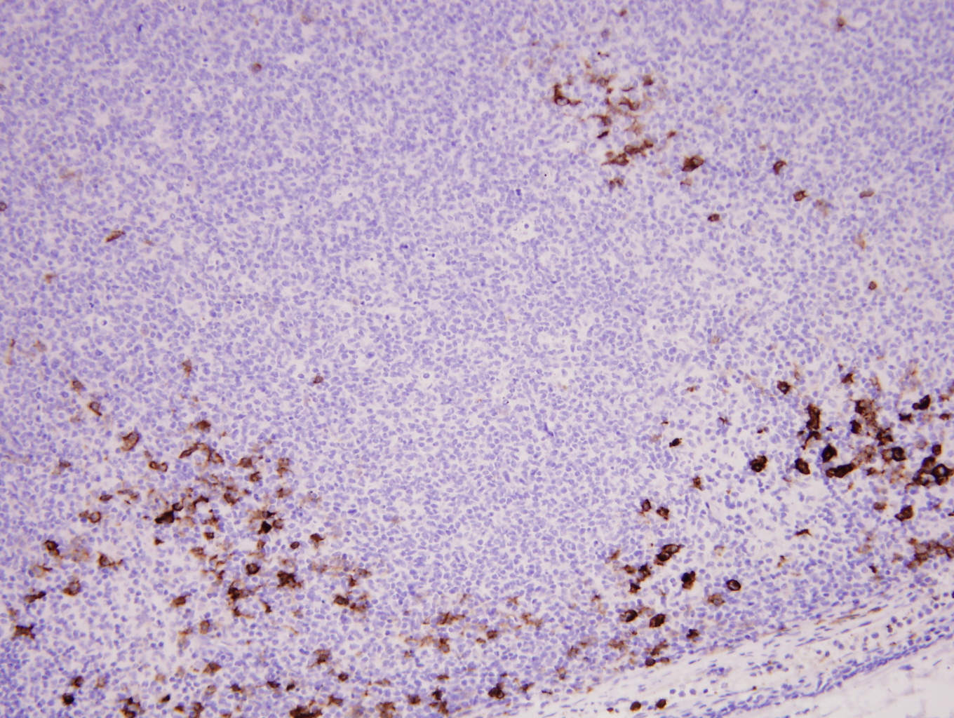 |
| Additional figure 6: CD1a IHC, 400x |  |