Institution: University of Minnesota
Session: Acute leukemias of ambiguous lineage
HISTORY
A 51 year old woman with a diagnosis of acute myeloid leukemia with monocytic differentiation is referred to a tertiary medical center for treatment. Though the medical center has an opportunity to review some of the outside material to confirm the diagnosis, the clinical team repeats a biopsy to document the immunophenotype and morphologic features “in house” prior to initiating and recommending therapy. The patient had received some treatment prior to obtaining this biopsy.
DETAILS
A biopsy and aspirate are performed on the left posterior inferior iliac crest. Blood is collected at the bedside for a smear. Bone marrow aspirate material collected without an anticoagulant is used to make a direct smear. EDTA-anticoagulated aspirate material is used to create concentrate smears. Blood, direct, and concentrate spears are Wright-Giemsa stained. The bone marrow core biopsy was fixed in AZF, decalcified, processed, and paraffin embedded, then sections were mounted and H&E stained. Aspirate material was submitted for flow cytometry and cytogenetics. On review of smears and sections, the peripheral blood is hallmarked by an excess of blasts, in a background of anemic blood with thrombocytopenia. Blasts predominate on the marrow smears and have a dimorphic appearance. The majority of blasts are larger with convoluted nuclei, prominent nucleoli, and moderate cytoplasm with fine granules. There is a minor population of small blasts with scant cytoplasm with occasional cytoplasmic budding. Using cytochemical stains, a majority of the blasts express alpha-naphthyl butyrate stain and non-specific esterase, and a minority express myeloperoxidase. The trephine core is effaced by blasts at greater than 90% cellularity.
IMMUNOHISTOCHEMISTRY AND FLOW CYTOMETRY
By IHC on the trephine core, the blasts are diffusely positive for lysozyme. A small subset of the blasts are positive for CD34, CD68, and CD79a. Scattered blasts are positive for nuclear TdT. Flow cytometry - based on CD45 versus side scatter, blasts comprise 90% of leukocytes. Most of the blasts express partial CD4, dim CD7, partial CD13, CD14, CD15, CD36, CD38, dim CD45, CD58, dim to absent CD117, partial HLA-DR, and partial cytoplasmic myeloperoxidase, but lack surface and cytoplasmic CD3, CD5, CD8, CD19, CD20, CD22, CD34 (predominantly negative), CD56, CD61, glycophorin A, nuclear TdT, and cytoplasmic CD79a. In addition, a subset of the blasts (5 to 7% of the blasts) have B lineage with the following immunophenotype: the blasts express CD19, partial CD22 (bright on a subset), CD34, CD38, dim CD45, CD58, nuclear TdT and cytoplasmic CD79a, but lack CD10, CD14, CD33, cytoplasmic CD3, surface T cell antigens, and cytoplasmic myeloperoxidase.
CYTOGENETIC FINDINGS
46, XX; negative for BCR/ABL1 gene fusion and MLL rearrangement by FISH
MOLECULAR FINDINGS
Not performed on this case. Additional unstained smears available for molecular testing, if needed.
INTERESTING FEATURES
This case was originally thought to be purely a myeloid leukemia with monocytic differentiation. However, using multicolor flow cytometry we were able to identify a subset of the blasts with a distinct immunophenotype of B lineage. This case is a good example of the utility of repeating the biopsy and ancillary studies at tertiary care centers. If selected for presentation, we would attempt to obtain original flow data to demonstrate the nuances of flow cytometric analysis of MPAL with integration of morphology and other ancillary studies. In addition, this case could be used to stimulate discussion - though the diagnosis criteria are typically applied to initial specimens obtained prior to any definitive therapy, large tertiary centers don't always evaluate and profile de novo cases, yet are asked to routinely characterize them as best as possible so that teams can make treatment decisions.
PROPOSED DIAGNOSIS
Mixed phenotype acute leukemia, B/myeloid, NOS
CONSENSUS DIAGNOSIS
Mixed phenotype acute leukemia, B/myeloid, NOS (bilineal)
| Bone marrow aspirate - Wright Giemsa and cytochemical stains |  |
| Core Biopsy and IHC |  |
| Flow cytometry 4 color panel part 1 | 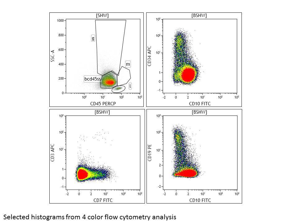 |
| Flow cytometry 4 color panel part 2 |  |
| Flow cytometry cytoplasmic stains | 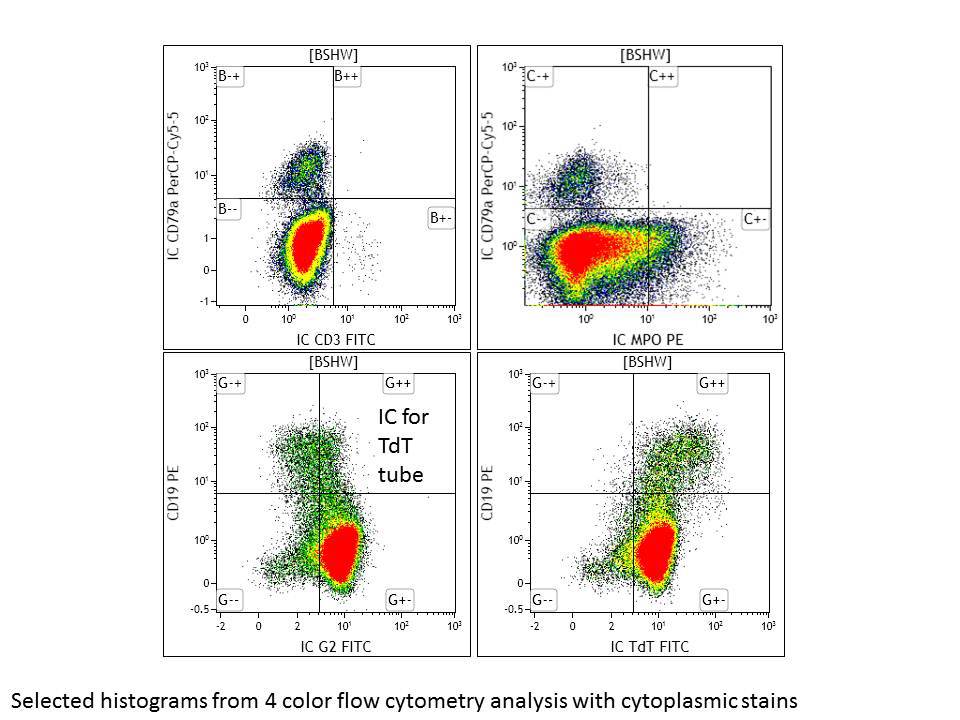 |
| Flow cytometry 8 color B lineage tube | 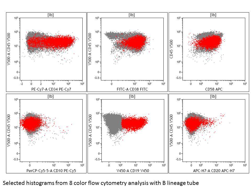 |