Institution: Hospital of the University of Pennsylvania
Additional authors:Darshan Roy, MD, Patti Cohen, MD, Adam Bagg, MD
Session: Acute leukemias of ambiguous lineage
HISTORY
Initial Presentation: A 36-year-old female was admitted for a cesarean section and immediately post-operatively developed a fever. She was discharged after receiving a two-day course of antibiotics for presumed endometritis. She had persistent fevers and was admitted two weeks later for a clinical work-up. At the time of admission, she had a peripheral smear that was reviewed by hematology-oncology and interpreted as having “atypical lymphocytes with some blast-like cells that look more viral/reactive than malignant.” She was subsequently discharged after an extensive infectious disease work-up failed to reveal a source of the fevers. She was re-admitted a week later with persistent fevers. Her CBC was significant for progressive cytopenias. Her peripheral blood was sent for flow cytometry and a bone marrow biopsy was performed.
Clinical Follow-up: The patient received dasatinib, HyperCVAD, and intrathecal methotrexate and cytarabine. She subsequently underwent an allogeneic stem cell transplant. Post-transplant chimerism analysis performed on the peripheral blood showed >99% donor cells. She has remained disease free for over a year, with the most recent (January 2013) RT-PCR for the BCR-ABL1 fusion mRNA remaining negative.DETAILS
Bone marrow biopsy; B5-fixed and decalcified following fixation; specimen measured 2.8 x 0.2 cm grossly.
Bone marrow trephine: H&E-stained sections showed a markedly hypercellular marrow (>90%) consisting almost exclusively of blasts. Some blasts were smaller with inconspicuous nucleoli and minimal cytoplasm while other blasts were large with prominent nucleoli and moderate amounts of cytoplasm. Trilineage hematopoietic elements were markedly decreased.Bone marrow core biopsy touch prep: The Wright-Giemsa stained touch prep showed an expanded population of blasts with large irregular nuclei, inconspicuous nucleoli, fine chromatin and variable amounts of cytoplasm. There was a morphologic variability in the blasts with cells ranging from small blasts with minimal cytoplasm to large blasts with moderate amounts of cytoplasm. No distinct Auer rods were identified. Cytoplasmic eosinophilic granules were seen in a minor subset of blasts. Erythroid and mature myeloid elements were markedly decreased.IMMUNOHISTOCHEMISTRY AND FLOW CYTOMETRY
Immunohistochemistry performed on the bone marrow core biopsy: The smaller blasts were predominantly positive for CD79a, TdT, and Pax5 while the majority of the larger blasts were positive for myeloperoxidase, lysozyme, and CD15. Both the smaller and larger blasts were strongly positive for CD10.
Cytochemical stains performed on the bone marrow touch prep: Many of the larger blasts were subset positive for myeloperoxidase and Sudan black B while the smaller blasts were negative for both. All blasts were negative for non-specific esterase.Flow cytometry performed from single cells teased from a fresh, unfixed bone marrow core biopsy demonstrated two discrete populations of blasts. One population was B-lymphoblastic (39%) with the following immunophenotype: CD10+ CD13+ CD19+ CD34+ cCD79a+ HLA-DR+ TdT+ with variable expression of cCD22 and IgM. These blasts were negative for CD4, CD11b, CD14, CD15, CD64, and MPO. The second population was myelo(mono)blastic (36%) with the following immunophenotype: CD4+ CD13+ CD15+ CD19+ CD33+ CD34+ CD64+ HLA-DR+ MPO+ with variable expression of CD10, CD11b, cCD79a, and TdT. These blasts were negative for cCD22 and IgM.Flow cytometry performed on the peripheral blood showed a population of blasts (12%) with an identical phenotype to the B-lymphoblastic population seen in the bone marrow. No myeloblast population was present in the peripheral blood.CYTOGENETIC FINDINGS
FISH performed on the decalcified bone marrow core biopsy demonstrated a BCR-ABL1 translocation in 156/200 (78%) of cells. Karyotypic studies were unable to be performed due to lack of growth.
MOLECULAR FINDINGS
Reverse transcription-polymerase chain reaction (RT-PCR) analysis performed on the peripheral blood was positive for a fusion mRNA transcript arising from a t(9;22)(q34;q11) translocation (e1a2 BCR-ABL1).
INTERESTING FEATURES
This is a classic cytogenetically defined subtype of the ambiguous leukemias consisting of a bilineal population of distinct lymphoblasts and myeloblasts. This is the most frequently seen recurrent cytogenetic abnormality in mixed phenotype acute leukemias, but it is extremely rare overall, comprising less than 1% of all acute leukemias. Some interesting features of this case are:
1) Both blast populations were detected in the bone marrow, but only the lymphoblast population was found circulating in the peripheral blood. Had only peripheral blood flow cytometry been performed, the myeloid component of the leukemia might not have been detected. 2) Although both blast populations met criteria for distinct B-cell and myeloid lineages, there was some “lineage infidelity” within each blast population. The B lymphoblasts expressed CD13 and the myeloblasts expressed CD10, CD79a, and TdT.PROPOSED DIAGNOSIS
Mixed phenotype acute leukemia with t(9;22)(q34;q11.2); BCR-ABL1
CONSENSUS DIAGNOSIS
Mixed phenotype acute leukemia (B/myeloid), with t(9;22)(q34;q11.2)
| Bone marrow core biopsy, 2.5x, H&E |  |
| Bone marrow core biopsy, H&E, showing greater than 90% blasts |  |
| CD10 5x |  |
| MPO 5x |  |
| TdT 5x |  |
| CD15 5x |  |
| CD79a 20x |  |
| Lysozyme 20x |  |
| Pax5 20x |  |
| MPO 20x |  |
| CD79a 40x, Smaller blasts are positive for CD79a | 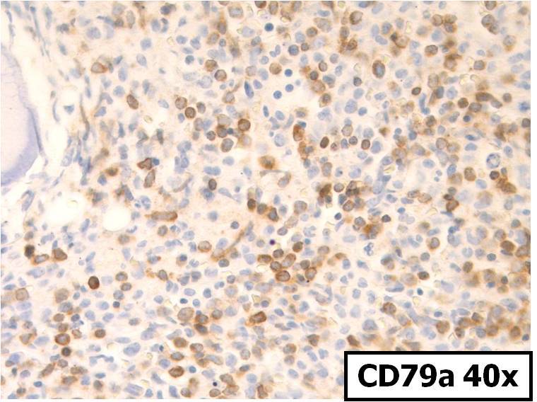 |
| CD79a 100x, Smaller blasts are positive for CD79a | 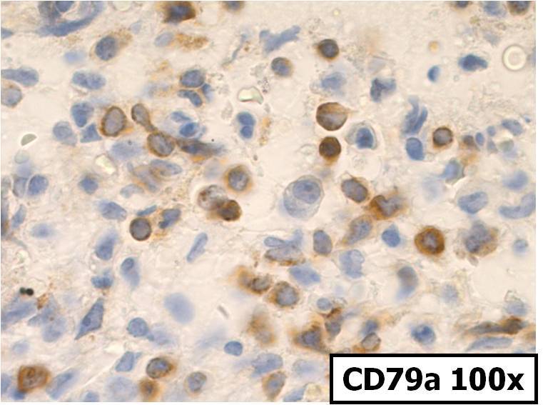 |
| MPO 40x, Larger blasts are positive for MPO |  |
| MPO 100x, Large blasts are positive for MPO |  |
| Bone marrow aspirate touch prep, Wright-Giemsa showing morphological spectrum of blasts |  |
| Bone marrow aspirate touch prep, Wright-Giemsa, 100x, showing subset of blasts with cytoplasmic granules |  |
| Bone marrow aspirate touch prep, MPO, 100x, showing larger blasts are MPO+ and smaller blasts are MPO- | 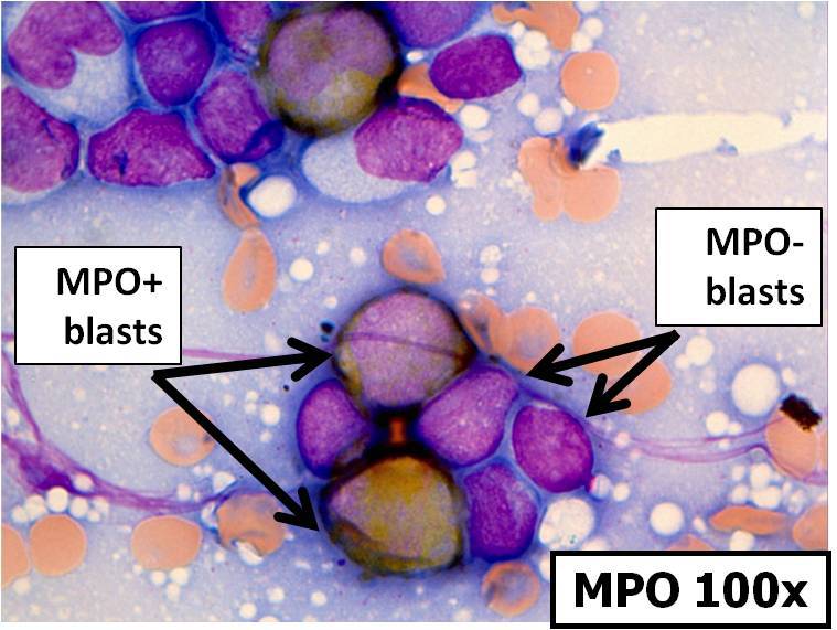 |
| Bone marrow aspirate touch prep, SBB, 100x, showing larger SBB+ blast and smaller SBB- blast | 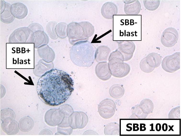 |
| Peripheral Blood Flow Cytometry showing 12% blasts. Only the lymphoblastic population is present in the blood. No myelobasts are seen. |  |
| Bone marrow flow cytometry showing two populations, a myelo(mono)blastic population (36%) and a lymphoblastic population (39%) |  |
| Bone marrow flow cytometry showing TdT+ MPO- CD4- CD3- lymphoblasts and TdT- MPO+ CD4+ CD3- myeloblasts | 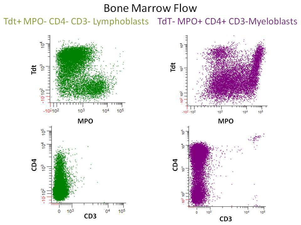 |
| Bone marrow flow cytometry showing CD19+ CD20- CD13+ CD33- lymphoblasts and CD19+ CD20- CD13+ CD33+ myeloblasts | 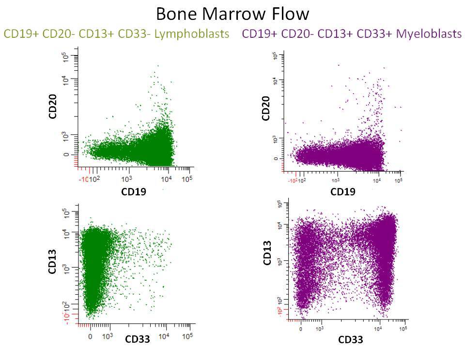 |
| FISH shows a +BCR-ABL1 translocation in 156/200 cells (78%) | 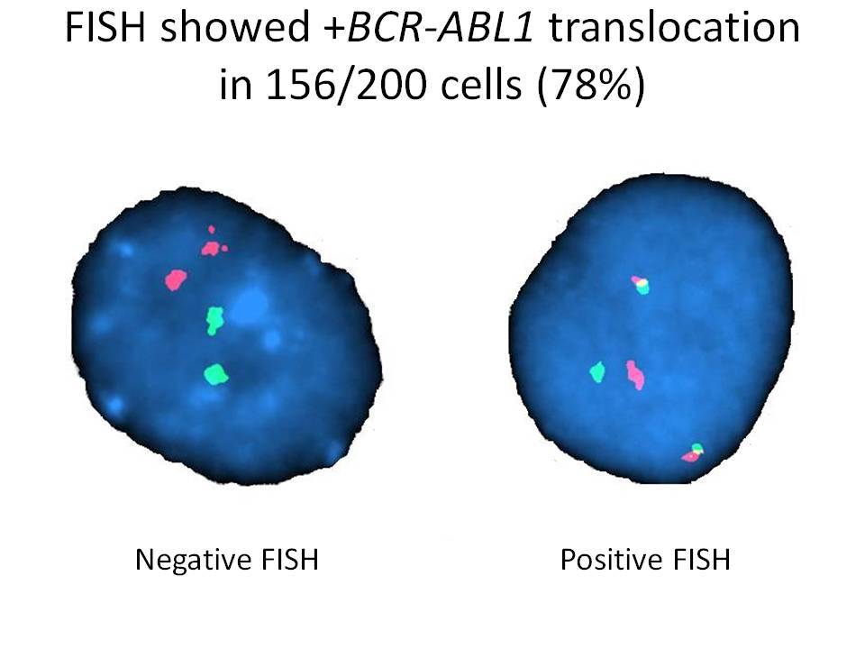 |