Institution: Hospital of the University of Pennsylvania
Additional authors:Adam Bagg
Session: Extramedullary manifestations of myeloid neoplasms
HISTORY
A 41-year-old man presented with pancytopenia and disseminated intravascular coagulation in June 2002. He was diagnosed with morphologically classical hypergranular t(15;17)(q24;q12)–positive acute promyelocytic leukemia (APL). Although metaphase cytogenetic studies revealed a normal 46,XY[20] karyotype in the bone marrow, the PML-RARA fusion was detected by both fluorescence in situ hybridization and qualitative reverse-transcription polymerase chain reaction (RT-PCR). All-trans-retinoic acid (ATRA) therapy was initiated, and a differentiation syndrome developed. Hematologic remission was attained 1 month after induction therapy with cytarabine, daunorubicin, and ATRA, with subsequent attainment of molecular remission. Consolidation chemotherapy consisted of idarubicin alternating with mitoxantrone. In 2004, the patient experienced a molecular-only relapse in the bone marrow, which was treated with arsenic followed by autologous stem-cell transplant. In 2005, he relapsed with overt morphologic peripheral blood and CNS involvement. This was treated with ATRA, intrathecal cytarabine, and CNS radiation. After the attainment of a second remission (by RT-PCR), he underwent nonmyeloablative 9/10 allele-matched unrelated-donor stem-cell transplantation in March 2006. In December 2006, he relapsed morphologically in the bone marrow. He was treated into molecular remission with ATRA and donor lymphocyte infusion. Molecular studies on bone marrow showed full engraftment for 15 months, as well as negativity for PML-RARA. In 2008, the patient presented with a small bowel obstruction (specimen submitted to workshop) and ascites (see PowerPoint). Tandem peripheral blood engraftment studies showed 100% donor DNA, and bone marrow examination showed no morphologic or cytogenetic evidence of disease. However, RT-PCR performed on peripheral blood was positive for a PML-RARA fusion.
DETAILS
a) Right hemicolectomy(Formalin fixed)
Gross: Segment of colon, cecum, small bowel, and appendix with a 3.5 cm green-blue mass located in the terminal ileum. The mass extended through the bowel wall into the serosal fat.
Microscopic: An expansion of blasts forms a mass involving the full thickness of terminal ileum, peri-appendiceal fat, and proximal small bowel resection margin. Blasts are large in size, with round to oval nuclear contours, conspicuous nucleoli, finely dispersed to granular chromatin, and moderate granular eosinophilic cytoplasm.
b) Ascites fluid: Numerous blasts/leukemic promyelocytes present. Blasts are large in size, with oval to irregular nuclear contours, conspicuous nucleoli, finely dispersed to granular chromatin, and moderate cytoplasm with prominent vacuolization.
IMMUNOHISTOCHEMISTRY AND FLOW CYTOMETRY
Flow cytometry performed on the mass showed the presence of CD33+, CD34-, HLA-DR-, MPO+ blasts/leukemic promyelocytes. Immunohistochemistry was not performed. Flow cytometry performed on the ascites fluid showed that the dominant population of CD45(dim) positive, high side scatter cells were positive for CD13, CD33, and MPO, and negative for CD34 and HLA-DR.
CYTOGENETIC FINDINGS
Not performed on current specimens.
MOLECULAR FINDINGS
RT-PCR was positive for a PML-RARA fusion (ascites fluid).
INTERESTING FEATURES
This case represents a unique site of extramedullary relapse of acute promyelocytic leukemia (APL).
The patient had a long history of APL treated with multiple modalities (including ATRA, arsenic, multiple chemotherapy agents, and stem cell transplant, both autologous and allogeneic) with multiple relapses, and presented with a small bowel obstruction representing an extramedullary relapse (EMR) of disease without bone marrow involvement. Of note, molecular studies at the time showed full donor engraftment.
Both ATRA and arsenic may be contributing to increasing EMR rates.
Several mechanisms may be involved; the increasing rates may simply be a result of patients surviving longer than they did in the pre-ATRA era, allowing for the increased development and observation of such relapses. Another hypothesis is that there may be sanctuary sites in which ATRA and arsenic do not reach therapeutic concentrations. Allied to this, there is evidence that ATRA and arsenic increase the expression of adhesion molecules in leukemic cells (eg, CD11b, CD11c, CD18, and CD56), which could allow those cells to access numerous different tissues. It has also been proposed that treatment with ATRA induces expression of multiple CC chemokines, which may result in increased chemotaxis into tissues. The presence of differentiation syndrome appears to be a risk factor for extramedullary relapse, as leukemic cells are theorized to infiltrate different organ tissues during this process.
Of note, survival appears to be worse in patients with extramedullary relapse as compared to isolated bone marrow relapse (6.7 v. 26.3 months). This patient succumbed to sepsis and renal failure soon after presentation.
In the era of ATRA and arsenic treatment for APL, increased vigilance for unusual EMR is warranted, even in the absence of obvious marrow or peripheral blood involvement.
PROPOSED DIAGNOSIS
Acute promyelocytic leukemia, isolated extramedullary relapse (small bowel) without bone marrow involvement.
CONSENSUS DIAGNOSIS
Acute promyelocytic leukemia with t(15;17)(q24.1;q21.2), PML-RARA, involving only small intestine (myeloid sarcoma) at relapse
| Extramedullary APL, small bowel (5X) |  |
| Extramedullary APL, small bowel (40X) | 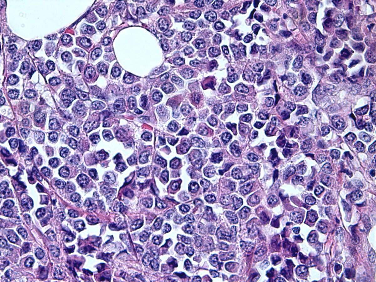 |
| APL, ascites fluid | 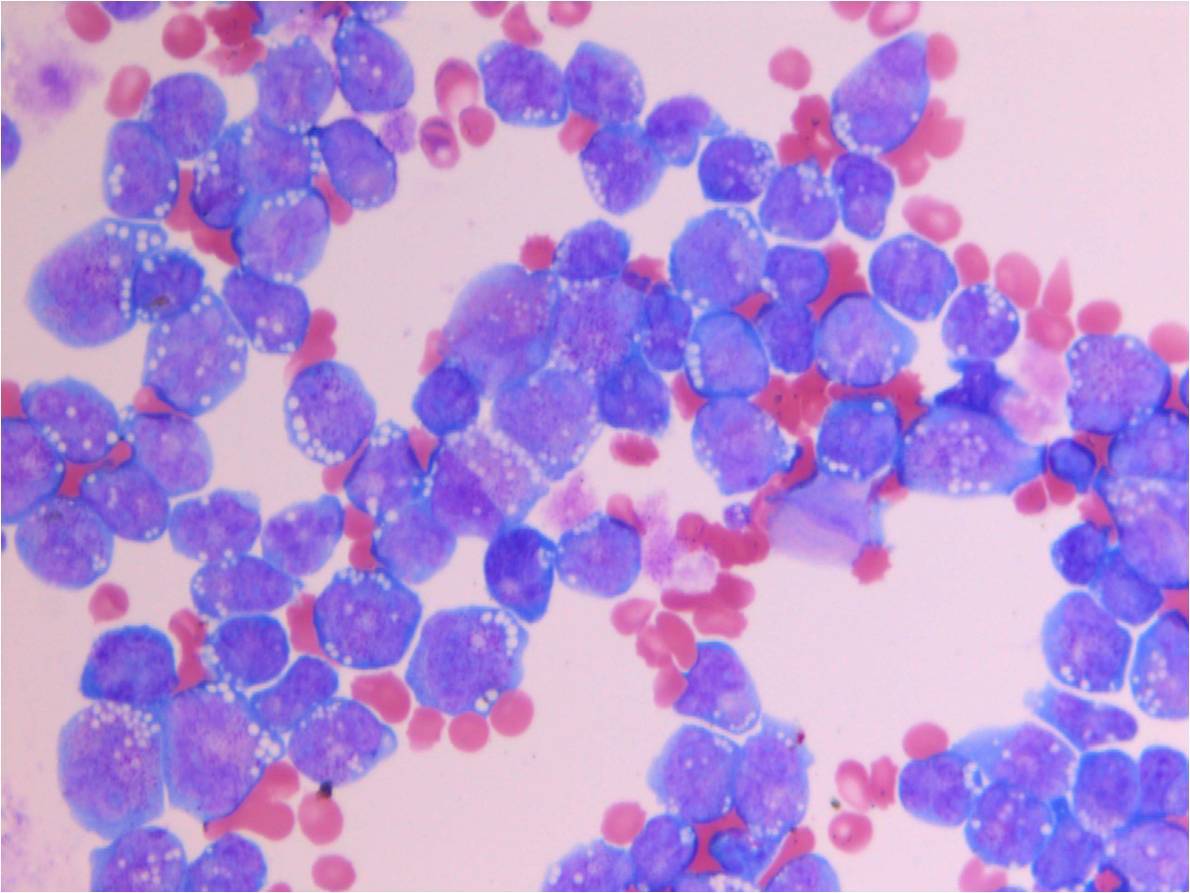 |
| Flow cytometry, ascites fluid | 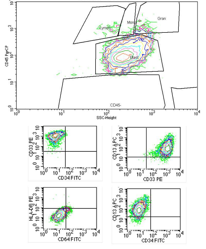 |
| Gel electrophoresis following analysis with PML-bcr3 primer, lane 5, arrowed |  |