Institution: University of Texas Health Science Center San Antonio
Additional authors:Kenneth N. Holder, M.D. Marsha C. Kinney, M.D.
Session: Therapy-related myeloid neoplasms
HISTORY
56 year old female with past medical history of stage IV poorly-differentiated lung adenocarcinoma was admitted for fatigue and leukocytosis with eosinophila. She was initially diagnosed with lung cancer in November 2011 after she was found to have a 9.3 x 6.4 x 9.4 cm lobulated mass in the left upper lobe of her lung. In January 2012 she developed left sided weakness. An MRI showed a 1.6 x 1.7 x 1.4 cm mass in the right parietal lobe with secondary mass effect upon the right lateral ventricle and midline shift of 5 mm. She underwent whole brain radiation therapy and radiation therapy to the lung mass, which was completed in January 2012. A CBC from approximately one week after brain and lung irradiation showed a normal white cell count of 7.5 K/uL with a mild absolute eosinophilia of 0.9 K/uL, hemoglobin of 8.9 G/dL, and normal platelet count of 230 K/uL. She was started on chemotherapy with carboplatin-taxol on February 2012 and completed cycle 4 in April of 2012.
In September 2012, the patient had progressive fatigue, and was discovered to have leukocytosis, anemia, thrombocytopenia and a positron emission tomography (PET) scan revealed marked progression in pulmonary disease and metastatic disease involving the liver, pancreas, and musculoskeletal system. A complete blood count (CBC) at this time showed a leukocytosis of 80.1 K/uL with a differential of 13% neutrophils, 27% bands, 9% metamyelocytes, 1% myelocytes, 5% lymphocytes, 1% monocytes, 44% eosinophils (absolute eosinophil count of 35.2 K/uL), and 0% basophils, a hemoglobin of 7.3 G/dL, and platelet count of 128 K/uL.DETAILS
Biopsy, bone marrow, right posterior iliac crest, B5 fixative.
A peripheral blood smear demonstrates leukocytosis with left shift and marked eosinophilia with abnormal eosinophil morphology including eosinophils with ringed nuclei, monolobate nuclei, rare hyperlobate nuclei, and eosinophils and neutrophils with hypogranular and/or vacuolated cytoplasm. Red cells are normocytic with anisocytosis and have a dual population of normochromic and hypochromic red cells. The platelets have normal morphology. Bone marrow biopsy: The bone marrow is hypercellular (80%-90%) with an increased M:E ratio of 4:1 due to granulocytic hyperplasia. The erythroid lineage shows mild nuclear to cytoplasmic dyssynchrony. A Prussian blue stain reveals increased storage iron with many ringed sideroblasts present. The granulocytic lineage shows orderly maturation with increased eosinophils. The megakaryocytes are present in adequate numbers; however they are large and atypical with some megakaryocytes having hyperlobation of their nuclei and rare widely-separated nuclei. In addition, metastatic adenocarcinoma, expressing Napsin A and TTF-1, consistent with the patient’s lung primary, was present. A reticulin stain demonstrates mild increase in reticulin fibrosis.IMMUNOHISTOCHEMISTRY AND FLOW CYTOMETRY
No abnormal lymphoid or blast population was detected by flow cytometric analysis.
CYTOGENETIC FINDINGS
Cytogenetics: 46,XX,add(7)(q32)[5]/46,XX[17]
The result is abnormal. Of the 22 cells analyzed, 17 were normal and 5 had additional material of unknown origin attached to chromosome 7q. Fluorescence in-situ hybridization (FISH): PDGFRA, PDGFRB, and FGFR1 are negative. Additional cytogenetic and/or FISH studies are being performed to identify the nature of the additional material on chromosome 7q.MOLECULAR FINDINGS
Molecular studies for BCR/ABL fusion transcripts and KIT (D816V) are negative.
INTERESTING FEATURES
This is an unusual case of a 56 year-old female patient with marked eosinophilia (absolute eosinophil count = 35.2 K/uL), anemia and thrombocytopenia with a clonal cytogenetic abnormality (gain of material on chromosome 7q32), arising with a relatively short latency period (7 months) after treatment with an alkylating agent and irradiation for lung cancer. Other cytogenetic/molecular abnormalities associated with eosinophilia were ruled-out. Bone marrow examination showed trilineage dysplasia. MDS with eosinophilia associated with chromosome 7 abnormalities (particularly unbalanced translocations involving chromosomes 1 and 7 and ring chromosome 7) has been rarely reported, including one patient with lung cancer, similar to this patient. Due to the marked eosinophilia and abnormal cytologic features and the short latency period we propose a MDS/MPN rather than the usual therapy-related MDS and suggest this process may be related to the specific genes involved on chromosome 7 and in the “extra material present”. We are attempting additional FISH studies to determine the origin of the extra material.
PROPOSED DIAGNOSIS
Myelodysplastic/myeloproliferative neoplasm (MDS/MPN) with marked eosinophilia, possibly therapy related, associated with concurrent metastatic poorly-differentiated adenocarcinoma of the lung.
CONSENSUS DIAGNOSIS
Therapy-related myeloid neoplasm, myelodysplastic/myeloproliferative, with eosinophilia, associated with concurrent metastatic poorly-differentiated adenocarcinoma of the lung.
| Peripheral Blood 600x Hypogranular and/or vacuolated cytoplasm in neutrophils and eosinophils and monolobate eosinophil |  |
| Peripheral Blood 600x Dysplatic, monolobate eosinophils |  |
| Peripheral blood 600X Dysplastic neutrophils and eosinophils including a hyperlobate eosinophil in upper right and eosinophil with ringed nucleus in lower left | 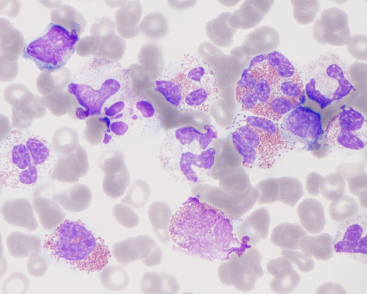 |
| Peripheral blood 600X Dysplastic eosinophil with ringed nucleus |  |
| Peripheral blood 600X Dysplastic eosinophil with ringed nucleus |  |
| Bone marrow biopsy 100x hypercellular marrow |  |
| Bone marrow 200x hypercellular marrow with eosinophilia and dysplastic megakaryocytes |  |
| Bone marrow 600x dysplastic hyperlobate megakaryocyte with separated nuclear lobes |  |
| Bone marrow 600x dysplastic monolobate megakaryocyte |  |
| bone marrow aspirate smear 600x dysplastic hyperlobate megakaryocyte |  |
| Bone marrow aspirate smear iron stain 1000x ring siderblasts |  |
| Bone marrow biopsy 600x mild reticulin fibrosis | 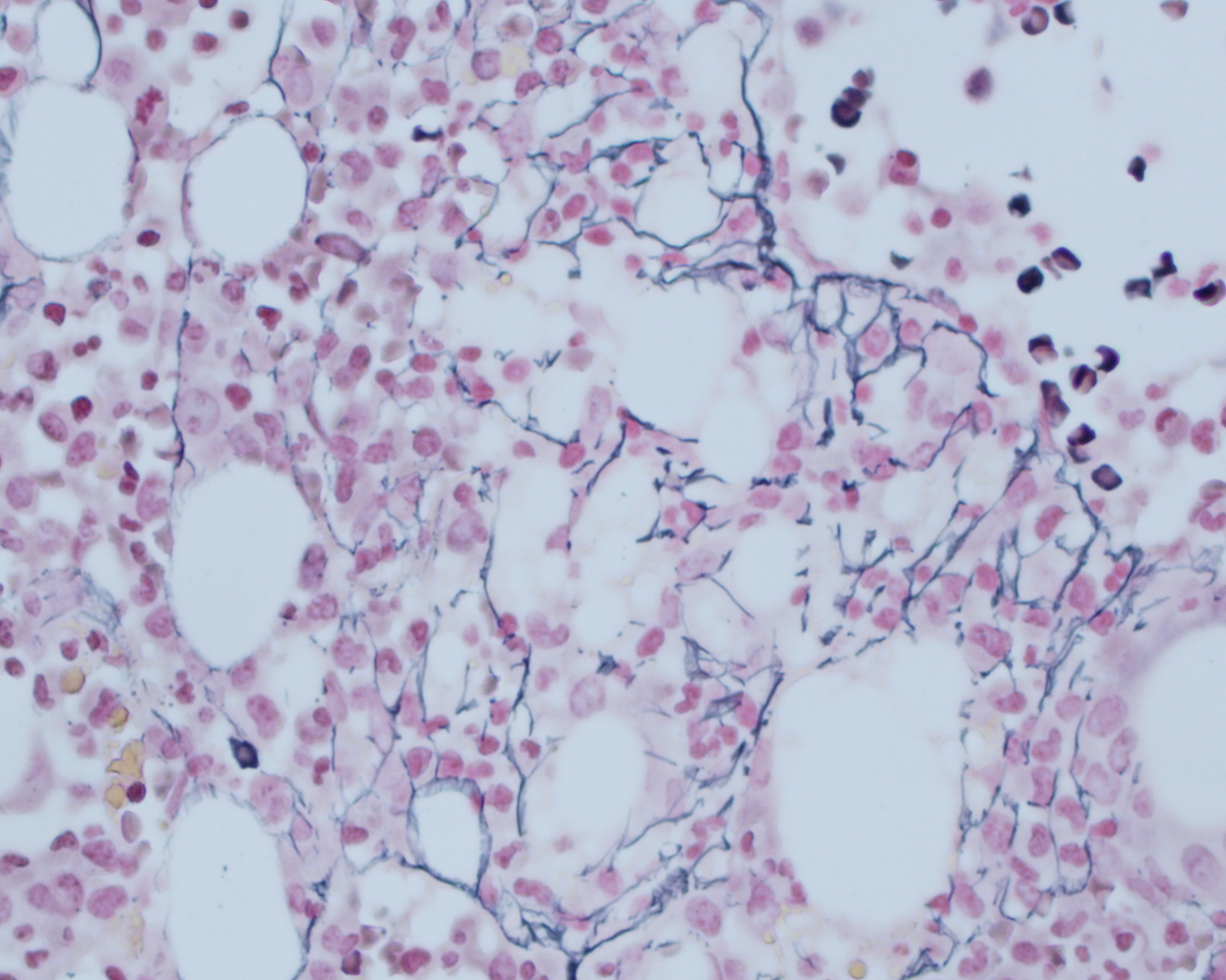 |
| Bone marrow biopsy 100x concurrent metastatic poorly differentiated adenocarcinoma of lung |  |
| Bone marrow biopsy 100x metastatic adenocarcinoma of lung TTF1 positive |  |
| Bone marrow biopsy 600x metastatic adenocarcinoma of lung Napsin A positive |  |
| Additional figure 1: FISH |  |