Institution: Pathology Associates of Albuquerque/Tricore Reference Laboratories
Additional authors:Clark Haskins MD
Session: AML with myelodysplasia-related changes
HISTORY
The patient is a 66 year old woman who was diagnosed with aplastic anemia at the age of 58. She underwent immunosuppression including cyclosporine A, followed by antithymocyte globulin, followed by tacrolimus. After 1.5 years she developed severe thrombocytopenia and myelodysplastic features. She has remained dependent on RBC and platelet transfusions as well as G-CSF PRN ever since. 7 years after initial diagnosis she experienced further progression to acute leukemia with 28% bone marrow blasts. She underwent a course of decitabine treatment without significant change of peripheral cytopenias. She remains alive with transfusion support and treatment of infectious complications as needed.
DETAILS
Multiple bone marrow biopsies, all posterior iliac crest, fixation with acetic zinc formalin (AZF)
3-3-2005: Peripheral blood: WBC 3.4, Hgb 12.8, MCV 100, plt 18, neutrophils absolute 1.3 Bone marrow biopsy: 5% cellular, mostly erythroid islands, no megakaryocytes4-27-2005: Peripheral blood: WBC 2.1, Hgb 8.9, MCV 99, plt 42 neutrophils absolute 0.08 Bone marrow biopsy: <5% cellular, single erythroid island, no megakaryocytes3-15-2006: Peripheral blood: WBC 3.8, Hgb 7.8, MCV 101, plt 78, neutrophils absolute 2.3 Bone marrow biopsy 60% cellular, M:E ratio 0.7, slight megakaryoctye dypsoiesis, 1% blasts10-23-2006: Peripheral blood: WBC 2.0, Hgb 11.2, MCV 112, plt 16, neutrophils absolute 1,3 Bone marrow biopsy: 50% cellular, M:E ratio 0.5, dyspoietic megakaryocytes, 1% blasts7-10-2012: Peripheral blood: WBC 2.0, Hgb 8.6, MCV 88, plt 8, neutrophils absolute 0.0 Bone marrow: 60% cellular, M:E ratio N/A, dyspoietic megakaryocytes, 28% blastsIMMUNOHISTOCHEMISTRY AND FLOW CYTOMETRY
Flow cytometry
3-3-2005 not performed4-27-2005 not performed3-15-2006 not performed10-23-2006 not performed7-10-2012 20% myeloblasts positive for CD34, CD117, CD13, CD33, dim CD45, HLA-DR, increased monocytic cells positive for CD33, CD13, CD4, subset CD56. Negative for PNH phenotype on WBC and RBC.CYTOGENETIC FINDINGS
3-3-2005 46,XX[20]
4-27-2005 46,XX[20]FISH analysis negative for aneuploidy of chromosome 7 or del7q313-15-2006 45,XX,-7[14],46,XX[10-23-2006 45,XX,-7[1],46,XX[9], incomplete study7-10-2012 49,XX,-7,+8,add(11)(p11.2),-18,+21, +22, +r[17] / 46,XX[3]MOLECULAR FINDINGS
None performed
INTERESTING FEATURES
Well documented clonal evolution of aplastic anemia to myelodysplasia to acute myeloid leukemia.
The patient’s history is remarkable for the long survival with severe cytopenias and myelodysplasia, primarly with immunosuppressive and supportive therapyPROPOSED DIAGNOSIS
Acute myeloid leukemia with myelodysplasias-related changes (WHO classification 2008)
arising from myelodysplasia with preceding aplastic anemiaCONSENSUS DIAGNOSIS
Acute myeloid leukemia with myelodysplasia-related changes, arising from myelodysplastic syndrome with preceding aplastic anemia
| Bone marrow Biopsy March 2005 | 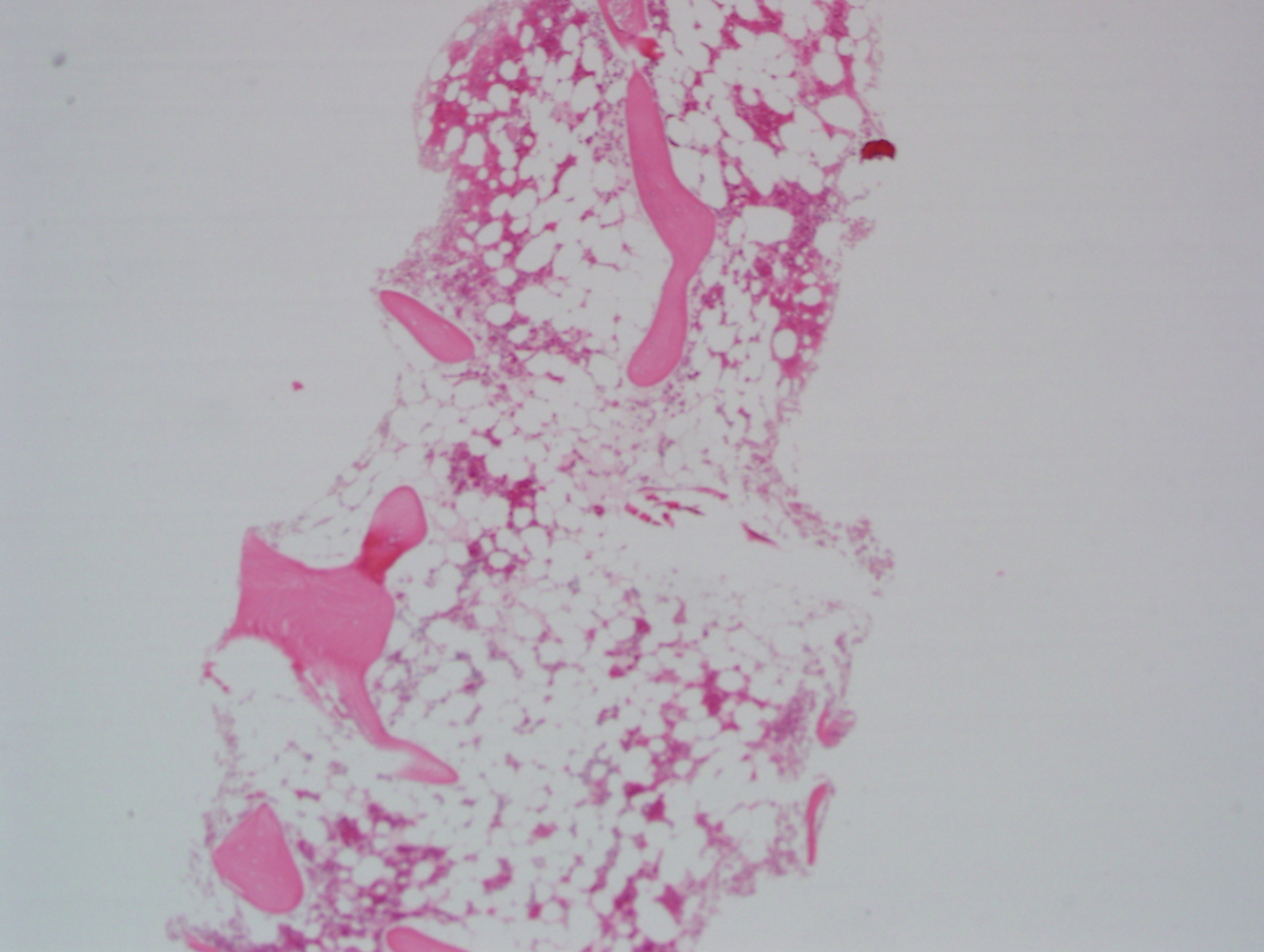 |
| Bone marrow Aspirate March 2005 | 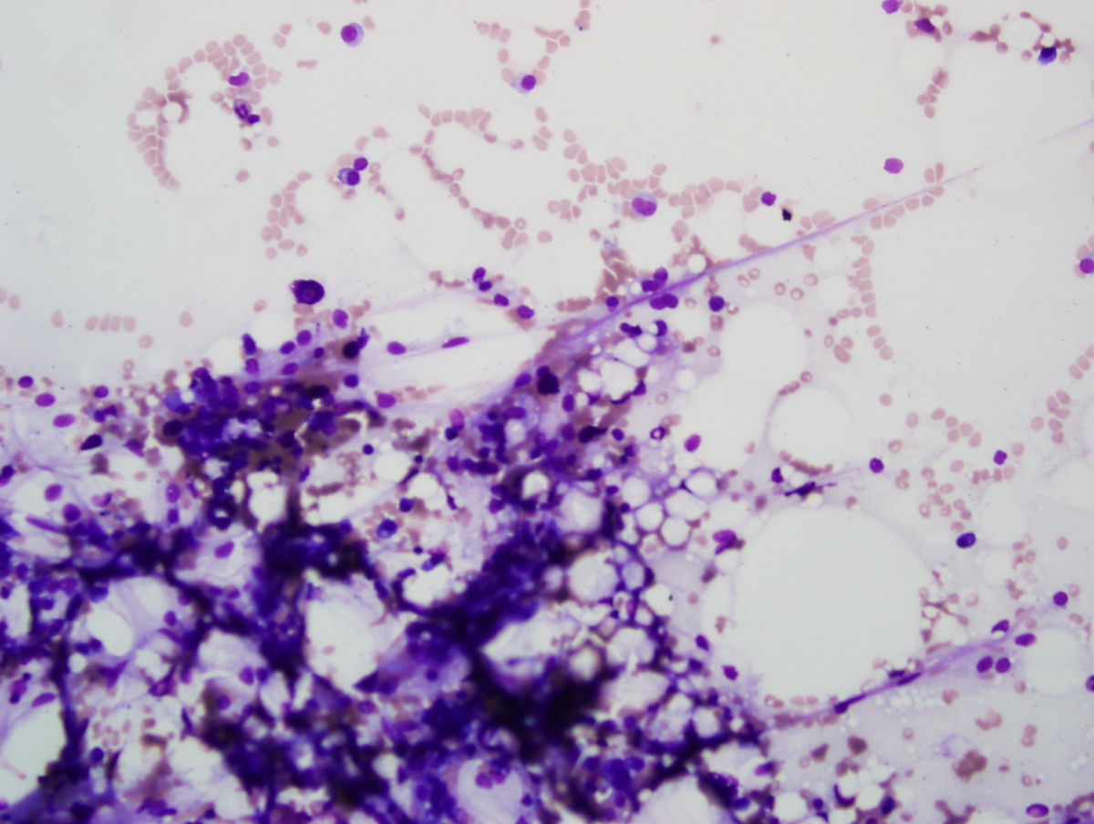 |
| Bone marrow Biopsy March 2006 | 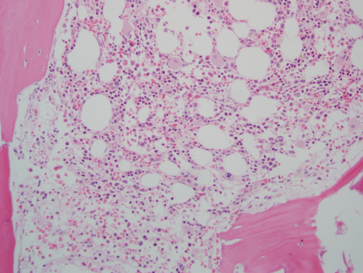 |
| bone marrow aspirate March 2006 |  |
| Bone marrow Biopsy July 2012 | 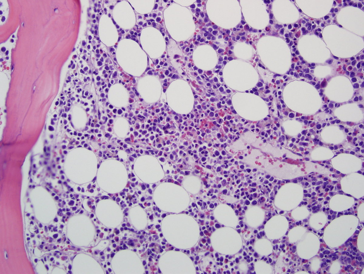 |
| Bone marrow Aspirate July 2012 | 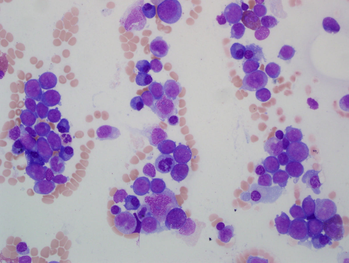 |