Institution: University of Maryland School of Medicine
Additional authors:Harold L. Evans, Nahid Nanaji
Session: T Lymphoblastic Leukemia/Lymphoma
HISTORY
The patient was a 56-year-old female who initially presented with abdominal epigastric pain in April, 2012, and was found to have a retroperitoneal mass measuring up to 7.5 by 10.5 cm. A laparoscopic biopsy was done in May of 2012. The pathology was consistent with Castleman disease, hyaline vascular variant. The patient was treated with rituximab and bendamustine. The mass however did not decrease in size. The patient underwent a laparoscopic retroperitoneal mass resection in November, 2012, and has been stable with no additional treatment on followup since that time.
DETAILS
The initial biopsy of the retroperitoneal mass showed typical morphology of hyaline vascular Castleman disease (please see images CD 1-3).
The second biopsy of the same mass was performed six month later after treatment with rituximab and bendamustine. The histologic sections show portions of lymph node with effaced architecture. The majority of the nodal tissue is replaced by vascular and fibrous proliferation with mixed small lymphocytes (this could be a post-treatment change). Focally there is a predominance of monotonous small to medium sized lymphocytes with relatively condensed chromatin. (please see images T-LBL 1-3). At the time of the second biopsy, no circulating blasts were identified and complete blood count indices were unremarkable.IMMUNOHISTOCHEMISTRY AND FLOW CYTOMETRY
Immunohistochmical stains show that the lymphocytes are positive for CD3, CD4, CD8, TdT, CD1a, CD99, and negative for CD20 and PAX-5; they have a high proliferative index (>90%) by Ki-67 staining. (please see immunostained images).
CYTOGENETIC FINDINGS
Not done.
MOLECULAR FINDINGS
PCR based TCR gene rearrangement studies failed to identify a clonal T cell population.
INTERESTING FEATURES
There is clear evidence of T-lymphoblastic proliferation with high proliferation index. However, there is no clonal TCR rearrangement. The patient is asymptomatic. There is no other lymphadenopathy or peripheral blood blasts. We feel this represents an indolent T-lymphoblastic proliferation.
Such cases have been described in oropharynx but is exceedingly rare elsewhere, in particular, in such a large size. The relationship between HV-CD and this lesion is unclear.It is unclear whether this represents a neoplasm or a hyperplasia. What is a best management?PROPOSED DIAGNOSIS
Indolent T-lymphoblastic proliferation.
CONSENSUS DIAGNOSIS
Castleman's disease, hyaline-vascular variant, with T-lymphoblastic proliferation of uncertain significance
| HV-CD 4X |  |
| HV-CD 10X |  |
| HV-CD 40X | 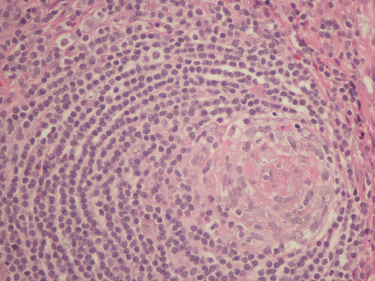 |
| T-LBL 4x |  |
| T-LBL 60x | 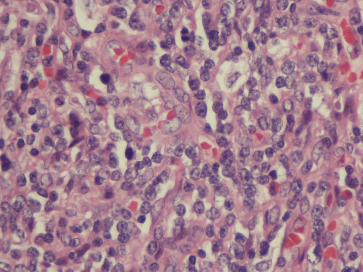 |
| T-LBL 60x lymphoid rich area |  |
| T-LBL CD3 |  |
| T-LBL CD1a |  |
| T-LBL TdT |  |
| T-LBL Ki-67 | 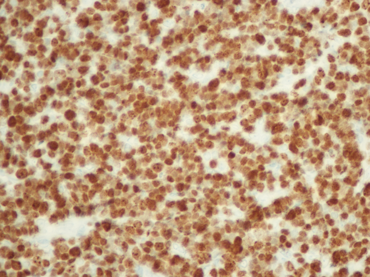 |