Institution: Henry Ford Hospital
Additional authors:Juan C. Gomez-Gelvez, Koichi Maeda, Kedar Inamdar
Session: B Lymphoblastic Leukemia/Lymphoma
HISTORY
A 31-year-old male patient presented with diffuse bone pain and productive cough of approximately 2 weeks of duration. Physical examination and initial laboratory work up revealed diffuse lymphadenopathy, leucocytosis (20.9 K/uL), mild anemia (11.3 g/dL) and thrombocytopenia (19 K/uL). Peripheral blood smear showed 60% blasts. The patient underwent bone marrow biopsy (see below) revealing a Mixed Phenotype (B/myeloid) Acute Leukemia with MLL gene rearrangement, trisomy 8 and 86% blasts. Cytologic and flow cytometric analysis of the cerebrospinal fluid demonstrated CNS involvement. The patient received treatment with Hyper-CVAD / HD MTX-araC chemotherapy as well as intrathecal cytarabine. Follow-up bone marrow biopsies done at 4 and 4.5 months after initial diagnosis showed persistent/relapsed disease with 10% and 75% blasts respectively. Due to these findings, the patient underwent salvage chemotherapy with clofarabine and subsequent allogeneic related peripheral stem cell transplant. However, follow-up bone marrow biopsy done 3 days after his transplant showed persistent/relapsed disease with 58% blasts. The patient is currently being managed with palliative care only.
DETAILS
Bone marrow from the right posterior superior iliac spine fixed in Bouin’s and decalcified with hydrogen chloride solution. Bone marrow smears show a predominant blasts cell population accounting for 86% in the differential counts. Dimorphism was apparent in the blast morphology in that there was a population of smaller blasts showing large nucleus with finely dispersed chromatin and high nuclear/cytoplasmic ratio, admixed with medium-to-large blasts showing frequently folded/clefted or grooved nuclei. Almost all blast cells exhibit presence of single prominent nucleoli and cytoplasmic vacuoles. No Auer rods are seen. Decalcified biopsy and clot section show an overall cellularity of 98-100% and sheets of immature appearing mononuclear cells with irregular folded or clefted nuclei, finely granular chromatin and occasional cells with prominent nucleoli. Markedly decreased trilineage hematopoiesis, rare scattered erythroid and megakaryocytic lineage cells are also seen. Prussian blue stain reveals decreased particulate iron, 56% sideroblasts and only rare ring sideroblasts. Sudan Black B is positive in 66% of blasts. Non-specific esterase is negative.
IMMUNOHISTOCHEMISTRY AND FLOW CYTOMETRY
Immunohistochemistry stains on the biopsy reveal that the blast cells are TDT positive, MPO weakly positive (subset), PAX5 positive, CD20 negative and CD3 negative.
Flow cytometry analysis of the blast cells shows that these are predominately B-lymphoblasts with a composite membrane/intracytoplasmic antigen profile of: CD2-, CD3-, CD5-, CD7-, CD10-, CD11b-, CD13-, CD14-, CD15+, CD16-, CD19+, CD20-, CD33-, CD34-, CD45+, CD56-, CD61-, CD117-, CD235a-, HLA-Dr+, cyMPO-, cyCD3-, cyCD79a+, CD23-, Kappa-, Lambda-.
CYTOGENETIC FINDINGS
There are two related abnormal clones identified in cytogenetic analysis. Approximately 95% of nuclei had 11q23 MLL gene rearrangement with t(4;11)(11q23), 65.5% of nuclei had trisomy 8.
Cytogenetic analyses of follow-up bone marrow biopsies demonstrated persistent/residual disease with similar findings.
MOLECULAR FINDINGS
Not performed
INTERESTING FEATURES
This case represents a B lymphoblastic lymphoma/leukemia with t(4;11)(11q23) MLL rearrangement with certain interesting features.
1. Morphologically 2 distinctive populations of blast cells were seen as shown in the pictures. Flow cytometric evaluation however identified single blast cell population with a phenotype consistent with B-lymphoblasts in the earliest stage of differentiation, so called early precursor B-ALL or pro-B-ALL (CD19+, CD79a+, CD10 Neg., and nuclear TdT+).
2. Another interesting feature in this case was that 66% of blasts were Sudan Black B positive. By immunohistochemistry, myeloperoxidase was weakly positive but only expressed by a subset of the blasts. Due to this finding and the possibility of a mixed phenotype (B/myeloid) acute leukemia was raised. Sudan Black B however, can be misleading in the assessment of acute leukemias since up to 2.7% of B-ALL can be positive, and according to the current scheme of the 2008 WHO classification of acute leukemias with ambiguous lineage Sudan B Black is not considered a surrogate for MPO [Ngan et al Mod Pathol. 1992 Jan;5(1):68-70)].
3. In addition to the MLL gene rearrangement and cytogenetic analysis also reported presence of trisomy 8. The significance of this finding is not well described in literature. These correspond to highly uncommon cytogenetic findings in B lymphoblastic lymphoma/leukemia, especially when they present within the same case.
PROPOSED DIAGNOSIS
B lymphoblastic lymphoma/leukemia with t(4;11)(11q23) MLL rearrangement and trisomy 8.
CONSENSUS GROUP: ADDITIONAL INFORMATION/STUDIES
Additional immunostains performed by conference consensus group:
MPO: Strongly positive in blasts
CONSENSUS DIAGNOSIS
B-acute lymphoblastic leukemia with t(4;11)(11q23), MLL rearrangement
| Bone marrow biopsy 40X | 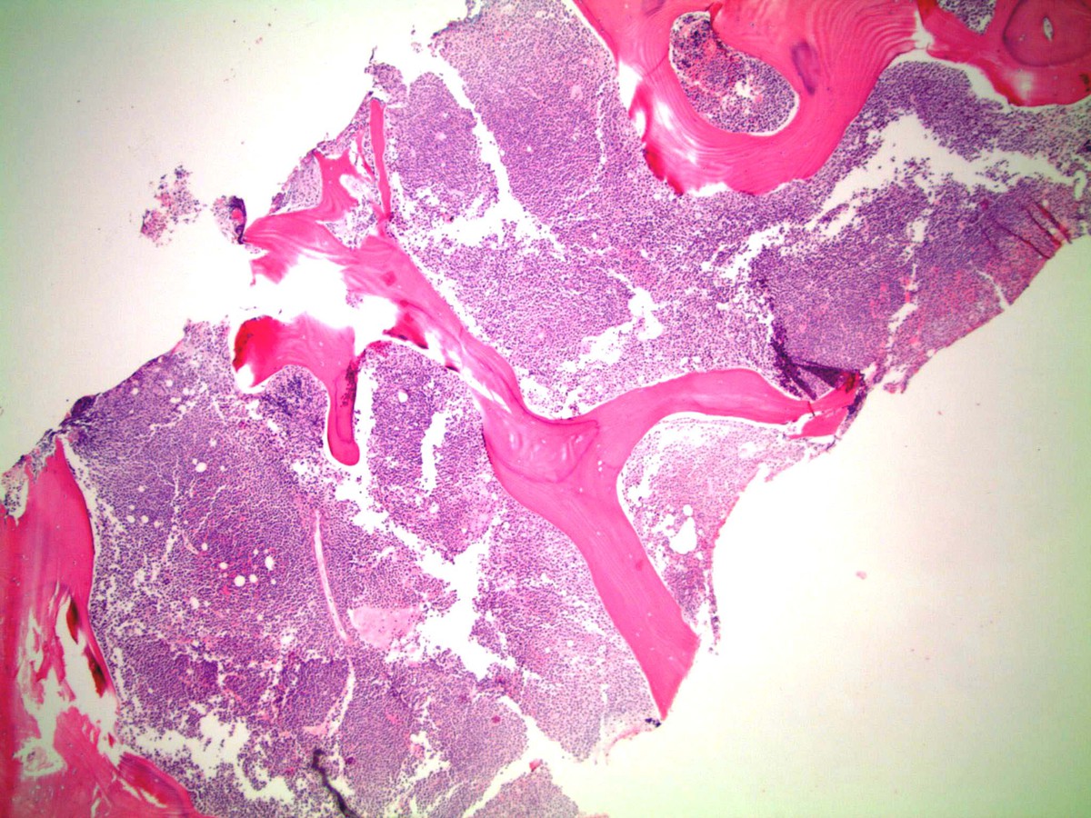 |
| Bone marrow biopsy 200X | 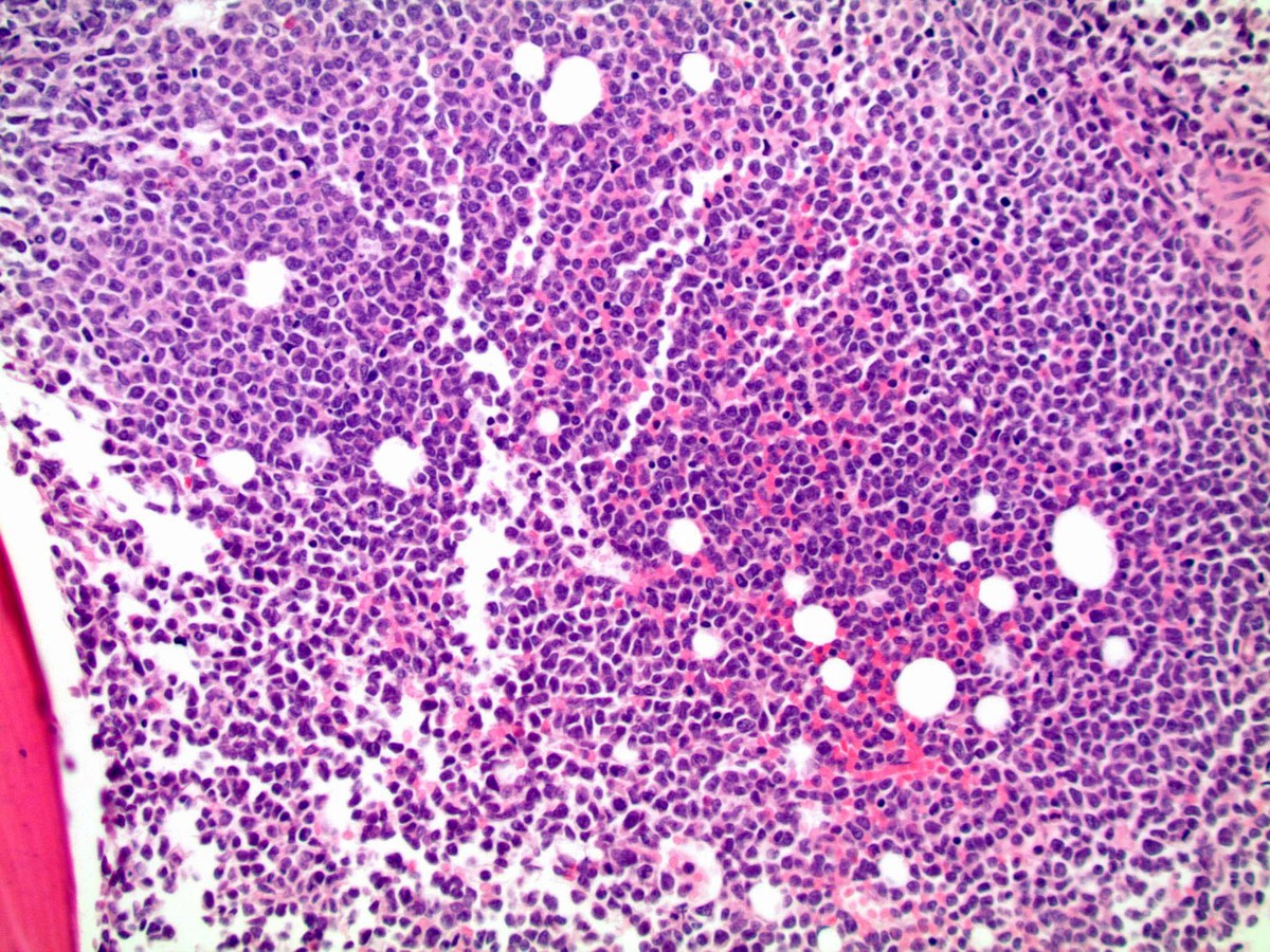 |
| Bone marrow biopsy 400X |  |
| MPO 200X |  |
| PAX5 200X |  |
| TdT 200X |  |
| SBB 1000X | 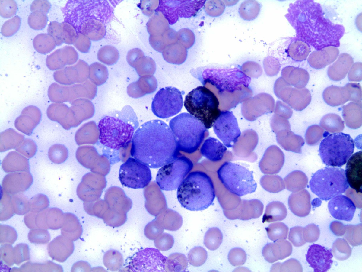 |
| Smear 500X |  |
| Smear A 1000X | 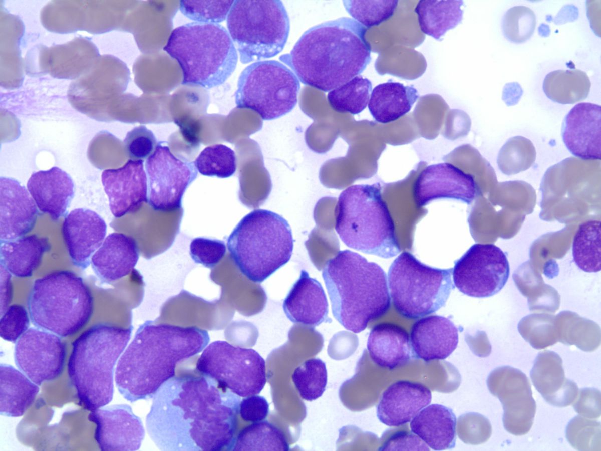 |
| Smear B 1000X |  |