Institution: Henry Ford Hospital
Additional authors:Juan C. Gomez-Gelvez, M.D, Kristin Hunt-Karner, M.D, Kedar Inamdar, M.D, Ph.D
Session: Therapy-related myeloid neoplasms
HISTORY
A 59-year-old female patient with past medical history of breast cancer, diagnosed and treated 9 years ago with Adriamycin and Cytoxan, presents with a chief complaint of fatigue for about 2-3 weeks. At evaluation, patient was found to be pancytopenic (Hb: 7.1 g/dL, WBC count: 1.2 K/uL, absolute neutrophils: 0.38 K/uL, and platelets: 42K/uL). The peripheral blood smear revealed 5% circulating blasts. Physical examination did not reveal organomegaly. The patient underwent bone marrow biopsy (see below). She was treated with cyclophosphamide and ifosfamide chemotherapy, and subsequently underwent allogeneic peripheral stem cell transplant. Follow up bone marrow biopsies at day 30th and 100th were consistent with remission, morphologically and cytogenetically, showing only slight fibrosis and normal blast counts. Platelet counts have been persistently low (between 10 and 112 K/uL, with last count of 69 K/uL). The patient is currently free of disease.
DETAILS
Bone marrow biopsy from the right posterior superior iliac spine fixed in Bouin’s and decalcified with hydrogen chloride solution. The bone marrow biopsy is 80-90% cellular and shows a marked megakaryocytic proliferation with morphologic atypia including hyperchromatic, pleomorphic nuclei and clustering. There is marked bone marrow fibrosis and no significant alterations of trabecular bone. There are also admixed lymphoplasmacytic infiltrates with focal aggregates of lymphoid cells. The myeloid and erythroid lineage cells are decreased showing left shifted myeloid maturation. The bone marrow smears show increased blasts (9.5% in differential count) with small-to-medium size, high N/C ratio, finely distributed chromatin and small nucleoli. Auer rods are not seen. The rest of the lineages are rather underrepresented due to hemodilution thus not allowing assessment for dysplasia.
IMMUNOHISTOCHEMISTRY AND FLOW CYTOMETRY
IHC: Immunohistochemistry stains reveal increased CD34+ cells without significant clustering. Reticulin stain reveals diffuse and dense increase in reticulin fibrosis with frequent intersecting fibers. Masson trichrome stain shows focal bundles of collagen thus consistent with grade MF-2 marrow fibrosis per WHO guidelines for semiquantitative grading of bone marrow fibrosis. Flow cytometric analysis demonstrated a blast population with myeloid immunophenotype as follows: Positive for CD34, CD117, CD13, CD33 and HLA-DR; Negative for CD2, CD3, CD5, CD7, CD10, CD11b, CD14, CD15, CD16, CD20, CD56, and CD235a. CD61 is indeterminate.
CYTOGENETIC FINDINGS
Cytogenetic analysis by fluorescent in situ hybridization (FISH) showed deletion 20q (31% of nuclei), deletion of 5q (29% of nuclei) and monosomy 7 (26% of nuclei). There were no anomalies detected on chromosomes 8 and 13.
MOLECULAR FINDINGS
There was no JAK2 mutation detected.
INTERESTING FEATURES
This case has interesting features in terms of morphologic findings of myeloproliferative neoplasm (MPN) along with strong genetic support for consideration of myelodysplastic syndrome (MDS) in the differential diagnosis. This case can be viewed as progression of MPN to MDS given the development of cytogenetic abnormalities that are classic for MDS. The patient's history of breast cancer and treatment with cytotoxic agents increased the risk of development of MDS in this possible MPN. The fact that the diagnosis of MPN was never established before makes the assessment difficult at this time with regards to the original classification of his myeloproliferative neoplasm. Blood counts prior to the patient's diagnosis and treatment of breast cancer were within normal limits and thus we cannot determine the nature of this myeloproliferative neoplasm The incidence of MDS or AML developing in PV, ET or primary myelofibrosis is rather low but previous cytotoxic therapy is a common risk factor for development of MDS in each of these entities.
PROPOSED DIAGNOSIS
Therapy-related MDS with fibrosis in the setting of MPN features
CONSENSUS DIAGNOSIS
Therapy-related myeloid neoplasm, myelodysplastic syndrome with myelofibrosis
| Bone marrow biopsy 40X | 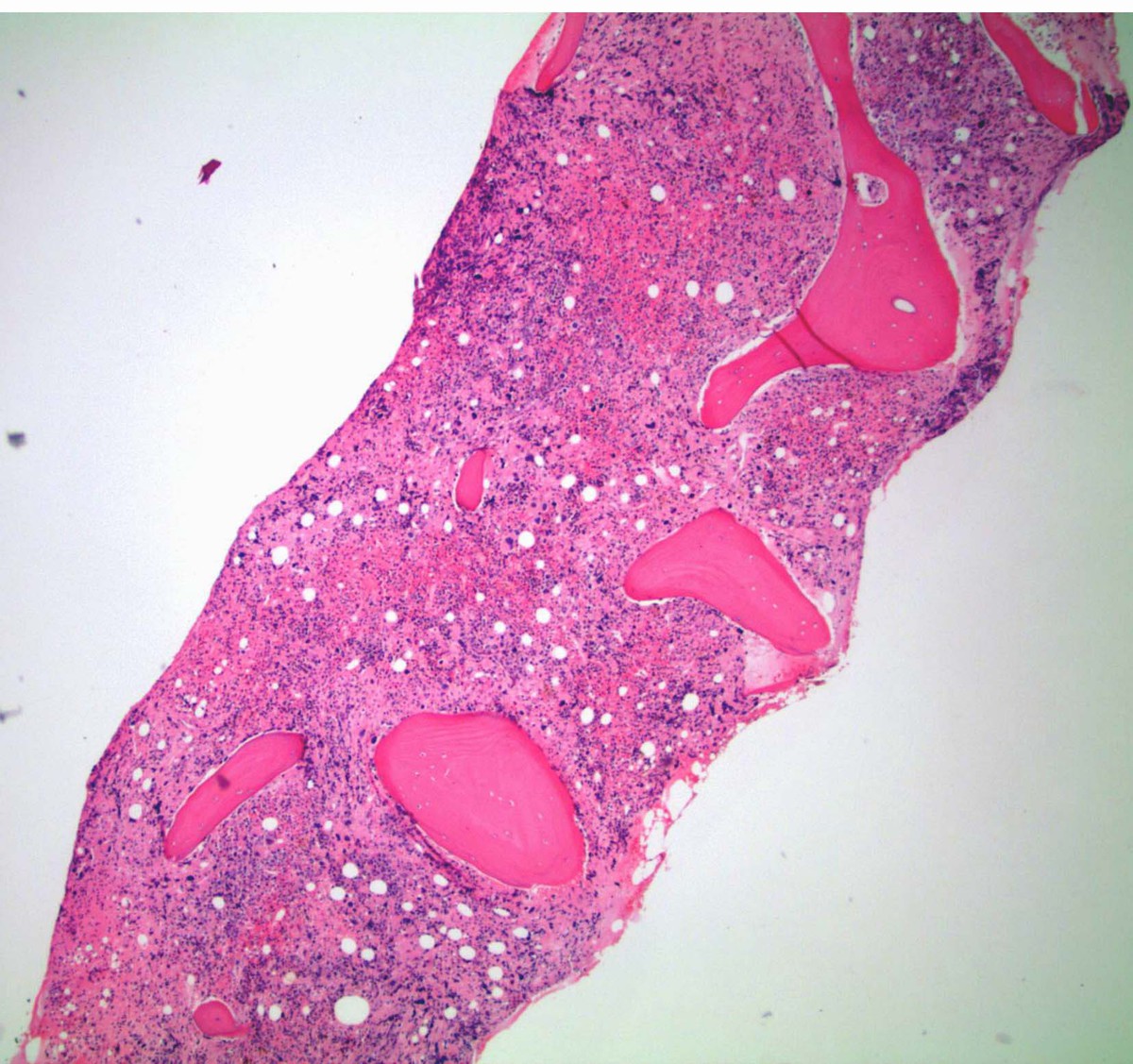 |
| Bone marrow biopsy 200X | 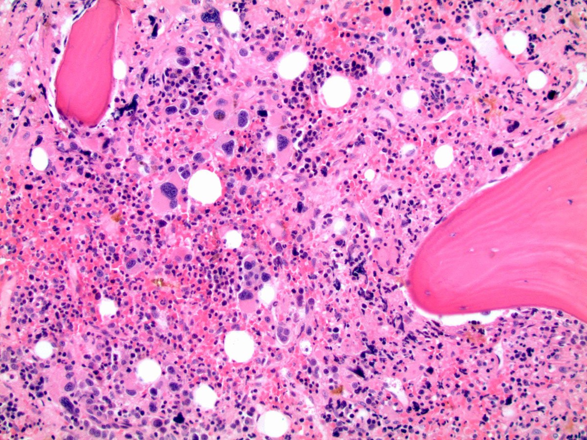 |
| CD34 200X | 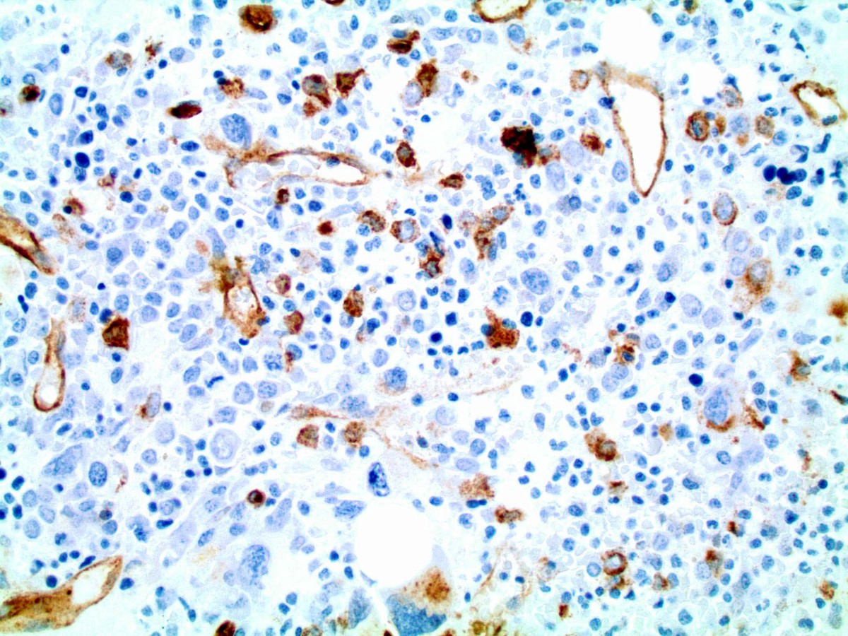 |
| Masson trichrome 200X | 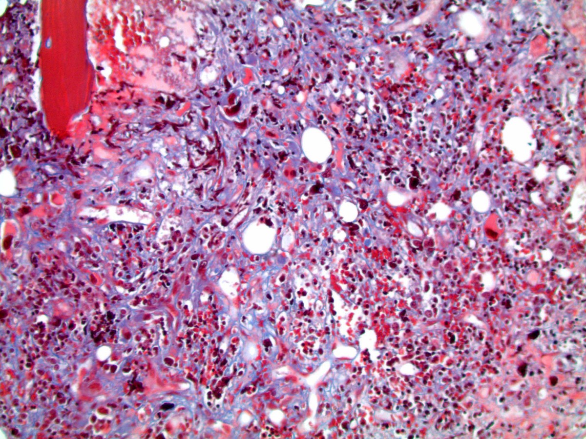 |
| Reticulin 100X | 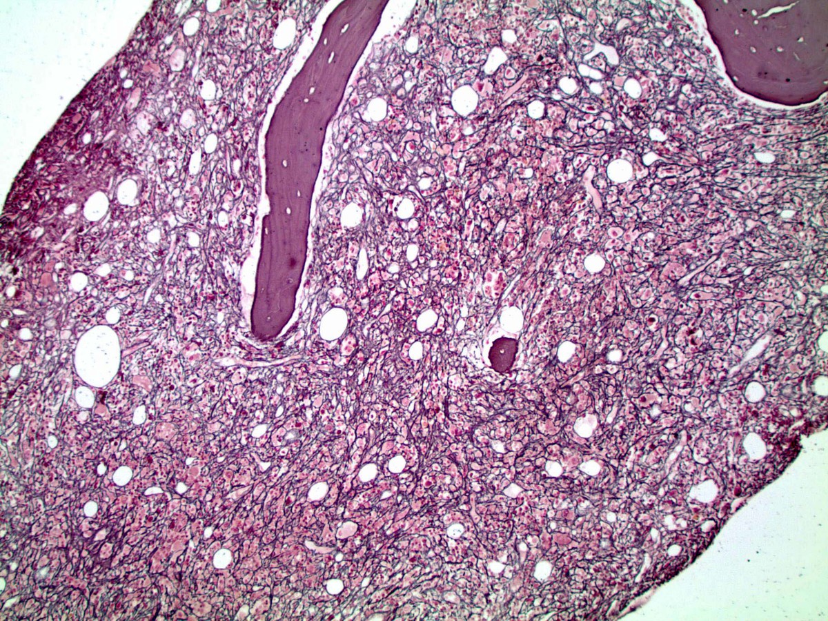 |
| Flow cytometry dot plots | 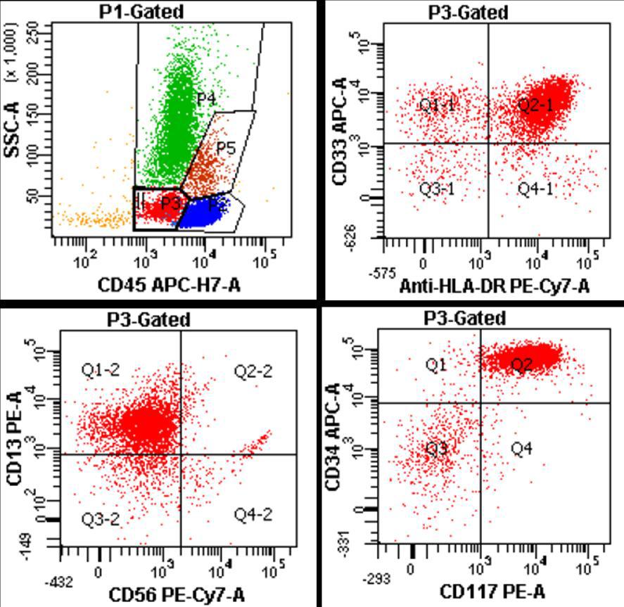 |