Institution: UCLA
Additional authors:Sophie X Song
Session: AML with recurrent genetic abnormalities Part I
HISTORY
A 27 year old Hispanic man with no significant past medical history presented with a 2 month history of fatigue, weight loss, and subjective fevers. Examination revealed hepatosplenomegaly, coagulopathy, pancytopenia, and elevated LFTs. A bone marrow biopsy was performed (see below). Subsequently the patient developed hypoxic respiratory failure necessitating intubation. Relevant labs: EBV PCR: 16opies Na 129 K 3.8 Cl 93 CO2 30 BUN 14 Cr 0.8 Glc 198 WBC 1.7 (N 64.3% L 28.1% M 6.5% E 1.0%) Hgb 9.4 Platelets 19 AST 148 ALT 229 Alk Phos 1,270 Direct Bili 7.1 Total Bili 8.3 Clinical followup: Following the diagnosis, the patient was started on treatment with L-asparaginase and Decadron as well as Acyclovir. However he continued to have an acute and rapidly progressive hepatic, renal and neurological decline suspicious for hemophagocytic syndrome, precluding full treatment with the SMILE regimen. The patient was transitioned to DNR/DNI status and subsequently expired.
DETAILS
A bone marrow biopsy and aspiration were performed. Wright Giemsa stains were applied to the aspirate smears; the core biopsy was submitted for B5 fixation and light decalcification; clot sections were submitted in formalin fixative. The peripheral blood demonstrated severe pancytopenia with numerous large atypical lymphoid cells with irregular to folded nuclei, mildly dispersed chromatin, small yet prominent nucleoli, and abundant basophilic cytoplasm containing occasional azurophilic granules. Bone marrow smears were aparticulate while touch preparations showed scattered large atypical lymphoid cells with irregular nuclei and granular cytoplasm. Core biopsy sections revealed an extensively necrotic marrow. Viable regions showed approximately 80% cellularity and extensive involvement (65% of marrow elements) by an atypical lymphoid infiltrate present in interstitial clusters and small aggregates as well as within sinusoids. The aforementioned cells were large in size and showed irregular nuclei, prominent nucleoli, and moderately amount granular cytoplasm. Histiocytes were increased and some demonstrated hemophagocytosis.
IMMUNOHISTOCHEMISTRY AND FLOW CYTOMETRY
Immunohistochemistry revealed that the large atypical lymphoid cells expressed CD2, CD3, CD7, TIA-1, Granzyme B, and EBV-EBER (in situ hybridization). There was no immunohistochemical expression of CD56 or CD57. Staining for CD68 highlighted background hemophagocytosis. Flow cytometric studies performed both on marrow aspirates as well as peripheral blood revealed a population of large and abnormal NK-cells which were positive for CD2 (bright), CD7 (dim), CD16 (bright), CD45, CD56 (partial), and HLA-DR. They were negative for CD3 (surface and intracellular staining), CD4, CD5, CD8, CD1a, TCR alpha/beta and gamma/delta, and CD57.
CYTOGENETIC FINDINGS
Chromosomal analysis revealed an abnormal composite male karyotype with additional material of unknown origin on chromosomes 3p, 18q, and 21p, and a deletion of 18q seen in 12/20 cells analyzed: 46,XY,add(3)(p25),add(18)(q21),del(18)(q11.2),add(21)(p12)[cp12]/46,XY[8] FISH analysis for TCRA/D and MLL was negative for MLL and TCRA/D rearrangements but demonstrated abnormal signal patterns consistent with +11q and +14q.
MOLECULAR FINDINGS
B- and T-cell clonality studies were indeterminate due to low and erratic PCR signals on multiple attempts. The quantity of recoverable material was insufficient for Southern blot analysis.
INTERESTING FEATURES
Arguably the single most rapidly lethal malignancy currently recognized in medicine with a recently described median survival time of 22 days, aggressive NK cell leukemia is a rare disorder for which immediate and accurate diagnosis is of paramount importance. This case highlights several salient findings including the atypical flow cytometric phenotype allowing distinction from a reactive NK cell process, classic peripheral blood large granular lymphocytic morphology, and typical fulminant clinical course with associated hemophagocytic syndrome.
PROPOSED DIAGNOSIS
Aggressive NK cell leukemia with associated hemophagocytosis
CONSENSUS DIAGNOSIS
Aggressive NK cell leukemia with associated hemophagocytosis
| Peripheral blood with circulating atypical large granular lymphoid cells (Wright Giemsa, 1000x) | 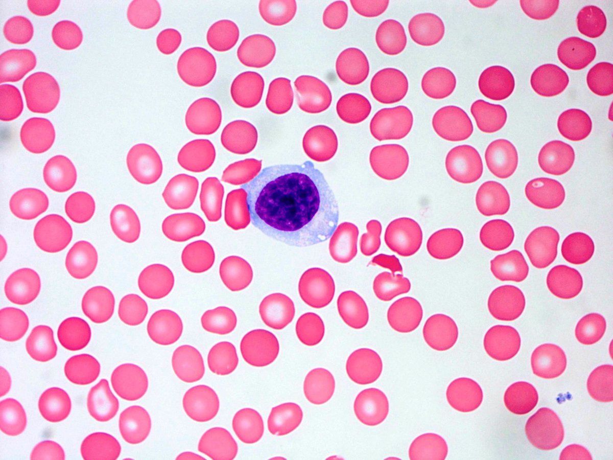 |
| Touch preparation showing atypical large granular lymphoid cell (Wright Giemsa, 1000x) | 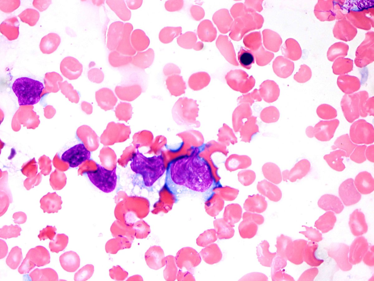 |
| Core biopsy section showing extensive necrosis (H&E, 100x) | 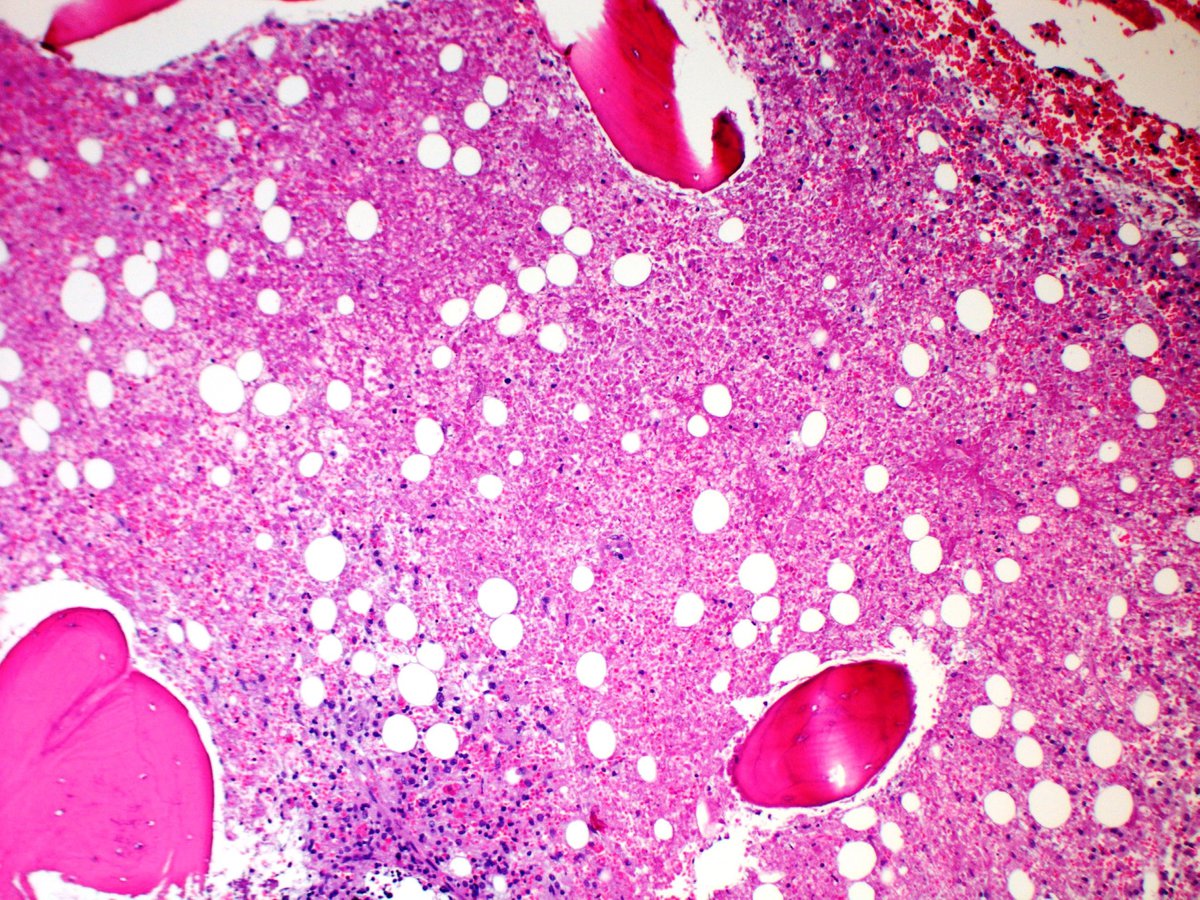 |
| Core biopsy section adjacent to necrotic area showing small clusters of atypical large lymphoid cells (H&E, 200x) | 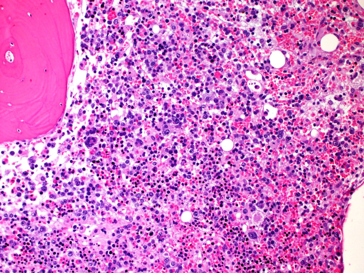 |
| Atypical lymphoid cells staining for CD2 (Immunoperoxidase, 400x) |  |
| Atypical lymphoid cells staining for CD3 (Immunoperoxidase, 200x) | 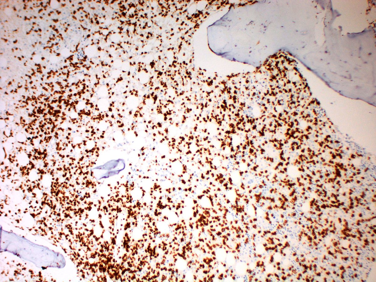 |
| Atypical lymphoid cells staining for CD3 (Immunoperoxidase, 1000x) |  |
| Atypical lymphoid cells staining for Granzyme B (Immunoperoxidase, 400x) | 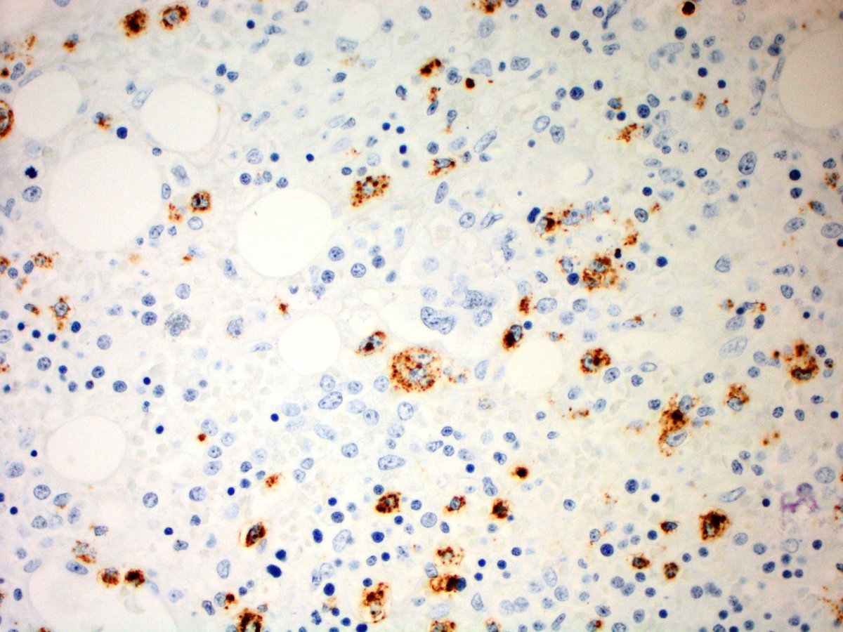 |
| Atypical lymphoid cells staining for TIA-1 (Immunoperoxidase, 200x) | 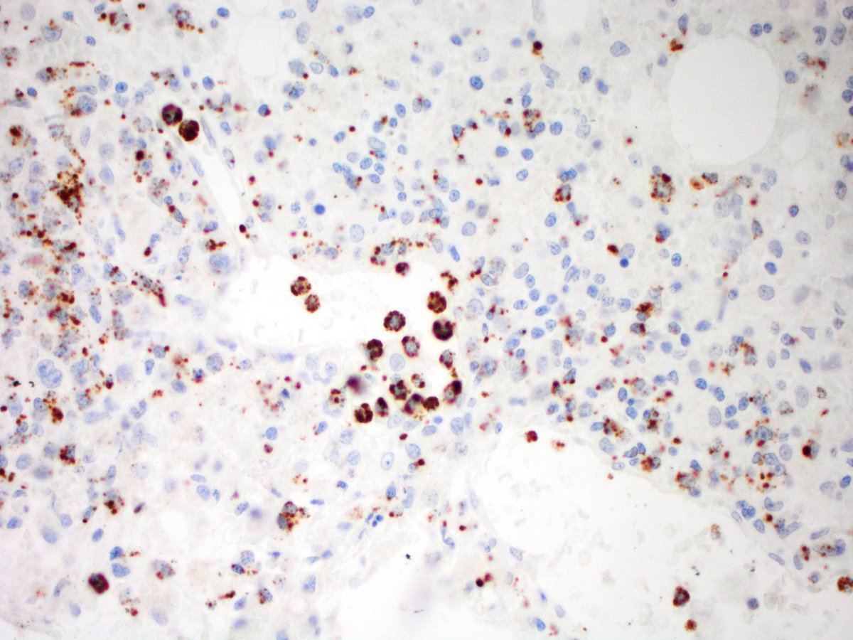 |
| Atypical lymphoid cells staining for EBV-EBER (In Situ Hybridization, 200x) |  |
| Atypical lymphoid cells are negative for CD56 (Immunoperoxidase, 200x) | 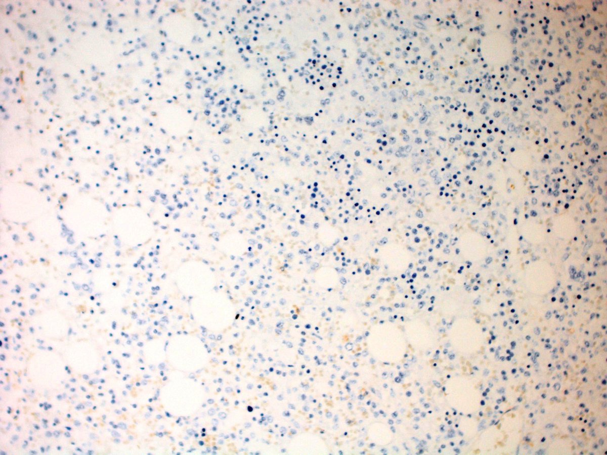 |
| Atypical lymphoid cells are negative for CD57 (Immunoperoxidase, 200x) |  |
| Macrophages demonstrating increased hemophagocytosis, highlighted on CD68 staining (Immunoperoxidase, 400x) | 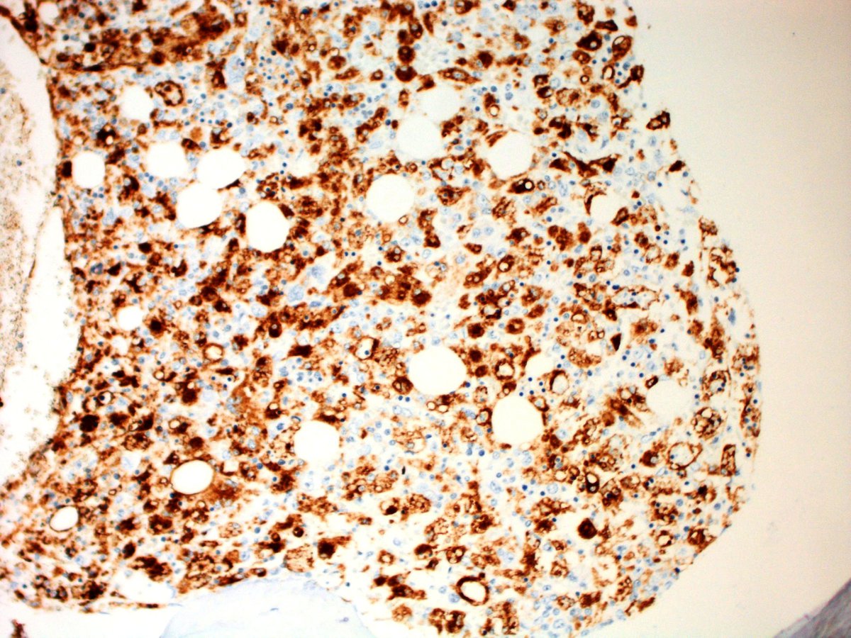 |
| Flow cytometry showing a population of atypical lymphoid cells with increased forward scatter and side scatter characteristics (green) | 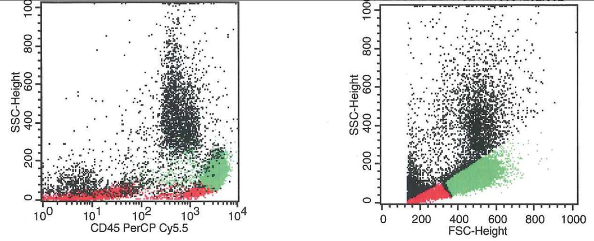 |
| Atypical lymphoid cells are positive for CD56 and negative for surface CD3 | 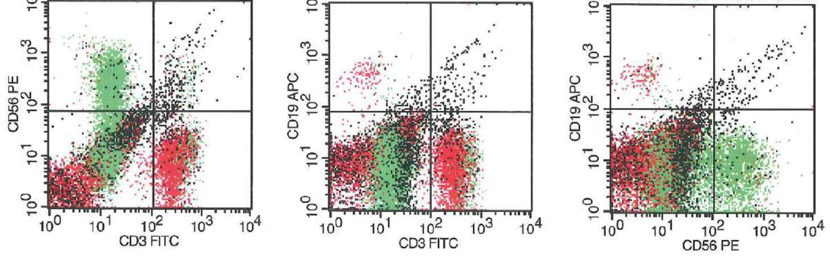 |
| Atypical lymphoid cells show expression of CD2 (bright) and CD7 (dim) but no expression of CD5 | 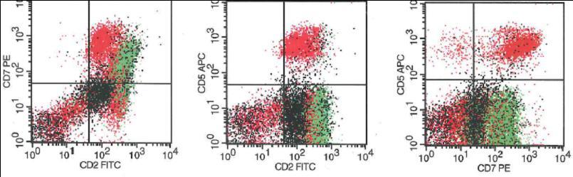 |
| Atypical lymphoid cells lack CD4, CD8, and CD25 |  |
| Atypical lymphoid cells lack both TCR alpha/beta and gamma/delta as well as CD1a | 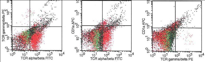 |
| Atypical lymphoid cells show bright expression of CD16 and CD2 but lack expression of CD57 |  |
| Core biopsy section showing large atypical lymphoid cells with irregular nuclear borders, prominent nucleoli, and vesicular chromatin; background small lymphoid cells and erythroid precursors for size comparison (H&E 1000x) |  |
| Chromosomal studies showing add(3)(p25), del(18)(q11.2), add(18)(q21), and add(21)(p12) |  |
| Dual color breakapart probes for 14q11 showing polysomy (+14q) | 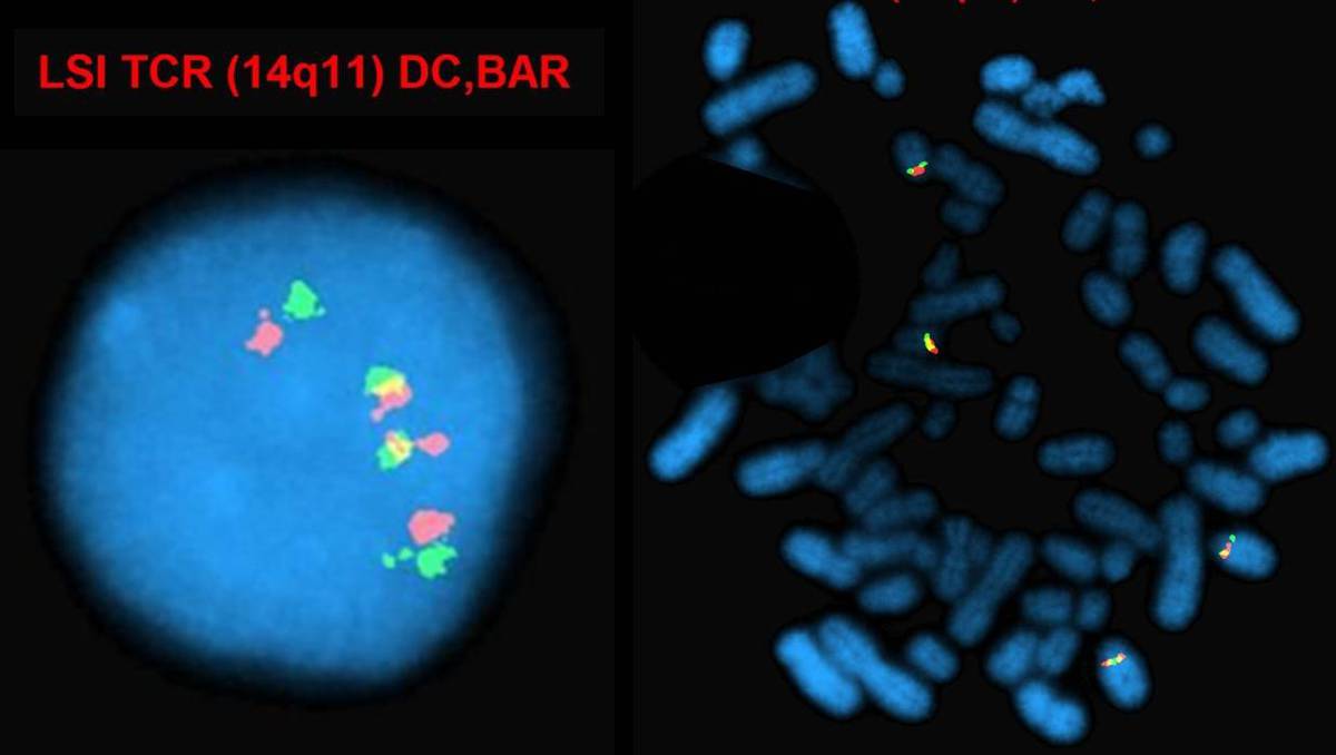 |
| Dual color breakapart probes for 11q23 showing polysomy (+11q) | 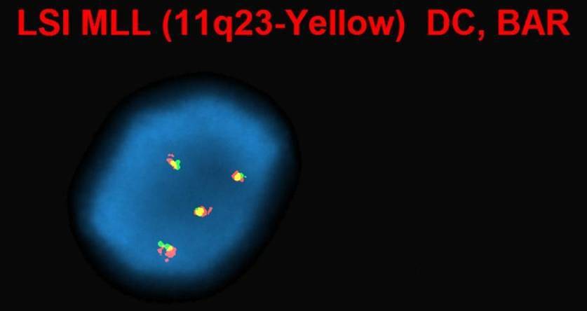 |