Institution: University of California Davis Medical Center
Additional authors:Mingyi Chen MD PhD and Joo Song MD
Session: Extramedullary manifestations of myeloid neoplasms
HISTORY
The patient is a 72 year-old male who presented with splenomegaly, abdominal pain and weight loss. His peripheral smear showed a pancytopenia. The bone marrow biopsy showed a hypercellular marrow (90% cellularity) with granulocytic and megakaryocytic hyperplasia and dysplasia with diffuse reticulin and collagen fibrosis (MF-3) and mild osteosclerosis. The dysplastic megakaryocytes were clustered and had hyperchromatic lobulated "cloud like" nuclei with an abnormal nuclear to cytoplasmic ratio. Immunohistochemistry (CD34 and CD117) showed no increase in immature myeloid precursors of blasts (1-2%). Cytogenetics showed a 47, XY, +8[20] karyoytype and no JAK2 V617F or MPL W515K/L mutations were detected using molecular studies. An ultrasound of the abdomen showed moderate hepatomegaly and splenomegaly. A subsequent splenectomy yielded an enlarged 3,150 gram spleen (28.0 x 22.0 x 9.5 cm) which microscopically showed diffuse extramedullary hematopoiesis (EMH) containing dysplastic megakaryocytes in a background of reticulin fibrosis. The patient was diagnosed with splenic EMH with an underlying myeloproliferative neoplasm (MPN) favoring primary myelofibrosis.
Three months later, the patient presented with severe shortness of breath for a week. A chest radiograph showed a marked left pleural effusion and a subsequent video assisted thoracic surgical (VATS) decortication was performed showing a fibrinous pleural thickening, pleural effusion and serosanguinous fluid.DETAILS
The specimen consisting of the fibrinous pleural thickening and pleural effusion was formalin fixed and paraffin embedded. Immunohistochemical stains were perfomed on the FFPE tissue using an automated immunostainer. Microscopically, the hematoxylin and eosin stained slides showed a myeloid predominant extramedullary hematopoiesis comprising the majority of the specimen (Figure 1).
IMMUNOHISTOCHEMISTRY AND FLOW CYTOMETRY
The myeloid predominant EMH was markedly positive for myeloperoxidase (MPO) and an increase in CD34+ myeloid cells were present (Figure 2, 3). Dysplastic megakaryocytes were highlighted with the von Willibrand factor stain. Calretinin was used to highlight the mesothelial cells present in the specimen. Flow cytometry was not performed on the specimen.
CYTOGENETIC FINDINGS
Interphase fluorescence in-situ hybridization (FISH) was perfomed on paraffin blocks and perfomed at Clarient Diagnostic Services (Aliso Viejo, CA) using the CEP8 probe to assess the pleural biopsy for the present of trisomy 8. Analysis of 100 interphase cells with each probe set showed an abnormal hybridization pattern, 42% of cells showed three copies of the CEP8 probe. The final result was nuc ish8p10(CEP8x3)[42/100], confirming the diagnosis of trisomy 8 in the immature myeloid cells (Figure 4).
INTERESTING FEATURES
Extramedullary hematopoiesis is defined by the proliferation of marrow elements in extramedullary sites. In adults, EMH is most common in the spleen, liver and lymph nodes, and has been reported in several other various sites. EMH can occur in the setting of a reactive hematological disorder due to an underlying hematological disease such as a hemoglobinopathy or can occur as a manifestation of a neoplastic myeloproliferative disorder. The EMH cells that are associated with a myeloproliferative disorder are morphologically, immunohistochemically and molecularly distinct from normal hematopoietic cells.
The most striking feature in this case is the involvement of EMH in the mediastinum and pleura. The presence of EMH in this location is a rare phenomenon with only a few published case reports. The malignancy of the EMH cells were confirmed by showing clonality with trisomy 8 by FISH and further supported by the patient's clinical presentation with his recent diagnosis of splenic EMH with an underlying MPN. Therefore in this case, the pleural EMH was a manifestation of a myeloproliferative process. The second most interesting feature of the case was the detection of the concurrent cytogenetic abnormality of trisomy 8 in both the previous bone marrow biopsy and of the pleural EMH, thus reinforcing the idea that EMH is "neoplastic" in nature. Lastly, the pleural biopsy showed increased CD34+/CD117+ immature myeloid cells thus possibly indicating an early phase of transformation to acute leukemia. In the pleura, leukemic transformation has been reported to occur in 10-20% of patients with myeloid predominant EMH, according to literature. Thus, in patients with underlying hematological conditions presenting with pleural effusion, EMH should be considered in the differential diagnosis and the presence of immature myeloid cells in the pleural lesion should be examined, as it may be indicative of a focus of a developing transformation of acute leukemic cells.PROPOSED DIAGNOSIS
Pleural extramedullary hematopoiesis associated with a myeloproliferative neoplasm
CONSENSUS DIAGNOSIS
Extramedullary manifestation of myeloproliferative neoplasm (pleural neoplastic extramedullary hematopoiesis)
| H&E of the pleural biopsy showing EMH | 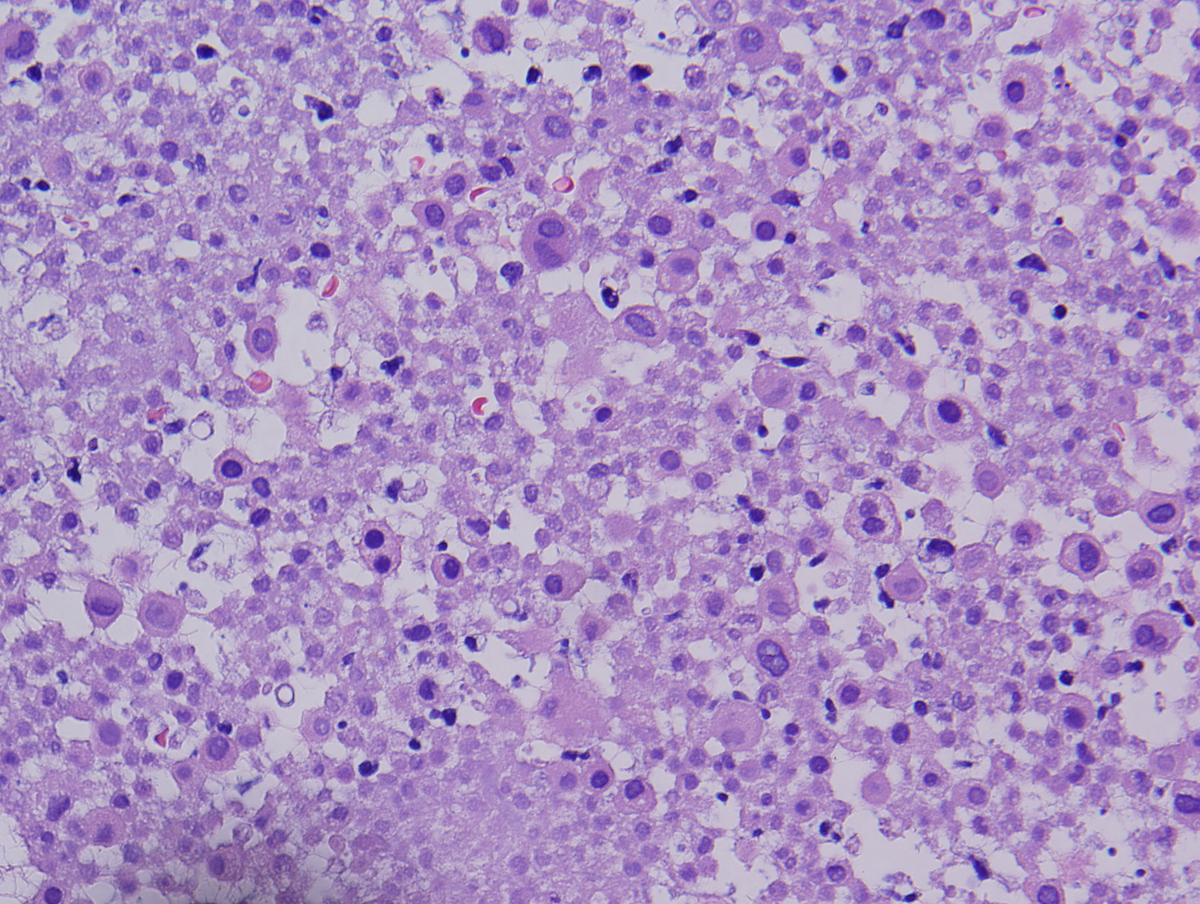 |
| EMH with MPO+ myeloid cells | 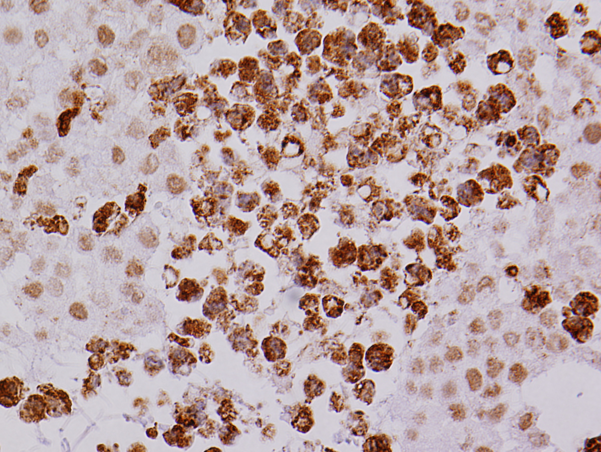 |
| EMH with CD34+ immature myeloid cells |  |
| FISH image showing trisomy 8 in immature myeloid cells in pleural EMH | 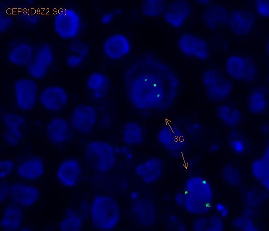 |