Institution: Clarient, Inc., Aliso Viejo, California, USA
Additional authors:Jianming Yin,Renuka Agrawal, Kate Grimm, Michele Kempik, Dennis O’ Malley
Session: AML with myelodysplasia-related changes
HISTORY
85 year old man, presents with pancytopenia and mild kidney dysfunction.
CBC: Hb: 8.9 g/dL, Hct: 26.9, MCV: 98 fL, WBC: 2.7 x 10³/μl [N:32%, L:61%, M:7%], PLT:112x 103/μlDETAILS
Bone marrow, aspirates and core (trephine) biopsy
Site: left posterior iliac crestMethod of fixation for trephine biopsy: Decalcification following by formalin fixationGross/microscopic pathology: Hypercellular marrow (75% cellularity); M:E Ratio: 1:1 Myeloid series: complete maturation, few pseudo Pelger-Huet forms, no increase in blasts Erythroid series: normoblastic and megaloblastoid maturation, binucleation, nuclear budding, cytoplasmic/ nuclear dyssynchrony Megakaryocytes: number increased, hypolobated forms and micromegakaryocytesNo increase in lymphocytes and plasma cellsIMMUNOHISTOCHEMISTRY AND FLOW CYTOMETRY
IHC not performed
Special stains::Reticulin stain: demonstrated no fibrosisIron stain: demonstrated normal storage iron, no ringed sideroblastsFlow cytometry: Slightly dyspoietic myeloid maturation, no increase in blastsCYTOGENETIC FINDINGS
Karyotype Analysis
46,XY,ider(20)(q10)del(20)(q11.2q13.3)[19]/46,XY[1] Abnormal karyotype characterized by an isoderivative chromosome composed of a mirror image duplication of the deleted long arm of chromosome 20 (20q) in 19 of 20 cells analyzed. One normal 46, XY male cell was also observed.CGH Arrayarr 20p13->p11.21::q11.22->q13.32(9993->25610059::31582338->57426889)x1; 20q11.21::q13.32->q13.33(29540343->30590729::57561657->62141109)x3Array CGH analysis was performed on this patient’s DNA sample referenced to a normal female control DNA sample. This test is comprised of 3519 BAC clones corresponding to genomic loci encompassing the 22 autosomes and the sex chromosomes including loci targeted to genomic regions known to be clinically significant in hematological malignancies. This analysis showed a single copy number loss of 83 loci on chromosome 20, including the entire short (p) arm and a large region on the long (q) arm from q11.2 to q13.3 and a gain of 13 loci on chromosome 20, including the q11.2 and q13.3 regions. An array profile consistent with a male sex chromosome complement was identified.MOLECULAR FINDINGS
FISH analysis demonstrated deletion 20q
INTERESTING FEATURES
This case of myelodysplastic syndrome (MDS) is associated with a rare cytogenetic abnormality, i.e. ider(20q) which results in loss of the short arm and duplication of the deleted long arm of chromosome 20. This event leads to loss and gain of genetic material.
This cytogenetic finding has been reported in a limited number of hematological malignancies including myelodysplastic syndrome (MDS), acute myeloid leukemia (AML), chronic myelomonocytic leukemia (CMML), acute lymphoblastic leukemia, and in two cases with minimal bone marrow morphological changes. Clinical significance of ider(20q) in MDS is yet unclear. We have five additional cases of ider(20q) in our files. These cases are associated with both myeloid proliferative neoplasm (MPN) and acute myeloid leukemia (AML).PROPOSED DIAGNOSIS
Refractory Cytopenia with Multilineage Dysplasia (RCMD)
CONSENSUS DIAGNOSIS
Refractory cytopenia with multilineage dysplasia
| Array CGH profile of isoderivative of deleted chromosome 20. | 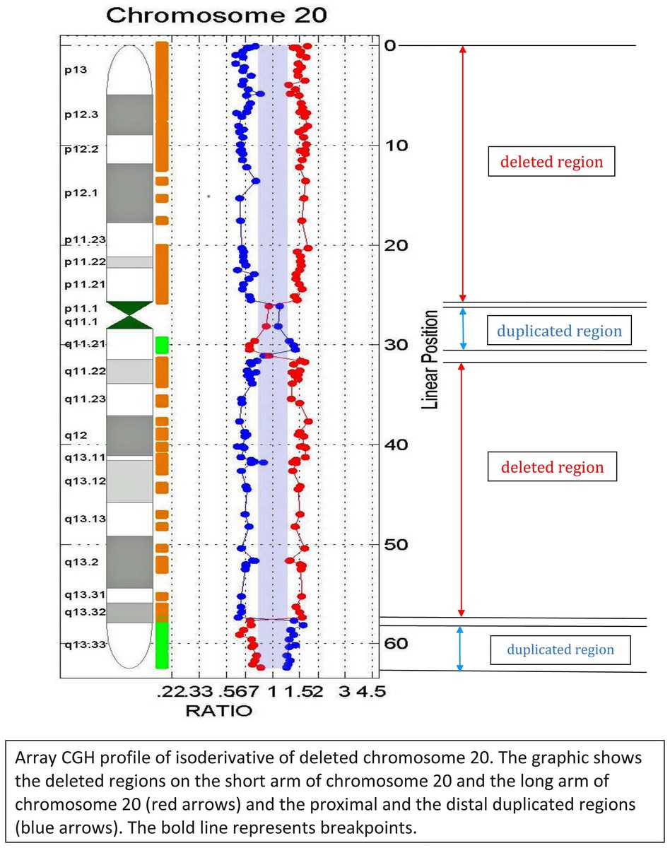 |
| Giemsa-banded karyogram showing ider(20) | 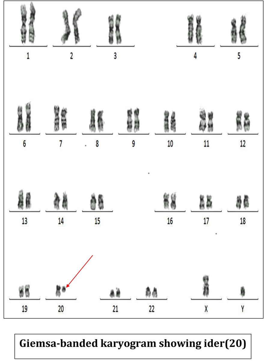 |