Institution: University of Minnesota
Additional authors:Robert McKenna
Session: AML secondary to myeloproliferative neoplasms and other types of disease progression in MPN
HISTORY
The patient is a 23 year old African American male who presented with approximately one month of fatigue, dyspnea on exertion, dizziness, headaches, fevers, and night sweats and was found to be profoundly anemic (hemoglobin 4.1 g/dL) and have leukocytosis (WBC 30.4x 10^9/L). He had a documented normal hemoglobin 3 years earlier. Work-up of the anemia showed a 2+ DAT with rare spherocytes on peripheral blood smear; however LDH was normal and a reticulocyte count was low (0.2% and 2.0 x 10^9/L). A bone marrow biopsy was performed (bone marrow 1) and showed a hypercellular bone marrow due to granulocytic hyperplasia. Over the next 10 months, he developed an increased WBC while he was on a trial of prednisone for possible hemolytic anemia. His anemia did not respond to prednisone and he was red cell transfusion dependent. He was treated with hydroxyurea intermittently. He developed thrombocytopenia approximately eight months after his initial presentation and his WBC count increased to over 250 x 10^9/L ten months after presentation. This was treated with high dose hydroxyurea which resulted in neutropenia. He underwent splenectomy at this time in preparation for a bone marrow transplant as he now had severe anemia and thrombocytopenia requiring regular transfusions. Splenectomy specimen showed splenomegaly at 855 grams, with red pulp expansion due to myeloid cells. Immunohistochemical stains for PAX5 and CD10 were performed a month later (after the diagnosis of B-lymphoblastic leukemia in the bone marrow) and showed no evidence of involvement by B-lymphoblastic leukemia. Approximately one month later he presented with fevers and increasing pleural effusion and was found to have numerous peripheral blood blasts (bone marrow biopsy 2) diagnosed as precursor B-lymphoblastic leukemia. He was treated with hyperCVAD but continues to have evidence of B-lymphoblastic leukemia.
DETAILS
Bone marrow biopsy 1:
Peripheral blood: WBC 22.3 x 10^9/L, hemoglobin 5.9 g/dL, platelets 150 x 10^9/L with a differential count of myelocytes 6%, metamyelocytes 6.5%, neutrophils and bands 73%, lymphocytes 4.5%, monocytes 1.5%, and eosinophils 8.5%. The neutrophils and precursors show increased granulation; however no Dohle bodies are seen. The bone marrow aspirate was limited as only 3 drops were obtained however both the direct smears and touch preparations showed a predominance of granulocytes and precursors with a granulocytic left shift. Differential count (500 cells): myeloblasts 0.4%, promyelocytes 23.6%, neutrophils/metamyelocytes/myelocytes 66.2%, erythroid precursors 2%, eosinophils and precursors 4.6%. No significant dysplasia is seen. The trephine core biopsy showed a hypercellular marrow (95-100%) with granulocytic hyperplasia and decreased but present erythropoiesis and megakaryopoiesis. Bone marrow biopsy 2: Peripheral blood: WBC 51.2 x 10^9/L, hemoglobin 6.8 g/dL, platelets 11 x 10^9/L with a differential of blasts 26%, promyelocytes 2.5%, myelocytes 7.5%, metamyelocytes 5%, neutrophils and bands 53%, lymphocytes 1%, monocytes 2%, eosinophils 3%. Neutrophils and precursor again showed increased granulation. Bone marrow aspirate differential count: blasts 42%, promyelocytes 2.8%, neutrophils and precursors 45%, erythroid precursors 0.2%, monocytes 3%, eosinophils 1%, basophils 1%, lymphocytes 6%. The blasts have basophilic cytoplasm, rare cytoplasmic blebs, sharply punched out vacuoles, and occasional delicate nuclear folds. Nucleoli vary from multiple small nucleoli to a single large nucleolus. Auer rods are not seen. The trephine biopsy shows 100% cellularity with a composition that reflects the aspirate differential.IMMUNOHISTOCHEMISTRY AND FLOW CYTOMETRY
Immunohistochemical stains on bone marrow biopsy 2 show the blasts to be positive for PAX5 and weak TdT and negative for EBER ISH, CD34, and bcl-2.
Flow cytometry on bone marrow biopsy 2 showed the blasts to be B cells expressing CD19, CD22, CD38, CD10, and bright CD45 (nearly equivalent to background T cells). These cells were negative for CD20, CD34, surface and cytoplasmic kappa and lambda immunoglobulin light chains, T-cell markers including cytoplasmic CD3, and myeloid markers including myeloperoxidase. Expression of nuclear TdT was equivocal by flow cytometry. The immunophenotype of the blasts was similar on follow up biopsies.CYTOGENETIC FINDINGS
The patient's myeloproliferative neoplasm had a non-constitutional t(7;9)(q22;p13). CDKN2A was translocated to the derivative chromosome 7 by FISH. There was no loss or gain of material at the translocation sites by array CGH and there was no other significant loss or gain. There was no evidence of PDGFRA;KIT (4q12), PDGFRB;TEL, or BCR;ABL1 by FISH. There were no abnormalities of SCFD2 or LNX by FISH.
Of note the patient had constitutional enlargement of the short arm of chromosome 22 (22pstkstk).Cytogenetic/FISH studies of bone marrow biopsy 2 showed two related clones. The first had only the t(7;9) consistent with persistent clonal myeloid neoplasm. The second clone had the t(7;9) plus two additional abnormalities: t(8;22)(q24.1;q11.2) that involved the MYC locus by FISH and der(19)(t(1;19)(q11;q13.4). This population appeared to be mononuclear cells by interphase FISH.MOLECULAR FINDINGS
The following studies were performed on various bone marrow biopsies involved by the patient's clonal myeloid neoplasm and were all negative/normal: PCR for BCR/ABL1 major and minor breakpoints, FLT3 ITD and D835V, MPL W515 and S505, JAK2 V617F, PML/RARA, sequencing of exons 2-18 of FGFR1.
INTERESTING FEATURES
The patient has a clonal myeloid neoplasm that does not fit well into the current 2008 WHO Classification of myeloid neoplasms. There is no evidence of BCR/ABL1 by cytogenetics, FISH, or PCR thus a diagnosis of CML cannot be made. There is also no evidence of PDGFRA, PDGFRB, or FGFR1 rearrangement. There are some features of chronic neutrophilic leukemia, however the left shift at diagnosis is too prominent: in the peripheral blood neutrophil precursors are slightly more than 10% and in the bone marrow there are a large number of promyelocytes. Atypical CML, BCR/ABL1 negative is another diagnostic consideration; however, there is not significant dysgranulopoiesis.
The second unusual feature is the transformation to B-lymphoblastic leukemia (BALL). Although transformation to a lymphoid leukemia is common in CML, other myeloid neoplasms invariably transform to acute myeloid leukemia. In addition, some features of the B-lymphoblastic leukemia are unusual including the presence of a MYC translocation (seen in 1-2% of BALL) and the presence of bright CD45 and lack of CD34 by flow cytometry. This combination of features raised the possibility of Burkitt lymphoma, however the presence of TdT (although weak) and lack of CD20 or light chain expression help to rule out this diagnosis.PROPOSED DIAGNOSIS
Clonal myeloid neoplasm either atypical chronic myeloid leukemia, BCR-ABL1 negative; chronic neutrophilic leukemia; or myeloproliferative neoplasm,unclassifiable with transformation to B-lymphoblastic leukemia
CONSENSUS DIAGNOSIS
Myeloid neoplasm, unclassifiable, with t(7;9) and B-lymphoblastic transformation with acquired t(8;22)(q24.1;q11.2)
| BM biopsy 1 peripheral blood 100x | 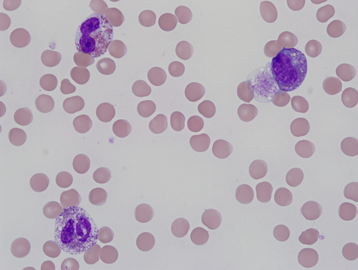 |
| BM biopsy 1 direct smear 100x | 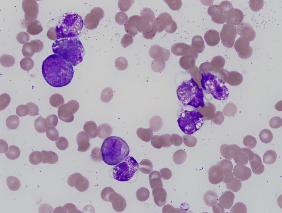 |
| BM biopsy 1 touch preparation 100x | 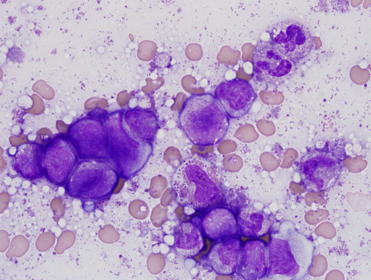 |
| BM biopsy 1 core 50x | 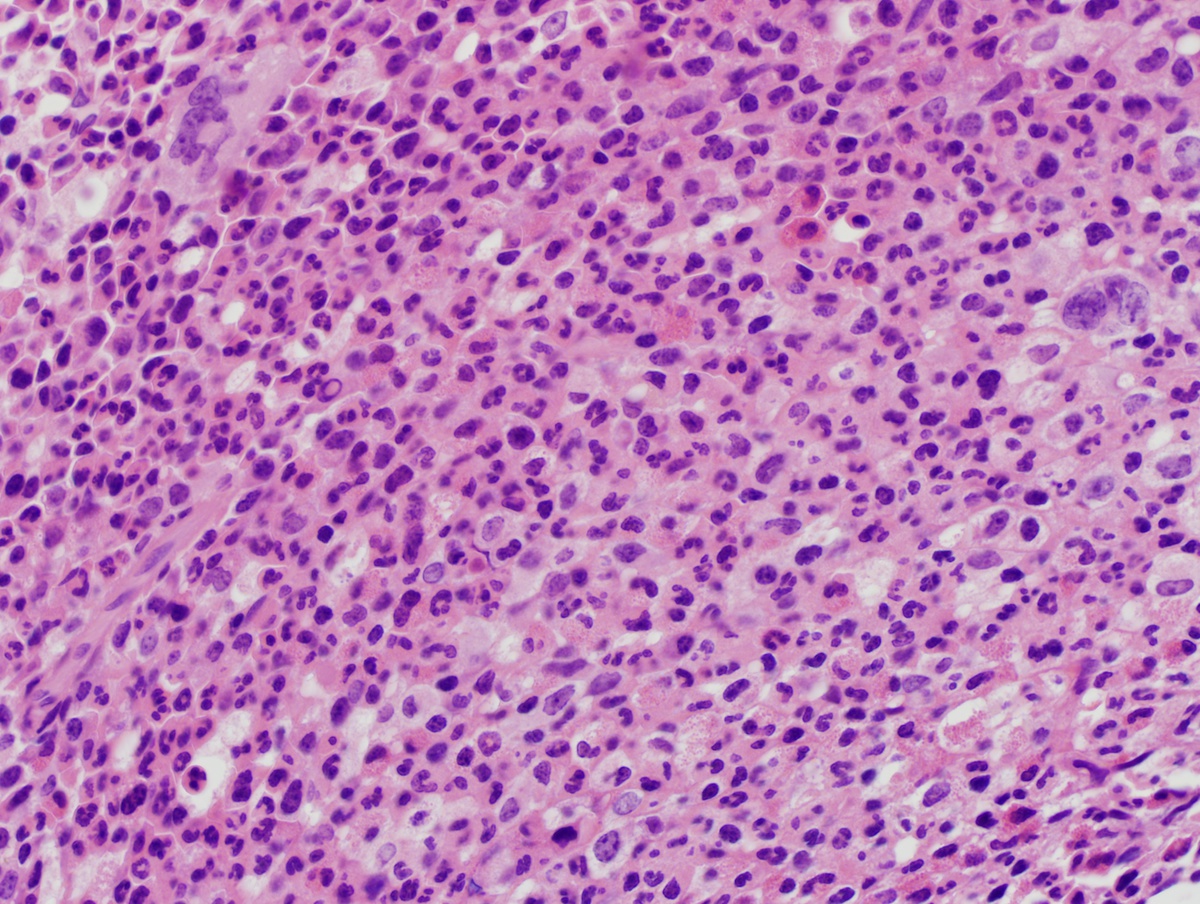 |
| BM biopsy 2 peripheral blood 20x | 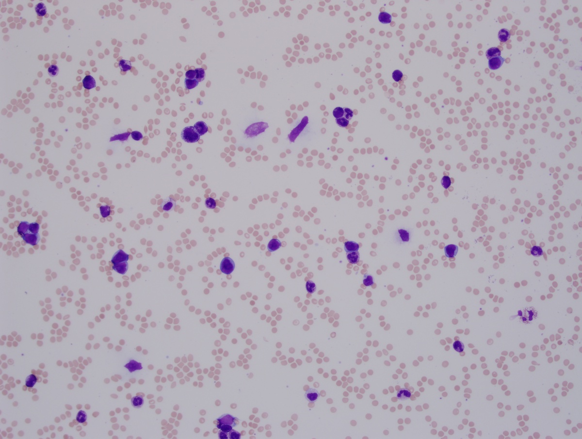 |
| BM biopsy 2 direct 100x | 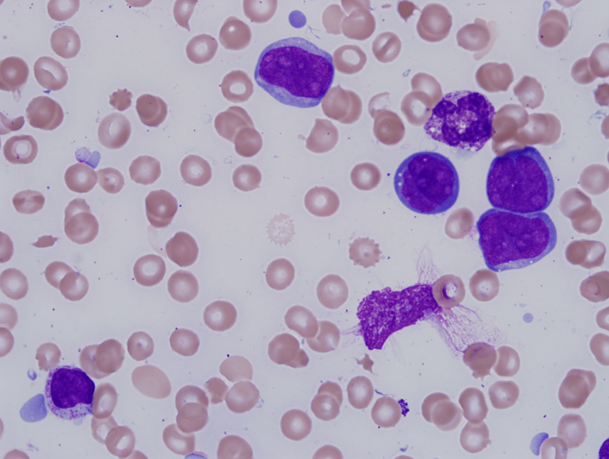 |
| BM biopsy 2 peripheral blood vacuoles 100x | 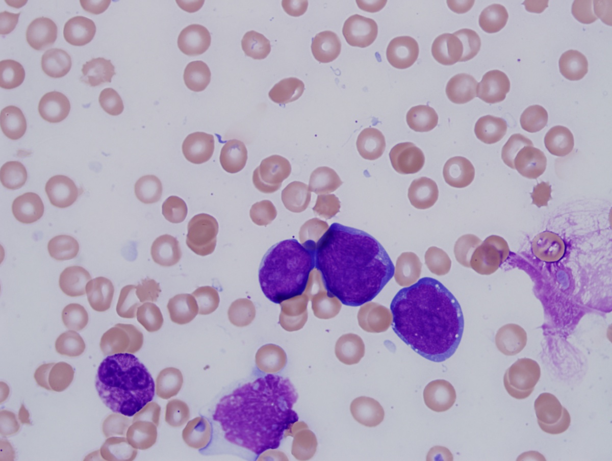 |
| BM biopsy 2 core 10x | 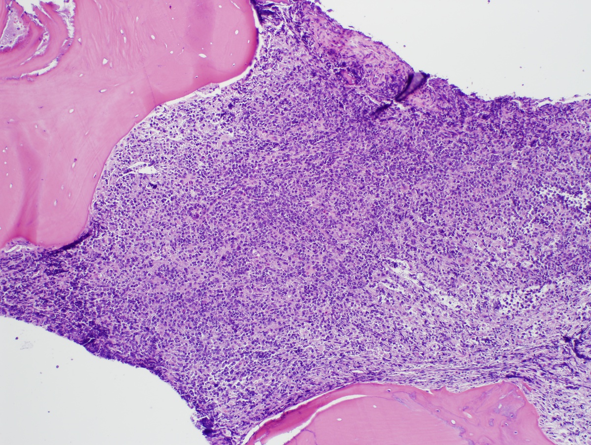 |
| BM biopsy 2 core 100x | 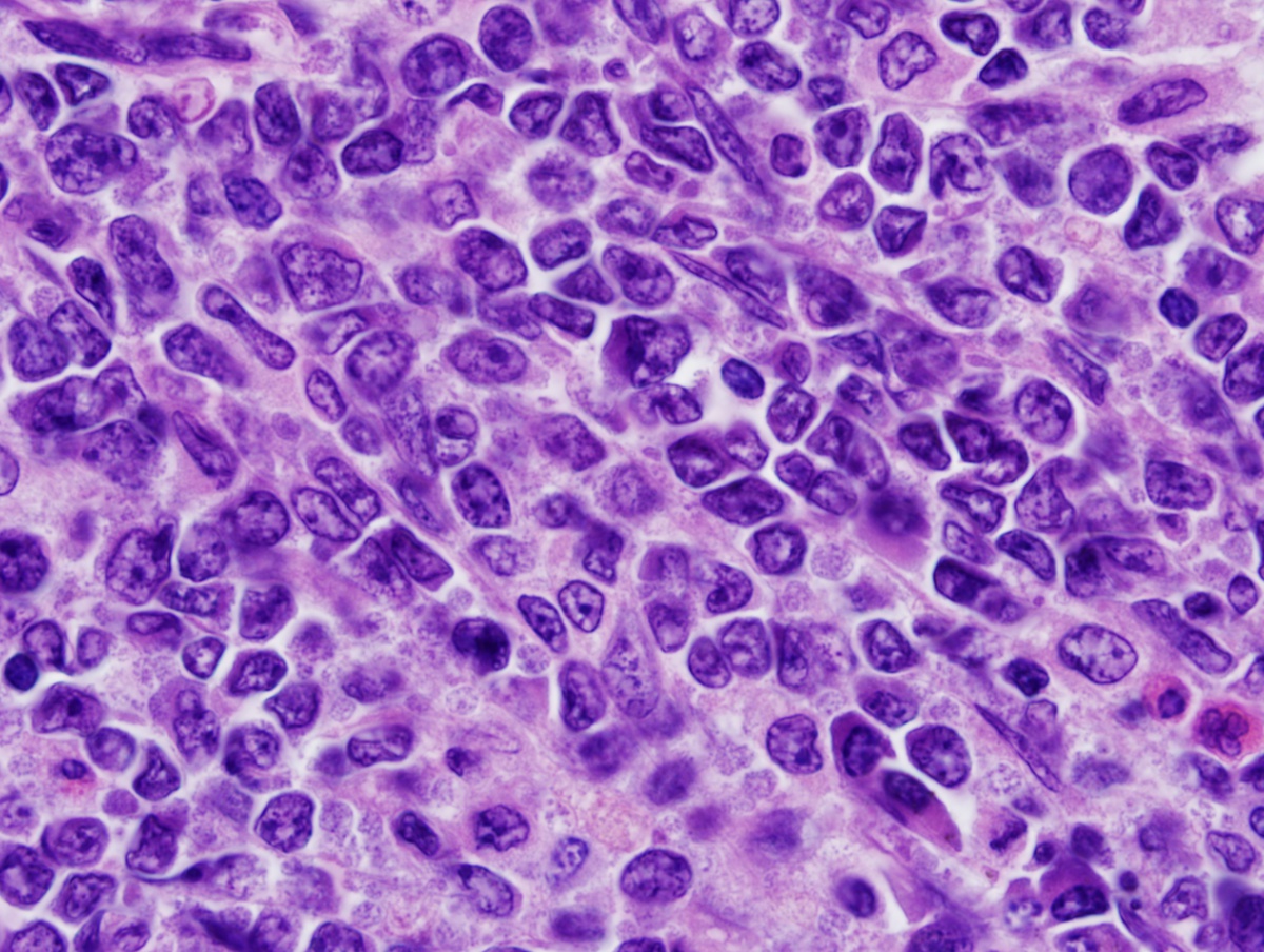 |
| BM biopsy 2 clot TdT 100x | 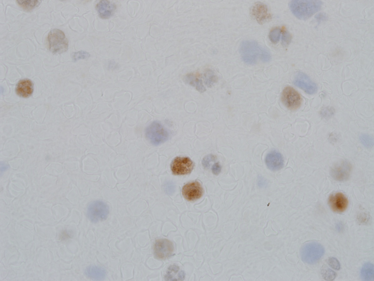 |
| Flow cytometry CD19 and CD10 (blasts in red) | 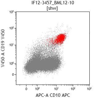 |
| Flow cytometry CD19 and CD20 (blasts in red) | 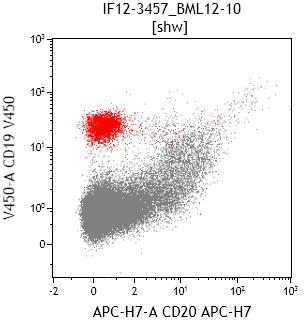 |
| Flow cytometry CD19 and CD34 (blasts in red) | 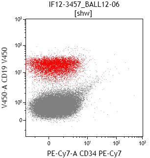 |
| Flow cytometry CD45 versus SSC | 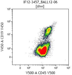 |