Institution: Pathology Department Hopital Necker-Enfants Malades, 149 rue de Sevres, 75015, PARIS France
Additional authors:Nicole Brousse,Thierry Molina, Vahid Asnafi
Session: T Lymphoblastic Leukemia/Lymphoma
HISTORY
Boy, 10 years old
One cervical lymph node appearing in August 2012 which decreased with anti-inflammatory treatment. Two months after, other cervical lymphadenopathies appeared associated with a mediastinal mass.
No systemic signs.
LDH= 220
DETAILS
Cervical biopsy of a lymph node (1.5cms). Formalin fixed tissue. Lymph node architecture partially preserved. Massive infiltration by pleiomorphic cells showing a mixture of medium cells and large cells sometimes very large. The nuclei were round or irregular. Mitosis and apoptosis were frequent. Myelogam showed rare abnormal cells (2%)
IMMUNOHISTOCHEMISTRY AND FLOW CYTOMETRY
The immunohistochemistry revealed that the abnormal cells were strongly positive for CD45, CD34, CD56, CD99 and Ki67 (90%+). They were negative for CD3, CD2, TdT, CD10, ALK, EMA, CD30, CD4, CD8, CD20 and CD79a.
The flow cytometry showed that the abnormal cells expressed CD45dim, CD34, CD56, CD33 with a weak expression of CD7 & CD3 and a partial expression of TdT and CD13. They were negative for CD1a, Cd2, CD5, CD4, CD8, CD19, CD20, CD10 & cMPO
CYTOGENETIC FINDINGS
Not done
MOLECULAR FINDINGS
Molecular analysis showed no detection of clonal rearrangement of IgH type DJ and no clonal rearrangement of TCR delta, gamma et beta in the bone marrow specimen and in the lymph node specimen.
INTERESTING FEATURES
Atypical morphology of the abnormal cells which were pleiomorphic with very large cells and aypical immunostaining (no usual markers of T-cell lymphoblastic lymphoma/eukemia and aberrant markers such as myeloid markers and CD56 staining).
PROPOSED DIAGNOSIS
Very immature atypical T-cell lymphoblastic lymphoma/leukemia
CONSENSUS GROUP: ADDITIONAL INFORMATION/STUDIES
Additional follow up per e-mail communication: The child had no bone marrow biopsy but had 2 myelograms: on the 1st one he had <1% of abnormal lymphoid cells and on the second <3 % of abnormal lymphoid cells. He had no lymphoblastic cells in his cerebrospinal fluid which was normal. He was treated following the protocol FRALLE 2000 of the lymphoblastic lymphomas in children.
CONSENSUS DIAGNOSIS
T-acute lymphoblastic leukemia/lymphoma, early T-cell precursor immunophenotype
| Cytologyx400 |  |
| Histology x400 | 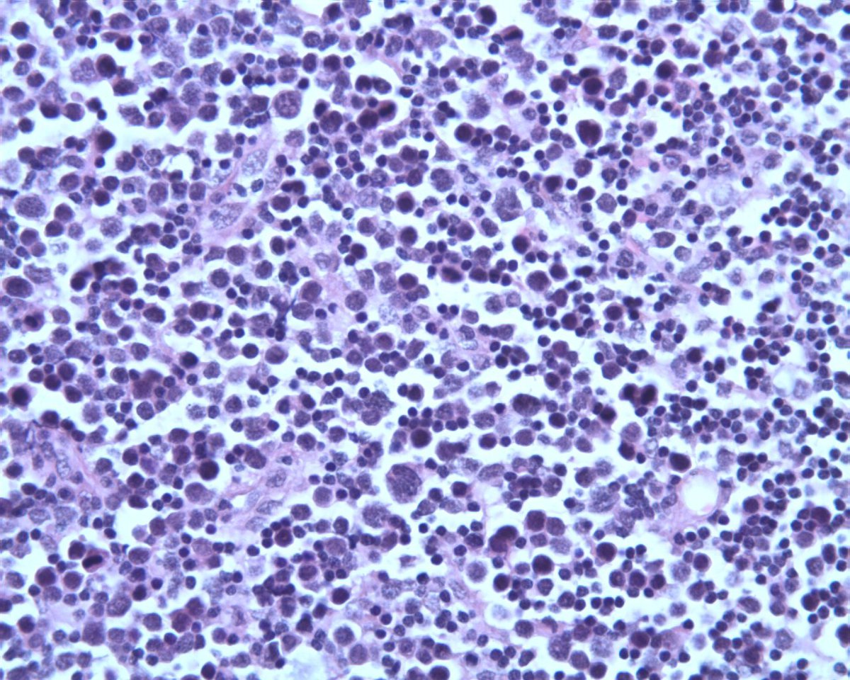 |
| Cd56 staining |  |
| CD34 staining |  |
| TdT negativex400 | 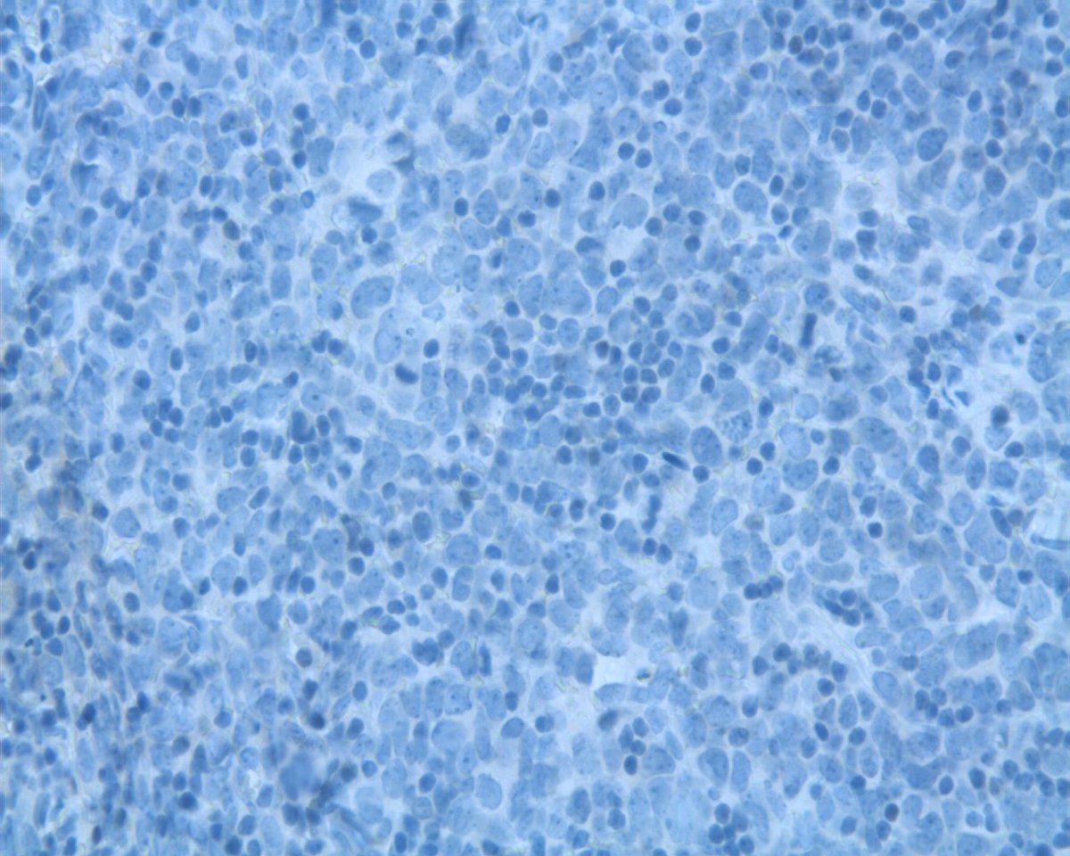 |
| Additional slide 1 - Lymph node flow cytometry |  |
| Additional slide 2 - T Lymphoblastic population cCD3+, CD3-, TdT+, CD34+, CD117+, CD2/7/5+ weak, CD34+, CD4+, CD8-, CD33+, CD56+ representing 20% of the cells of the lymph node analyzed |  |