Institution: Henry Ford Hospital
Additional authors:Kristin Hunt Karner M.D., Kedar V. Inamdar M.D. Ph.D.
Session: AML with recurrent genetic abnormalities Part I
HISTORY
The patient is a 25-year-old African American female with history of fever and neck pain for which she came to the emergency department. She was found to have a peritonsillar abscess, and also a WBC of 39.6 K/uL with 89% blasts in the peripheral blood, hemoglobin of 10.6 g/dl and a platelet count of 108 K/uL. A bone marrow biopsy done on the same day showed acute myeloid leukemia with t(8;21)(q22;q22); RUNX1-RUNX1T1 with KIT mutation. She was started on induction chemotherapy (Cytarabine with Idarubicin) four days later and finished her course in seven days. A few days later she developed a fever and was started on broad-spectrum antibiotics. Cultures performed were negative. On day 22 of induction chemotherapy, the bone marrow biopsy showed up to 22% blasts but FISH studies for t(8:21) were negative. A persistent disease versus regeneration was considered in the differential diagnosis.
A bone marrow biopsy was performed one week later and now showed 5% blasts. FISH studies for the t(8;21) were again negative, while RT-PCR showed a minimal level of positivity. The case was reviewed again with tumor board and it was recommended to proceed with consolidation chemotherapy. The transplant team was consulted but no complete match was found so the decision was made to complete four cycles of consolidation chemotherapy The patient completed four cycles of HiDAC along with supportive treatment of Neupogen. Four weeks after completion of chemotherapy a bone marrow biopsy was performed which revealed morphologic remission with both FISH cytogenetics for the t(8;21) and a qualitative PCR assay for KIT Asp816Val mutation being negative.DETAILS
The first biopsy was 1.2 cm long and fixed in Bouin's fluid along with 2.5 cm of fragmented clot section. The initial biopsy showed cellularity of 90% with a blast count of 76%. The blasts showed fine chromatin with sometimes delicately folded nuclei and occasionally prominent nucleoli. They also had pale blue cytoplasm with granules and occasional Auer rods.
On day 22 status post induction chemotherapy, the 1.3 cm long core biopsy with 3.3 cm clot section fixed in Bouin's fluid showed 22% blasts. The blasts showed morphology similar to the initial bone marrow biopsy but occasional blasts exhibited blebbing of cytoplasmic membrane. The overall cellularity was 40-50% with an M:E ratio of 42:1. The cellular elements in the core biopsy consisted of infiltrates of immature mononuclear cells as predominant cell population amidst decreased trilineage hematopoietic lineage cells and minor populations of plasma cells and lymphocytes. There was a left shift in the myeloid maturation and erythroid lineage cells were decreased. Megakaryocytes were present and mostly unremarkable but there were occasional atypical megakaryocytes with nuclear hypolobation. Immunostains on the biopsy and clot did not reveal a significant population of CD34+/CD117+ blasts and no significant positivity for CD56 was seen on the blasts. On the bone marrow aspirate smears the myeloid series was left shifted with increase in blasts. Auer rods were not obvious. The blasts had fine chromatin with sometimes delicately folded nuclei and occasionally a prominent nucleolus. They had pale blue cytoplasm with granules. Occasional blasts exhibited blebbing of cytoplasmic membrane. Some of the maturing myeloid cells exhibited dyspoiesis with nuclear cytoplasmic dyssynchrony and cytoplasmic hypogranularity while others exhibit cytoplasmic hypergranularity. Megakaryocytes were adequately represented with mostly unremarkable morphology but there were a few small and hypolobated forms noted. Erythropoiesis was markedly decreased. The day 30 marrow showed a hypercellular bone marrow (80-90% cellularity and M:E ratio of 1.6:1) with no definite evidence of residual leukemia. CD34 stains were negative and smears revealed slightly increased blasts (4.5%). The blasts were significantly decreased in comparison to the previous biopsy. Although minimal residual leukemia was included in the differential diagnosis, findings were suggestive of regeneration. After completion of consolidation chemotherapy (five months later) a bone marrow biopsy was performed which revealed 3% blasts in a patchy 50% cellular bone marrow with an M:E ratio of 1.4:1. The final diagnosis was a normocellular to mildly hypocellular marrow with trilineage hematopoiesis, no dysplastic changes and consistent with morphologic remission.IMMUNOHISTOCHEMISTRY AND FLOW CYTOMETRY
Initial Flow:
The whole sample revealed a predominant blast cluster (57% of the total flow sample) with a composite antigen profile of: CD13+, CD33+, CD34+, CD45+ (dim), CD56+, CD117+, HLA-DR+ & cyto. MPO+.Second flow:The second flow was performed on the marrow from Day 22 and showed 8% myeloblasts but this time we did not detect clearly aberrant CD56 expression.CYTOGENETIC FINDINGS
On the initial marrow, FISH detected t(8;21) gene rearrangement in 96.5% of interphase cells and a karyotype revealed 46,XX,t(8;21)(q22;q22)[19]/46,XX[2]. Cytogenetic tests performed on the marrow at day 22, day 30 and after the full dose of chemotherapy showed normal FISH and normal karyotype results.
MOLECULAR FINDINGS
A peripheral blood sample was tested for KIT Asp816Val and N822K mutation analysis (Qualitative PCR) and the results were reported as positive. A bone marrow aspirate sample was sent five months after the intial diagnosis for KIT Asp816Val gene mutation analysis and was reported as negative.
The day 30 bone marrow was sent for detection of t(8;21) by quantitative RT-PCR. The RNA quality was reported as good and an AML1/ETO t(8;21) fusion transcript was detected. The ratio of the fusion gene transcripts to control gene transcripts was 0.0019.INTERESTING FEATURES
The marrow specimen on Day 22 demonstrated persistence of blasts that represented up to 22% of cellularity. While this number of blasts in the setting of recently diagnosed AML would morphologically favor a persistent/residual leukemia, interestingly fluorescence in situ hybridization analysis for t(8;21) was negative. Thus an unusual, brisk regenerative response was considered in the differential diagnoses. After correlation with conventional cytogenetics and clinical presentation a final interpretation of regenerating marrow was favored. The latest bone marrow biopsies done five months after initial diagnosis showed morphologic remission with normal cytogenetics and negative for t(8;21) and the KIT mutation.
PROPOSED DIAGNOSIS
Acute myeloid leukemia with t(8;21)(q22;q22);RUNX1-RUNX1T1 with KIT mutation, at day 22 with 22% blasts consistent with regenerating marrow and negative for evidence of residual disease.
CONSENSUS DIAGNOSIS
Acute myeloid leukemia with t(8;21)(q22;q22);RUNX1-RUNX1T1 with KIT mutation
(at day 22 with 22% blasts consistent with regenerating marrow and negative for evidence of residual disease)| Initial core biopsy | 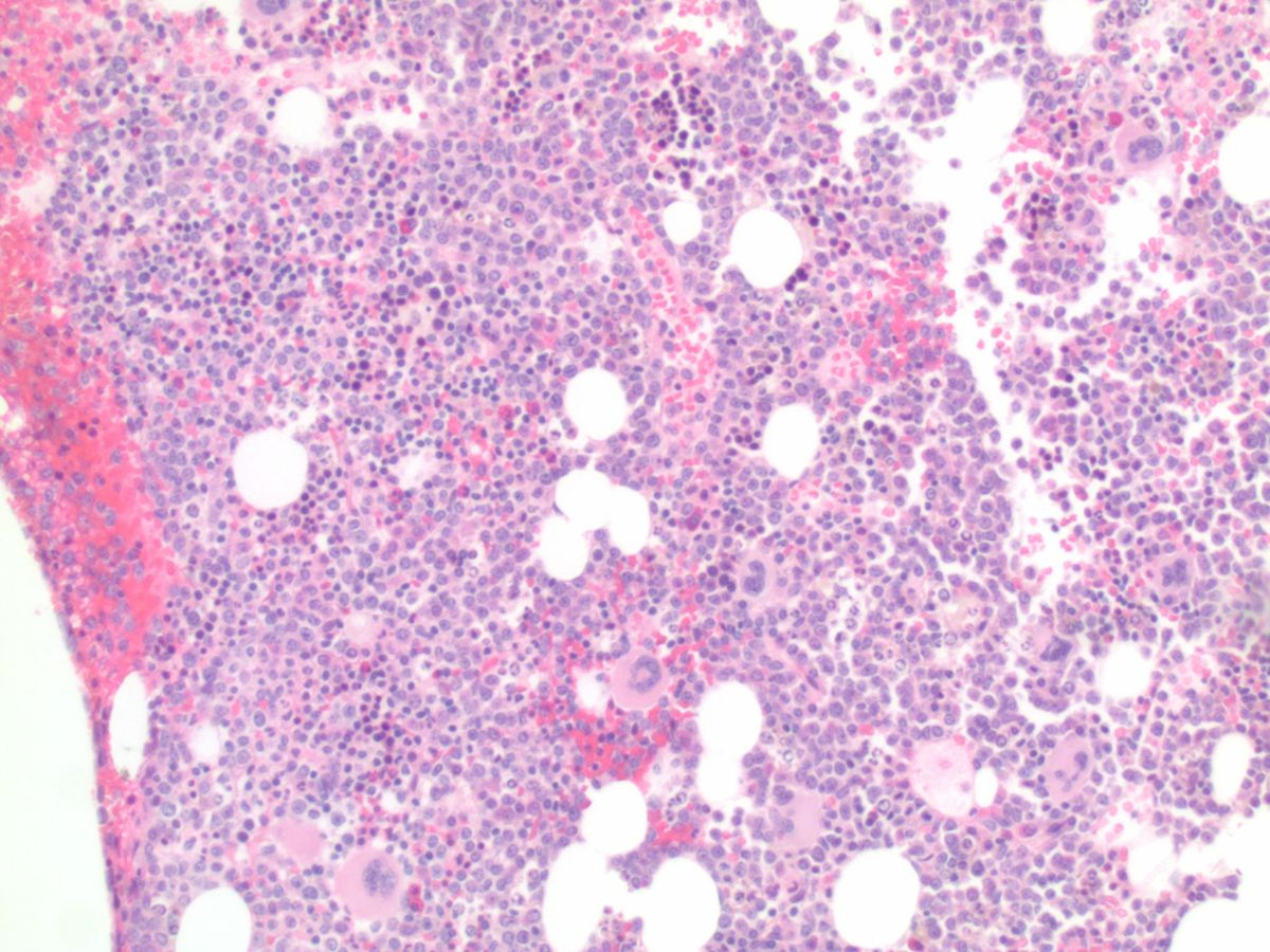 |
| Initial bone marrow aspirate |  |
| Initial bone marrow aspirate #2 |  |
| Day 22 bone marrow core biopsy low power | 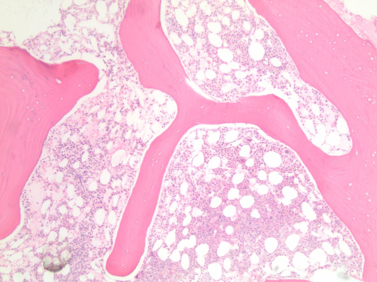 |
| Day 22 bone marrow biopsy high power | 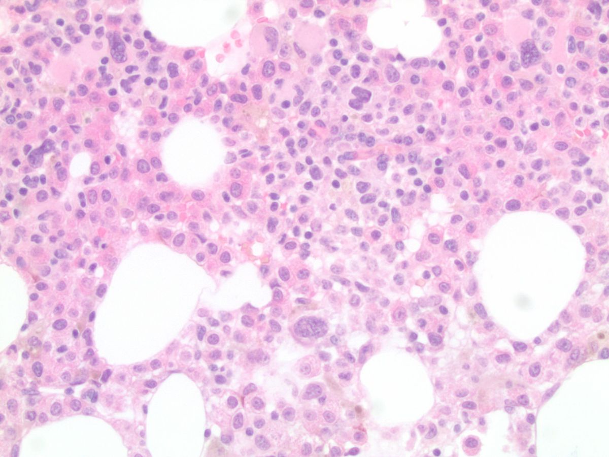 |
| Day 22 bone marrow aspirate | 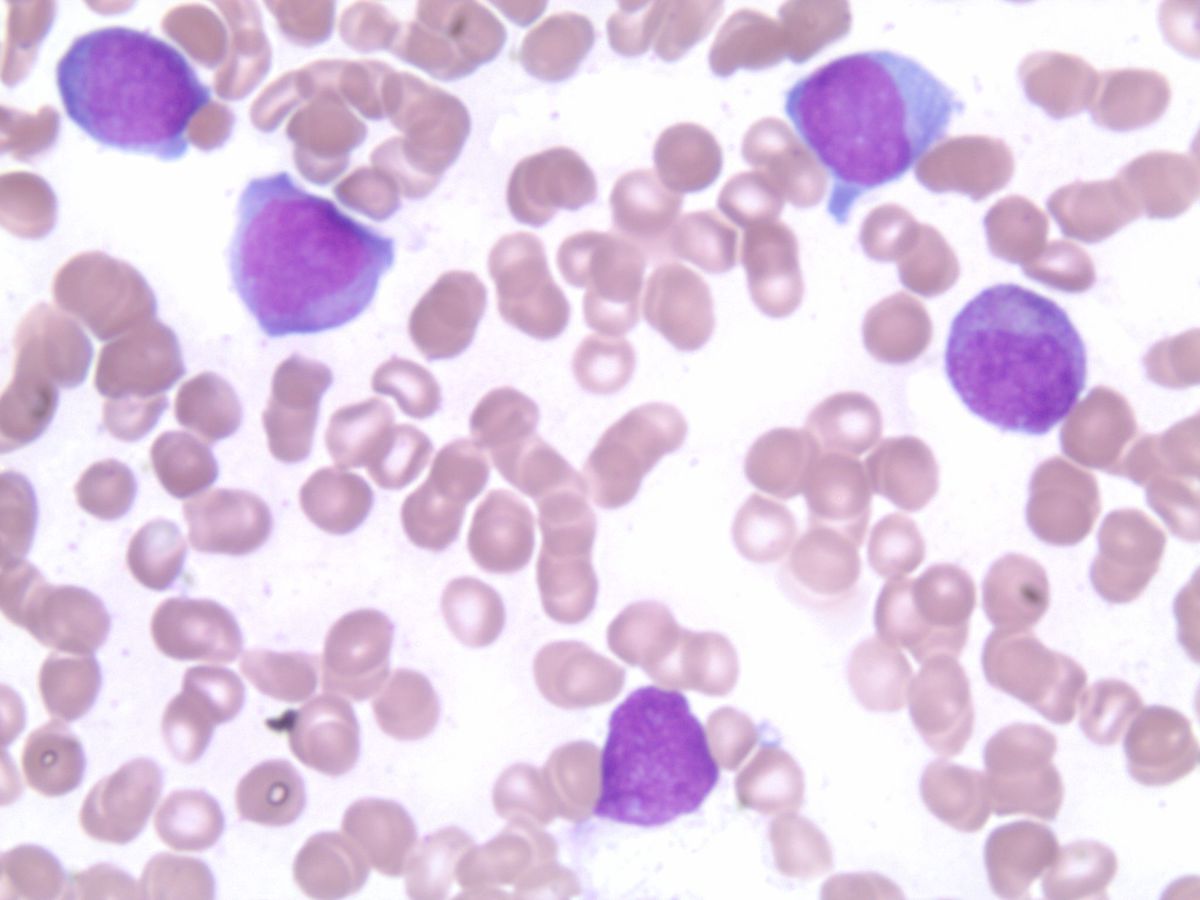 |
| Day 22 bone marrow aspirate #2 | 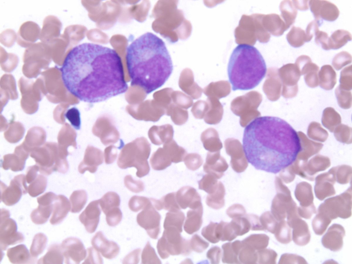 |
| t(8;21) positive; initial bone marrow biopsy |  |
| t(8;21) negative; Day 22 bone marrow |  |